4
Scalp and Face
The scalp and face are two interconnected regions on the superior, lateral, and anterior surfaces of the skull. The strong, layered structure of the scalp, which includes hair-bearing skin, helps protect the skull and brain.
The face is positioned on the anterior surface of the skull. It contains openings for sight, smell, respiration, and intake of nutrients through the orbits, nose, and mouth. Small changes in the muscles of facial expression convey different emotions and expressions.
The scalp is supported by the bones of the neurocranium (see Chapter 3), and the face is supported by some of the smaller, more complex bones of the viscerocranium (see Chapter 3).
Scalp
The scalp extends from the supra-orbital margin of the frontal bone (superciliary arch) on the anterior aspect of the skull (see Chapter 3) to the superior nuchal line on the posterior aspect of the skull (see Chapter 27). Laterally, it extends to the level of the zygomatic arches. The five layers of the scalp can be remembered by the acronym SCALP:
- Skin—containing the hair follicles, sebaceous glands, and sweat glands.
- Connective tissue—a layer of strong collagen fibers mixed with small amounts of fatty tissue and containing blood vessels and superficial nerves.
- Aponeurosis—a thick sheet of collagen fibers that extends between the frontalis and occipitalis muscles. These two muscles are responsible for the voluntary ability to slide the scalp back and forth across the skull and to wrinkle the forehead.
- Loose connective tissue (“danger zone”)—a layer of collagen fibers mixed with large amounts of fatty tissue. It contains the emissary veins, which are special valveless veins that transport blood from within the skull to the veins of the scalp, thus providing some of the venous drainage for the brain and potentially allowing spread of infection.
- Pericranium (periosteum)—a richly innervated covering composed of dense, interweaving collagen fibers. It is loosely attached to the surface of the skull, except at the suture lines, where it passes between the skull bones and contributes to their joints. The periosteum is continuous with the periosteal layer of the dura mater within the skull.
- Connective tissue—a layer of strong collagen fibers mixed with small amounts of fatty tissue and containing blood vessels and superficial nerves.
NERVES
Motor innervation to the scalp muscles is provided by branches of the facial nerve [VII], which emerges from the stylomastoid foramen. At the level of the ear, the sensory innervation to the scalp divides into anterior cutaneous innervation and posterior cutaneous innervation (Fig. 4.1). Anterior to the ear, the scalp is innervated mostly by branches of the divisions of the trigeminal nerve [V]:
- supratrochlear and supra-orbital nerves (from the ophthalmic nerve [V1])
- zygomaticotemporal nerve (from the maxillary nerve [V2])
- auriculotemporal nerve (from the mandibular nerve [V3])
- zygomaticotemporal nerve (from the maxillary nerve [V2])
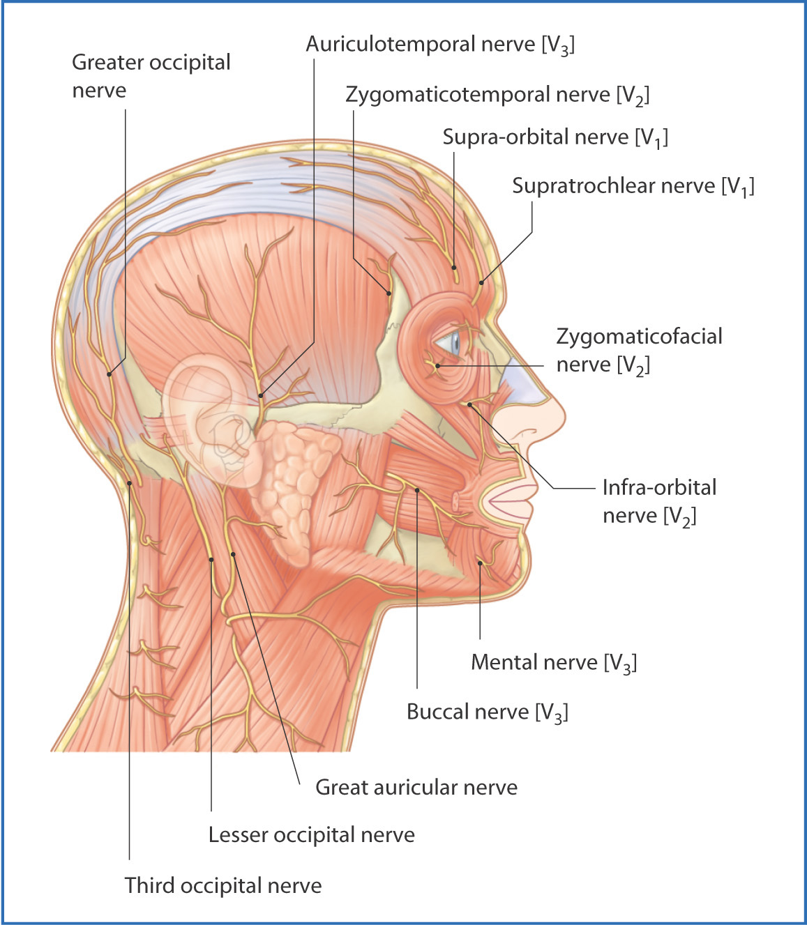
FIGURE 4.1 Nerves of the scalp and face (lateral view).
Posterior to the ear, the scalp receives cutaneous innervation from the spinal cutaneous nerves that originate in the neck (C2, C3):
ARTERIES
Blood is supplied to the scalp by four small arteries—the supratrochlear and supra-orbital arteries, which are branches of the ophthalmic artery (itself a branch of the internal carotid artery), and the superficial temporal artery and occipital arteries, branches of the external carotid artery.
VEINS AND LYMPHATICS
Venous drainage is along the venae comitantes of the arteries:
- The supra-orbital and supratrochlear veins unite at the medial canthus of the eye to form the facial vein.
- The superficial temporal vein joins the maxillary vein to create the retromandibular vein just posterior to the neck of the mandible.
- The posterior auricular vein originates behind the ear and channels venous blood from the posterior of the scalp toward the external jugular vein.
- The superficial temporal vein joins the maxillary vein to create the retromandibular vein just posterior to the neck of the mandible.
Lymph drains from the scalp to the superficial horizontal ring of superficial lymph nodes at the junction between the head and neck. Some lymph also drains directly to the deep cervical lymph nodes (see Chapter 12).
Face
MUSCLES AND NERVES
The face extends laterally from ear to ear and from the chin to the hairline on the forehead. The skin of the face is thick and vascular. Beneath the skin is the subcutaneous fascia, which contains the muscles of facial expression, blood vessels, and nerves (Fig. 4.2). The face contains the organs of sight—the eyes—and the proximal portions of the respiratory and digestive systems—the nose and mouth, respectively.
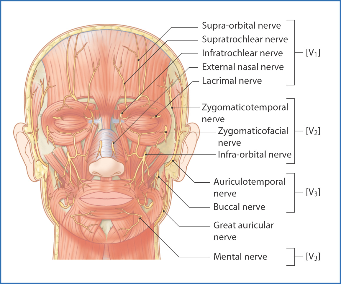
FIGURE 4.2 Sensory innervation of the face: trigeminal nerve [V].
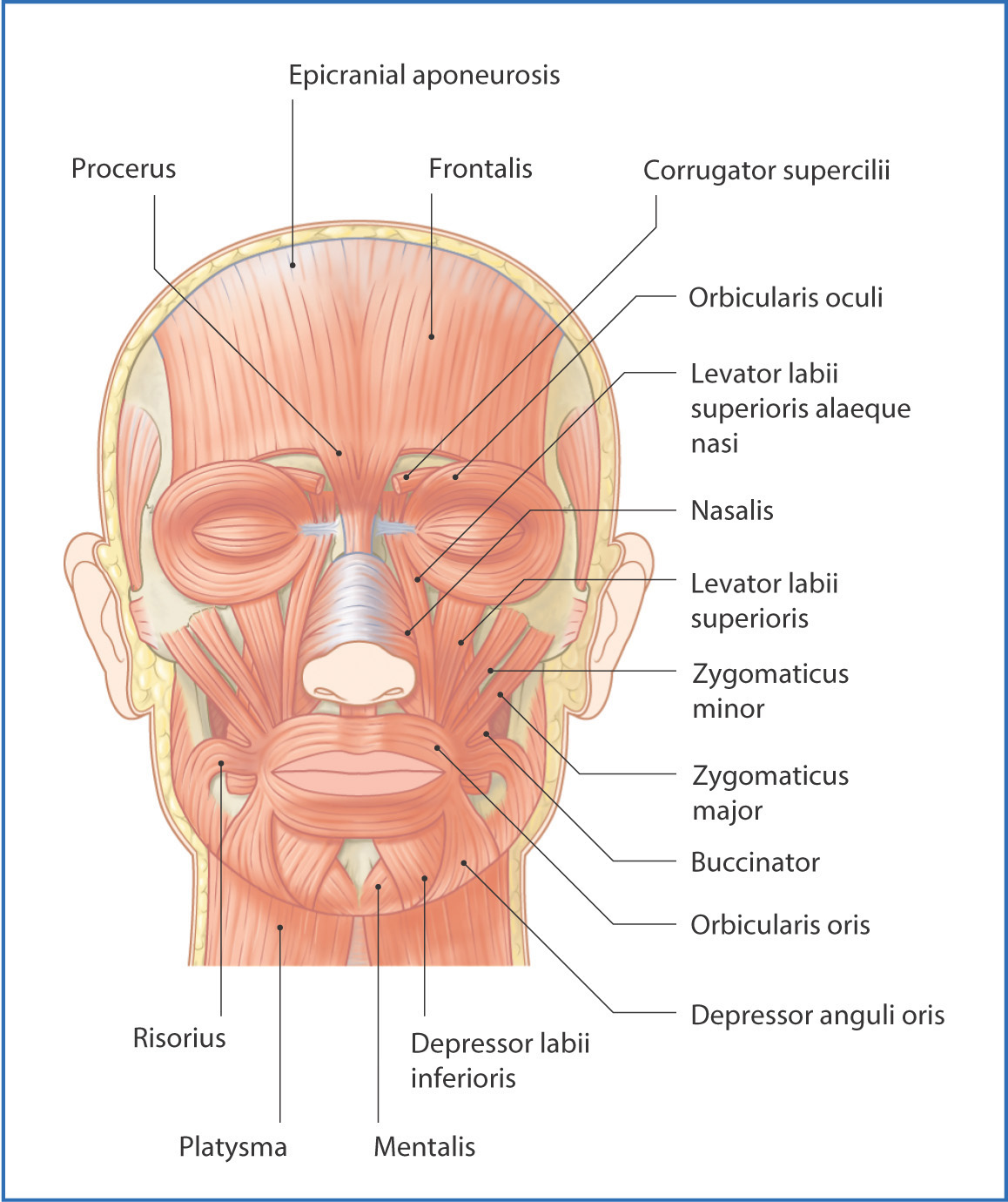
The muscles of the face insert into the skin (Fig. 4.3), which allows them to move the skin of the face in complex ways. The facial nerve [VII] innervates the muscles of facial expression (Fig. 4.4). It has five main branches. From superior to inferior these branches are the
Stay updated, free articles. Join our Telegram channel

Full access? Get Clinical Tree


