• No association with tuberous sclerosis (unlike cardiac type)
• Some fetal rhabdomyomas associated with nevoid basal cell carcinoma syndrome (Gorlin syndrome)
Clinical Issues
• Fetal type: Usually infants and children
• Adult and genital types: Most common in adults
• Genital type shows strong female predilection
• Adult and fetal types most common in head and neck region
• Genital lesions mostly in vagina, occasionally vulva or cervix
• Excellent prognosis after complete excision
Macroscopic
• Median: ~ 3 cm
Microscopic
• No significant atypia or necrosis; mitoses usually absent
• Fetal rhabdomyoma
Classic (immature) type: Bland spindle cells in abundant myxoid stroma
Intermediate (juvenile) type: Increased cellularity with more skeletal muscle differentiation
• Adult rhabdomyoma
Large polygonal cells with eosinophilic cytoplasm
Intracytoplasmic cross striations or rod-like inclusions
• Genital rhabdomyoma
Hypocellular proliferation of bland spindle cells and mature rhabdomyoblastic elements
Lacks cellular, subepithelial cambium layer
Ancillary Tests
• Desmin (+), myogenin (+), MYOD1(+)
Top Differential Diagnoses
• Carcinoma or melanoma
• Granular cell tumor
• Hibernoma
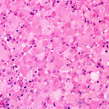
Adult Rhabdomyoma High magnification shows adult rhabdomyoma composed of large polygonal cells with copious eosinophilic cytoplasm (varying in staining intensity) and small round nuclei with uniform nucleoli.
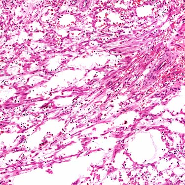
Fetal Rhabdomyoma Intermediate magnification shows fetal rhabdomyoma of classic (immature) type. Slender spindle cells form loosely organized fascicles in myxoid stroma. Note the absence of pleomorphism and necrosis.
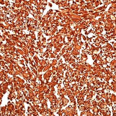
Desmin Expression in Rhabdomyoma Desmin stain in an adult rhabdomyoma shows strong, diffuse positivity throughout the lesion. This is a typical finding in rhabdomyoma and can also highlight cross striations.
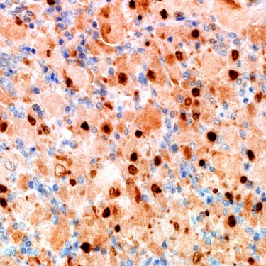
Myogenin Expression in Rhabdomyoma Positive myogenin in an adult rhabdomyoma shows immunoreactivity in the nuclei of many of the lesional cells. This is diagnostic of skeletal muscle differentiation. Cytoplasmic staining is sometimes seen, but it is nonspecific. MYOD1 is also useful, although less sensitive.
TERMINOLOGY
Definitions
• Benign tumor with skeletal muscle differentiation
• Extracardiac rhabdomyoma includes adult, fetal, and genital types
ETIOLOGY/PATHOGENESIS
Developmental Anomaly
• No association with tuberous sclerosis (unlike cardiac type)
• Some fetal rhabdomyomas associated with nevoid basal cell carcinoma syndrome (Gorlin syndrome)
PTCH1 gene mutations
CLINICAL ISSUES
Epidemiology
• Incidence
Rare
• Age
Fetal type
– Infants and children (particularly in 1st year of life)
Adult and genital types
– Most common in adults (mean: 50 years)
• Sex
Adult and fetal types: Male predilection
Genital type: Strong female predilection
Site
• Rarely cutaneous lesion
• Adult and fetal types are most common in head and neck region
Larynx, oropharynx, mouth, neck
• Genital lesions are subepithelial
Mostly in vagina, occasionally in vulva or cervix
Rarely in paratesticular region or epididymis
Presentation
• Slowly growing, painless mass
• Often solitary
Only gold members can continue reading. Log In or Register to continue