TABLE 52.1
Cells of the Immune System
| Cell Type | Synonyms | Primary Immune-Related Actions |
| MAJOR CELL TYPES | ||
| B lymphocytes | B cells | |
| CTLs | Cytolytic T cells, cytotoxic T cells, CD8 cells | |
| Helper T lymphocytes | Helper T cells, CD4 cells | |
| Macrophages | ||
| Dendritic cells | ||
| ACCESSORY CELLS | ||
| Mast cells | ||
| Basophils | ||
| Neutrophils | Polymorphonuclear leukocytes | |
| Eosinophils | ||
B Lymphocytes (B Cells)
B lymphocytes have the job of making antibodies. Hence, B cells mediate humoral immunity. As discussed later, antibody specificity is determined by the structure of highly specific receptors found on the surface of B cells. Like all other lymphocytes, B cells circulate in the blood and lymph. B cells are so named because in chickens, where B cells were discovered, these cells are produced in the bursa of Fabricius, a structure not found in mammals. In humans and other mammals, B cells are produced in the bone marrow.
Cytolytic T Lymphocytes (Cytolytic T Cells, CD8 Cells)
Cytolytic T cells are key players in cellular immunity. These cells do not produce antibodies. Rather, they attack and kill target cells directly. Specificity of attack is determined by the presence of antigen molecules on the surface of the target cell and specific receptors for that antigen on the surface of the T cell. Cytolytic T cells are also known as CD8 cells and cytotoxic T cells. The designation “CD8” refers to the presence of cell-surface marker molecules known as cell differentiation complex 8. The “T” in T cell stands for thymus, the organ in which cytolytic T cells and helper T cells mature. Like B cells, cytolytic T cells circulate in the blood and lymph.
Helper T Lymphocytes (Helper T Cells, CD4 Cells)
Helper T cells contribute to the immune response in three ways: (1) they have an essential role in antibody production by B cells, (2) they release factors that promote type IV sensitivity reactions, also known as delayed-type hypersensitivity (DTH), and (3) they participate in the activation of cytolytic T cells. Specificity of helper T cells is achieved through highly specific cell-surface receptors that recognize individual antigens. Like other lymphocytes, helper T cells circulate in the blood and lymph. Helper T cells carry CD4 (cell differentiation complex 4) marker molecules on their surface and hence are referred to as CD4 cells.
The term helper is somewhat misleading in that it connotes a useful but dispensable role. Nothing could be further from reality. Helper T cells are not simply nice to have around, they are absolutely required for an effective immune response. The critical nature of their contribution—and the grim consequences of their absence—are manifested in people with HIV/AIDS: helper T cells are the immune cells that HIV attacks. Because of helper T-cell loss, AIDS patients are at high risk for death from opportunistic infection.
Macrophages
Macrophages begin their existence in the bone marrow, enter the blood as monocytes, and then infiltrate tissues, where they evolve into macrophages. Macrophages are present in all organs and tissues.
The primary function of macrophages is phagocytosis (i.e., ingestion of microbes, other foreign material, and cellular debris). In their role as phagocytes, macrophages are the principal scavengers of the body. Although their major job is phagocytosis, macrophages also have an important role in specific acquired immunity, natural immunity, and inflammation.
In specific acquired immunity, macrophages have three functions: (1) they are required for activation of T cells (both helper T cells and cytolytic T cells), (2) they are the final mediators of DTH, and (3) they phagocytize cells that have been tagged with antibodies. Of these three immune-related roles, activation of T cells is arguably the most critical. When performing this function, macrophages are referred to as antigen-presenting cells (APCs). Because antigen presentation is an absolute requirement for specific immune responses (see later), you can appreciate how important macrophages are.
Dendritic Cells
Dendritic cells perform the same antigen-presenting task as do macrophages. However, unlike macrophages, dendritic cells do not also serve as scavengers. Dendritic cells are found in lymph nodes and other lymphoid tissues.
Mast Cells and Basophils
These cells mediate immediate hypersensitivity reactions. Mast cells, which are derived from basophils, are concentrated in the skin and other soft tissues. Basophils circulate in the blood. Both cell types release histamine, heparin, and other compounds that cause the symptoms of immediate hypersensitivity. Release of these mediators is triggered when an antigen binds to antibodies on the cell surface. The role of mast cells and basophils in allergic reactions is discussed in Chapter 61.
Neutrophils
Neutrophils, also known as polymorphonuclear leukocytes, phagocytize bacteria and other foreign particles. As discussed later, neutrophils avidly devour cells that have been tagged with antibodies of the immunoglobulin G (IgG) class. Accordingly, neutrophils can be viewed as important effectors in humoral immunity. Neutrophils are also major contributors to inflammation.
Eosinophils
Eosinophils attack and destroy foreign particles that have been coated with antibodies of the IgE class. Their usual target is helminths (parasitic worms). Eosinophils also contribute to tissue injury and inflammation associated with immediate hypersensitivity reactions.
Antibodies
Antibodies are a family of structurally related glycoproteins that mediate humoral immunity. The most characteristic feature of antibodies is their ability to recognize and bind with specific antigens. Alternative names for antibodies are immunoglobulins and gamma globulins.
All antibodies are produced by B lymphocytes. Some of the antibodies that B cells produce are retained on the surface of the B cell, where they serve as the receptors whereby B cells recognize specific antigens. However, most of the antibodies that B cells produce are secreted from the cell, after which they bind to their specific antigen, thereby initiating the effector phase of humoral immunity. The process of antibody production is discussed in detail later.
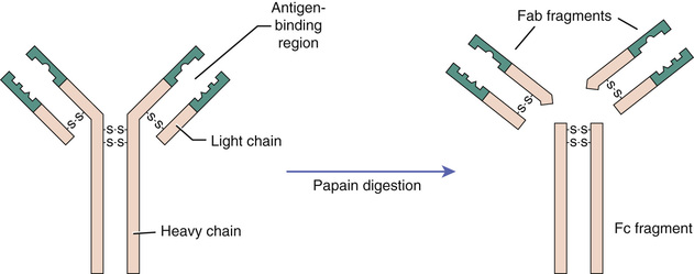
All antibodies are composed of units that have the same basic structure. As shown in Fig. 52.2, antibodies have four chains: two heavy chains and two light chains. Disulfide bridges connect the four chains to form a unit. Each heavy chain and each light chain has two regions, one in which the sequence of amino acids is constant and one in which the sequence is highly variable. The variable regions form the antigen-binding site.
There are five classes of antibodies (immunoglobulins), known as IgA, IgD, IgE, IgG, and IgM. All are constructed from the same basic parts just described. However, the heavy chains differ for each class. Primary functions of the five classes are shown in Table 52.2.
TABLE 52.2
Functions of Antibody Classes
When antibodies are subjected to digestion by papain in the laboratory, they break down into three pieces (see Fig. 52.2). Two of the pieces retain the ability to bind antigen and hence are called Fab fragments (fragment, antigen binding). The third piece does not bind antigen and tends to form crystals in the test tube and hence is called the Fc fragment (fragment, crystalline).
Stay updated, free articles. Join our Telegram channel

Full access? Get Clinical Tree


