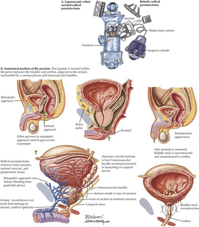
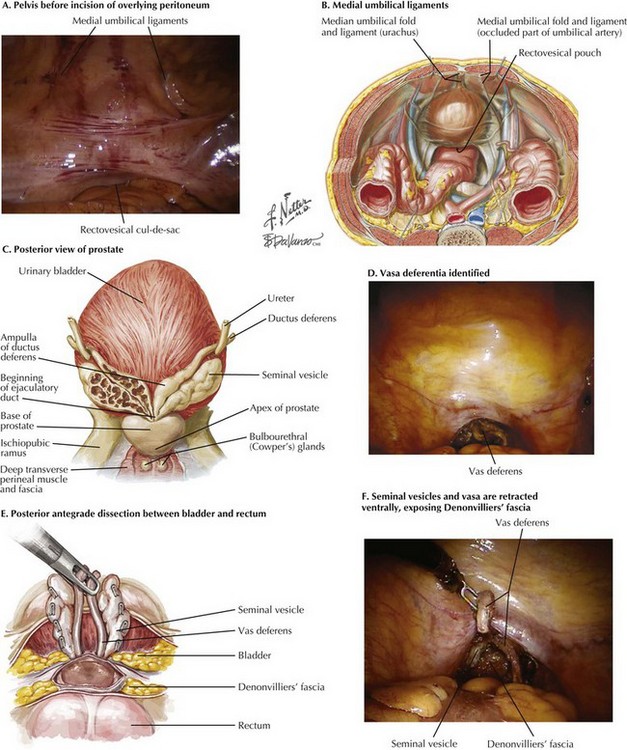
Radical Prostatectomy

Chapter 54


Radical Prostatectomy


image


Introduction


Radical prostatectomy for the treatment of prostate cancer has been performed for more than 100 years. The location of the prostate deep in the pelvis challenged surgical extirpation and caused significant morbidity. Radical prostatectomy is a “gold standard” in the treatment of localized prostate cancer, and level 1 evidence confirms that it offers a survival advantage over the absence of treatment in men with this disease. With increased understanding of the anatomy of the prostate and surrounding structures, along with improvements in instrumentation, the morbidity associated with radical prostatectomy has decreased substantially. Currently, urinary continence is achieved in approximately 90% of patients, and erectile function is preserved in most men with good preoperative function.


Traditionally, radical prostatectomy has been performed in the open retropubic or perineal approach. In the last decade, however, use of minimally invasive approaches has dramatically increased, particularly laparoscopic robot-assisted radical prostatectomy (Fig. 54-1, A). This approach has gained popularity because of its greater technical ease for the surgeon, especially with laparoscopic suturing, which allows for more precise suture placement during the vesicourethral anastomosis.


Prostate Cancer: Therapeutic Principles


Once the diagnosis of prostate cancer is made on biopsy, staging is obtained depending on the patient’s Gleason score, prostate-specific antigen (PSA), clinical stage, and life expectancy. Staging can include a bone scan and pelvic CT or MRI scan. If the malignancy has a high probability of being localized to the prostate, treatment options are discussed with the patient, including active surveillance, radiation therapy, or surgery, depending on disease factors, life expectancy, and patient preference. Radical prostatectomy is considered in men with life expectancy greater than 10 years who have prostate cancer at significant risk of progression.


The variation in prostate size and shape, as well as its location deep in the pelvis between the bladder and urethra and adjacent to the rectum, make surgical extirpation challenging (Fig. 54-1, B). Additionally, the prostate is surrounded by a venous plexus, and the neurovascular bundles responsible for erection run alongside the prostate. Therefore the surgical dissection required during radical prostatectomy should be performed meticulously by a surgeon with detailed knowledge of the anatomic relationships of the prostate.


Surgical Approach


Posterior Prostatic Dissection


Initial inspection of the pelvis is performed to identify the relevant landmarks: the medial umbilical ligaments and the vasa deferentia. The rectovesical cul-de-sac (pouch of Douglas) is approached, and the courses of the vasa are identified through the peritoneal layer (Fig. 54-2, A-D).


The overlying peritoneum is incised, and the ampulla of the vas deferens is isolated and transected. Retraction on the vas deferens ventromedially allows for identification and dissection of the ipsilateral seminal vesicle. The artery to the seminal vesicle is either clipped or controlled with bipolar electrocautery. The contralateral vas deferens and seminal vesicle are then dissected in a similar manner.


The seminal vesicles and vasa are retracted ventrally, exposing Denonvilliers’ fascia (Fig. 54-2, E and F). This fascia is incised sharply just dorsal to the base of the prostate. This approach allows entry into a plane containing perirectal fat, and the surgeon can then carefully dissect the prostate off of the rectum in an antegrade direction to the prostatic apex.

Only gold members can continue reading. Log In or Register to continue

Stay updated, free articles. Join our Telegram channel

Aug 12, 2016 | Posted by in ANATOMY | Comments Off on Radical Prostatectomy

Full access? Get Clinical Tree

Get Clinical Tree app for offline access