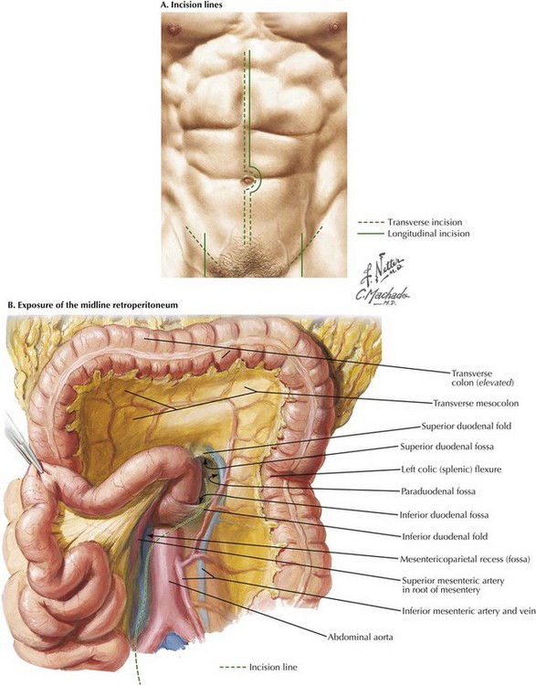
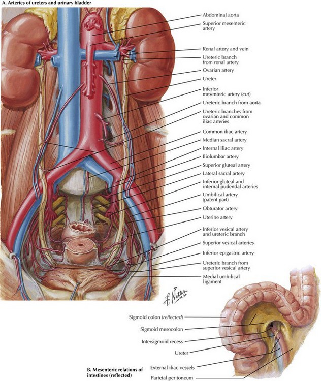
Aortic Exposure from the Midline Abdomen

Chapter 33


Aortic Exposure from the Midline Abdomen


image


Surgical Planning


A midline incision is usually made from the xiphoid to pubic symphysis if a juxtarenal or infrarenal AAA is being repaired. A shorter, lower midline incision ending just below the umbilicus is used if an aortobifemoral bypass (AF2) is being performed.


Disease of the aortoiliac arteries can manifest as aneurysmal or occlusive disease. Aneurysmal disease is mainly of the juxtarenal or infrarenal aorta and may or may not involve the common iliac arteries. The external iliac arteries are usually spared, so an extensive midline abdominal incision gives all the exposure necessary. If the disease process is occlusive, it can take multiple forms or patterns. The external iliac arteries are frequently involved or become involved over time, so an AF2 is the best surgical option. This procedure requires a smaller abdominal incision, usually ending just below the umbilicus, and two groin incisions, transverse or longitudinal, depending on surgeon preference. If there is a combination of aortic aneurysm and aortoiliac disease, a full abdominal incision (xiphoid to pubis) with groin incisions is necessary.


Although angiography was once the mainstay of preoperative planning, it has a more limited role today. A computed tomographic angiogram (CTA) with 3-mm cuts and reconstructions provides the needed information. The CTA can be supplemented with sagittal and coronal cuts and three-dimensional (3D) reconstruction. This type of CTA is also used to plan endovascular aortic repair (EVAR).


Incisional Anatomy


To begin the operation, the patient is placed on the operating room table in a supine position. A midline abdominal incision is made from the xiphoid process to below the umbilicus for an appropriate distance (Fig. 33-1, A). An AF2 or AAA not involving the common iliac arteries, and for which a predetermined tube graft will be performed, is sufficiently exposed with a shorter abdominal incision. If groin incisions will be used, the groin is opened first in the patient with no previous abdominal incision, with minimal difficulty predicted in exposing the abdominal aorta.


In patients with previous abdominal surgery, the author usually begins with the abdominal incision because of a higher likelihood of enterotomy during lysis of adhesions, and the groin would remain unscarred. In patients with extensive prior surgery, especially involving infection with peritonitis, a retroperitoneal or low thoracoabdominal approach may be warranted.


The choice of groin incision is also predetermined by looking at the patient and the CTA. A straightforward anastomosis to the common femoral artery or the anastomosis, including the first centimeter of superficial femoral artery or profunda femoris (deep femoral) artery, can be exposed with a transverse incision. Complexity in the groin necessitating more extensive or expansile exposure of the superficial femoral artery, or more often the profunda femoris artery, requires a longitudinal incision, the length of which can be modified to suit the situation.


A midline incision is made as illustrated in Figure 33-1. The small bowel is moved to the right and superiorly in the abdomen, and the sigmoid colon is gently retracted to the left. These maneuvers expose the midline retroperitoneum, and depending on the patient’s body mass index (BMI), retroperitoneal structures can be easily identified or obscured by retroperitoneal fat (Fig. 33-1, B).


Iliac Exposure


Palpation of the aortic bifurcation identifies the midline, and the author incises the pelvic retroperitoneum either in the midline or slightly to the right. This approach allows the surgeon to dissect the right common iliac bifurcation, retracting the peritoneum and its attached fat to the right. Remember that the ureter crosses the iliac vessels anteriorly and at the level of the common iliac bifurcation bilaterally (Fig. 33-2, A). Depending on the level of bypass, vessel loops can be placed around the right external and internal iliac arteries or around the distal common iliac artery, respecting the intimate relationship between the iliac arteries and veins. The most common atherosclerotic pattern demonstrates disease at the distal common iliac artery, so vessel loops around the external and internal iliac arteries are preferred. The vessels are usually soft at this location and will provide the most flexibility in constructing the anastomosis.


Stay updated, free articles. Join our Telegram channel

Aug 12, 2016 | Posted by in ANATOMY | Comments Off on Aortic Exposure from the Midline Abdomen

Full access? Get Clinical Tree

Get Clinical Tree app for offline access