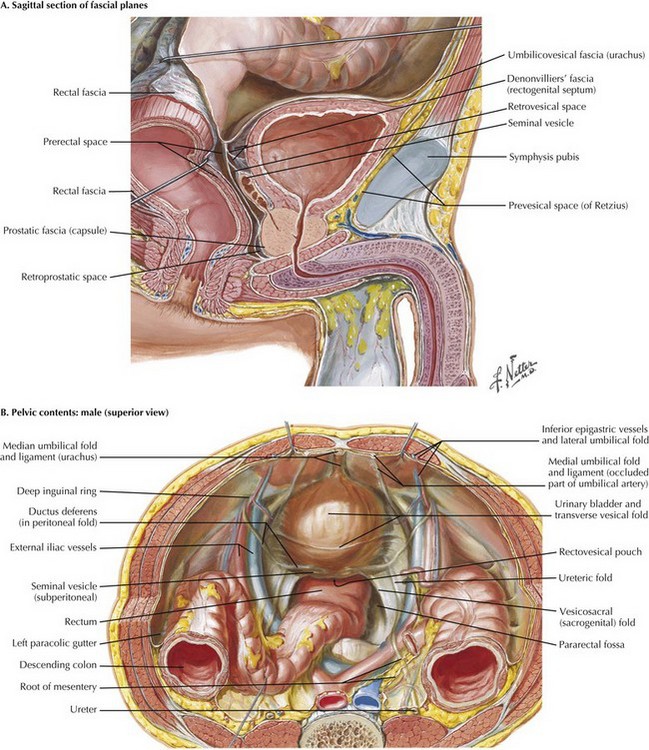
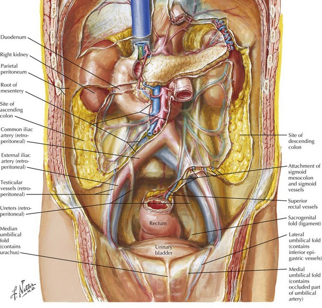
Radical Cystectomy

Chapter 55


Radical Cystectomy


Introduction


In the United States, bladder cancer is the fourth most common type in men and the ninth most common type of cancer in women. Non–muscle-invasive bladder cancer can often be treated with transurethral resections of bladder tumors and possible intravesical immunotherapy or chemotherapy with cystoscopic surveillance. Unfortunately, for muscle-invasive bladder cancer or for tumors with high-grade features and invasion into the lamina propria (T1) without metastatic disease, radical cystectomy with urinary diversion remains the cornerstone of treatment. Other indications for cystectomy include bacille Calmette-Guérin (BCG)–refractory carcinoma in situ (CIS), and as a palliative procedure for patients with severely symptomatic, metastatic bladder cancer necessitating acute hospital care and readmissions.


The first reported total cystectomy was in 1887 by Bernhard Bardenheuer. Radical cystectomy in men classically involves the removal of the bladder, distal ureters, prostate, and seminal vesicles. In women who choose not to maintain their fertility and who have extensive disease, anterior pelvic exenteration is traditionally performed, including cystectomy, salpingo-oophorectomy, hysterectomy, urethrectomy, and resection of the anterior one third of the vaginal wall. For radical cystoprostatectomy, advances in surgical technique have included nerve-sparing procedures to preserve potency in men and prostate-sparing procedures to accompany orthotopic bladder substitution. Recent data also challenge anterior pelvic exenteration in female patients and support more limited resection in those without extensive disease, preserving the anterior vaginal wall, uterus, ovaries, and pelvic supporting ligaments. Pelvic lymphadenectomy is performed regardless of gender.


Surgical Approach


Cystectomy has traditionally been performed through a low abdominal midline incision. Advances in laparoscopic technique now offer patients the option of laparoscopic or robotic-assisted cystectomy. However, despite reports of successfully completed intracorporeal laparoscopic construction of urinary diversions, the postcystectomy method of urinary reservoir formation is generally still completed with open techniques.


Bladder Mobilization


The midline incision length is determined on the basis of surgeon preference and experience, individual patient anatomy, extent of disease, and planned urinary diversion. Inferiorly, the incision should extend to the symphysis pubis to obtain adequate exposure. After incision of the skin and anterior and posterior layers of anterior abdominal wall fascia, the extraperitoneal space is entered first, and the space of Retzius is developed to mobilize the bladder away from the symphysis pubis anteriorly and to expose the external iliac artery and vein laterally.


The peritoneum located at the proximal midline incision is opened and extended laterally from the medial umbilical ligament to the internal inguinal rings. The urachus is divided high near the level of the umbilicus and subsequently used for retraction. Anatomically, an avascular plane of fibroareolar connective tissue can be followed, between the posterior rectus sheath and the peritoneum and along the medial umbilical ligaments. Care should be taken to avoid injury to the inferior epigastric arteries, which course in this plane and serve as the primary blood supply to the rectus abdominis muscle.


With the medial umbilical ligaments traced inferiorly and the space of Retzius open, a self-retaining retractor can be placed to facilitate exposure. Extra care is taken when applying traction on the most inferior aspect of the wound; injury to the femoral and genitofemoral nerves can result from prolonged stretch or compression. The symphysis should be visible with the retractor in place (Fig. 55-1, A).


The sigmoid colon is often adherent laterally to the side wall and occasionally to the bladder. The sigmoid colon is mobilized medially at this point by opening the peritoneum at the white line of Toldt. The small bowel can be placed in a damp, countable towel and pushed cephalad, and the sigmoid laid in the midline, with gentle retraction holding this exposure. These maneuvers provide excellent exposure and facilitate dissection.


The peritoneal wings previously left in place by developing the space of Retzius and opening the peritoneum from the umbilicus to the inguinal ring are easily seen and divided with electrocautery to the level of the vas deferens in the male patient. The vas deferens is divided with the understanding that vascular structures are associated with both vasa. The authors prefer to use ties to allow later identification of the seminal vesicles (Fig. 55-1, B).


Dissection of Ureters


After bilateral ligation of the vas deferens, the immediate posterior peritoneum along the bladder and above the sigmoid is opened. This approach allows identification of the ureters as they cross the iliac vessels and anterior mobilization of the bladder off the proximal rectum to the level of the prostate and seminal vesicles. This maneuver can be done with a gentle blunt movement; extra care should be taken because this loose plane of connective tissue becomes dense at the level of the prostate and cannot always be easily bluntly dissected. Forceful blunt dissection here risks rectal injury.


The left ureter is identified posterior to the sigmoid colon, in a position often more medial than expected. The retroperitoneal space behind the sigmoid colon at the level of the sacral promontory is opened to allow the ureter to be passed to the other side after its division. The right ureter is found by dividing the visible peritoneal fold overlying it (Fig. 55-2).


The ureters are mobilized cephalad approximately 5 cm and caudad to the ureterovesical junction. Care must be taken to preserve soft tissue around the ureter. The periureteral blood supply is enveloped in this layer, and excellent vascularity will aid in a successful, patent anastomosis for the urinary diversion. The distal 5 mm of each ureter can then be divided and sent as a frozen section to ensure adequacy of the margin. In some cases of CIS, numerous frozen sections may need to be sent before an adequate margin is achieved. However, this practice has come under debate in recent years, with contradictory conclusions regarding the value of a negative margin in reducing risk of upper tract cancer recurrence.


This dissection allows for clear visualization of the common, external, and internal iliac arteries and prepares the surgeon to commence with pelvic lymphadenectomy.

Stay updated, free articles. Join our Telegram channel

Aug 12, 2016 | Posted by in ANATOMY | Comments Off on Radical Cystectomy

Full access? Get Clinical Tree

Get Clinical Tree app for offline access