Pseudocyst on CT CT shows a unilocular cyst located in the head of the pancreas, consistent with a pancreatic pseudocyst .
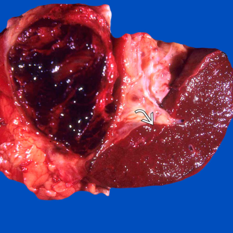
Pseudocyst With Hemorrhagic Fluid Gross photograph shows a pseudocyst in the tail of the pancreas. The cyst is filled with hemorrhagic fluid. Note the adjacent spleen .
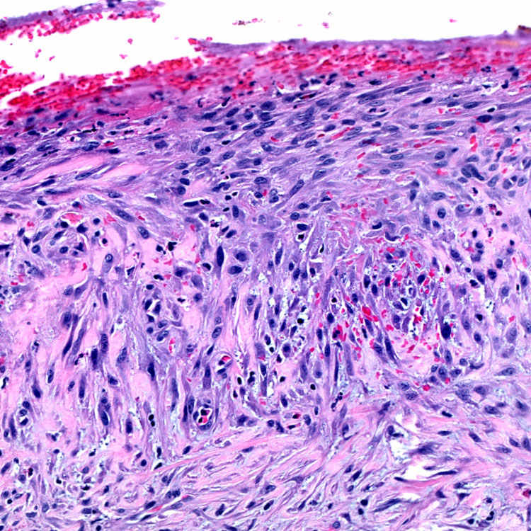
Pseudocyst Wall With Fibroblasts Pseudocysts lack a true epithelial lining. The cyst wall shows an exuberant fibroblastic proliferation.
Hemosiderin and Fibrosis in Wall The pseudocyst wall is composed of fibrosis and few scattered inflammatory cells. Note the hemosiderin in the lumen of the cyst.
TERMINOLOGY
Definitions
• Cystic collection of pancreatic or peripancreatic fluid rich in pancreatic enzymes
ETIOLOGY/PATHOGENESIS
Risk Factors
• Acute or chronic pancreatitis
• Can occur with biliary disease, surgery, or other trauma
• Rarely, may develop adjacent to pancreatic mass lesion, including adenocarcinoma
CLINICAL ISSUES
Epidemiology
• Incidence
Historically thought to represent 80% of all pancreatic cysts
– High-resolution imaging now suggests neoplastic pancreatic cysts more common than pseudocysts
• Age
Young to middle-aged adults
• Sex
Female predominance associated with gallstone-related pancreatitis
Male predominance associated with alcohol-related pancreatitis
Only gold members can continue reading. Log In or Register to continue
 .
.
 .
.
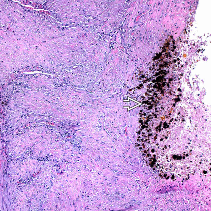
 in the lumen of the cyst.


