10
Pharynx and Larynx
The pharynx and larynx are tube-like structures in the upper part of the neck. The pharynx is part of the digestive system and is a pathway for food and air; the larynx connects the lower part of the pharynx to the trachea. Commonly referred to as the “windpipe,” the trachea channels air into the lungs.
Pharynx
The pharynx is a fibromuscular tube that connects the nasal cavity, oral cavity, and larynx. Accordingly, it is subdivided into the nasopharynx, oropharynx, and laryngopharynx (Fig. 10.1). The pharynx extends from the base of the skull to the level of the cricoid cartilage (at vertebral level CVI), at which point the pharynx joins the esophagus. Structural support is provided by bony, cartilaginous, and ligamentous elements associated with the skull, hyoid bone, and laryngeal cartilages. The pharynx is a channel for swallowing and respiration.
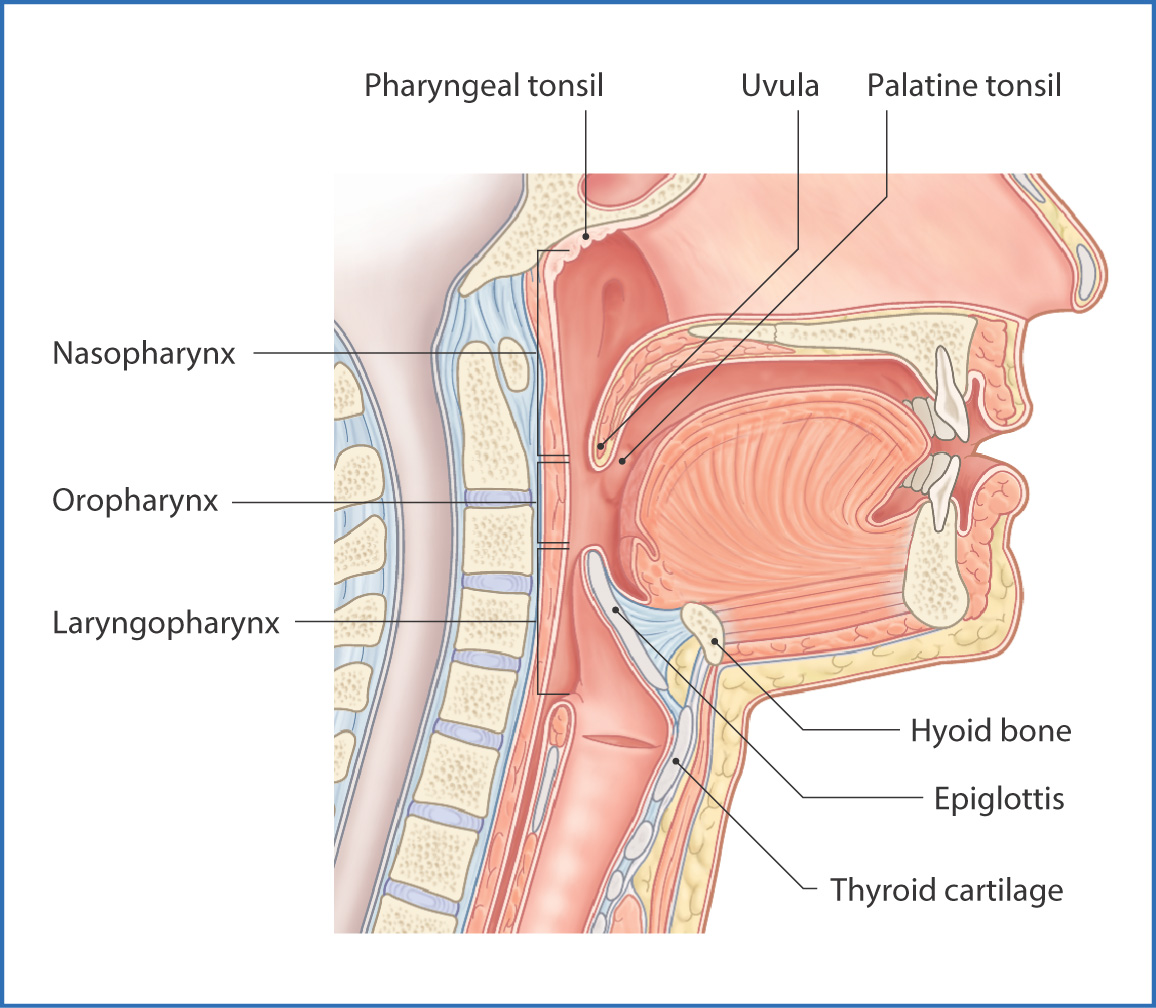
FIGURE 10.1 Divisions of the pharynx.
The nasopharynx—the superior part of the pharynx—communicates anteriorly with the nasal cavity through the choanae and extends inferiorly to the soft palate. The pharyngeal opening for the pharyngotympanic tube is visible next to the torus tubarius, a cartilaginous elevation on the lateral wall of the pharynx. The pharyngotympanic tube connects the nasopharynx to the middle ear and equalizes air pressure on both sides of the tympanic membrane (see Chapter 7). This can be a route for spread of infection from the nasopharynx to the middle ear. The pharyngeal tonsil (adenoid) is on the superior wall of the nasopharynx.
The oropharynx lies between the soft palate and the tip of the epiglottis. It communicates anteriorly with the oral cavity through the oropharyngeal isthmus, which is formed by the tongue inferiorly and the palatoglossal and palatopharyngeal arches laterally. The palatoglossal (anterior) and palatopharyngeal (posterior) folds on the lateral walls of the oropharynx are formed by the palatoglossus and palatopharyngeus muscles, respectively.
The palatine tonsil lies between the palatoglossal and palatopharyngeal folds. Throat infections in children often result in enlarged palatine tonsils, which can interfere with breathing and speech (see Chapter 9). The palatine tonsil is innervated by branches of the glossopharyngeal nerve [IX]. Blood is supplied by tonsillar branches of the facial, ascending palatine, lingual, descending palatine, and ascending pharyngeal arteries. Venous drainage is to the pharyngeal plexus of veins. Lymphatic drainage is mainly to the jugulodigastric node of the deep cervical lymph node group (see Chapter 12 and Fig. 2.5).
The laryngopharynx extends from the tip of the epiglottis to the level of the cricoid cartilage (vertebral level CVI), where it joins the esophagus. Anteriorly, it communicates with the larynx through the laryngeal inlet (aditus). During swallowing, the epiglottis bends posteriorly and the laryngeal structures are pulled superiorly. As a result, the laryngeal inlet is partially closed by the epiglottis, which prevents food from entering the trachea. The piriform fossae are located on both sides of the laryngeal inlet and channel swallowed substances to the esophagus.
MUSCLES
The muscular layer of the pharynx (Figs 10.2 and 10.3) comprises the semicircular superior, middle, and inferior constrictor muscles and the longitudinal palatopharyngeus, salpingopharyngeus, and stylopharyngeus muscles. The constrictors propel food toward the esophagus; the longitudinal muscles elevate the pharynx and larynx during swallowing and phonation.
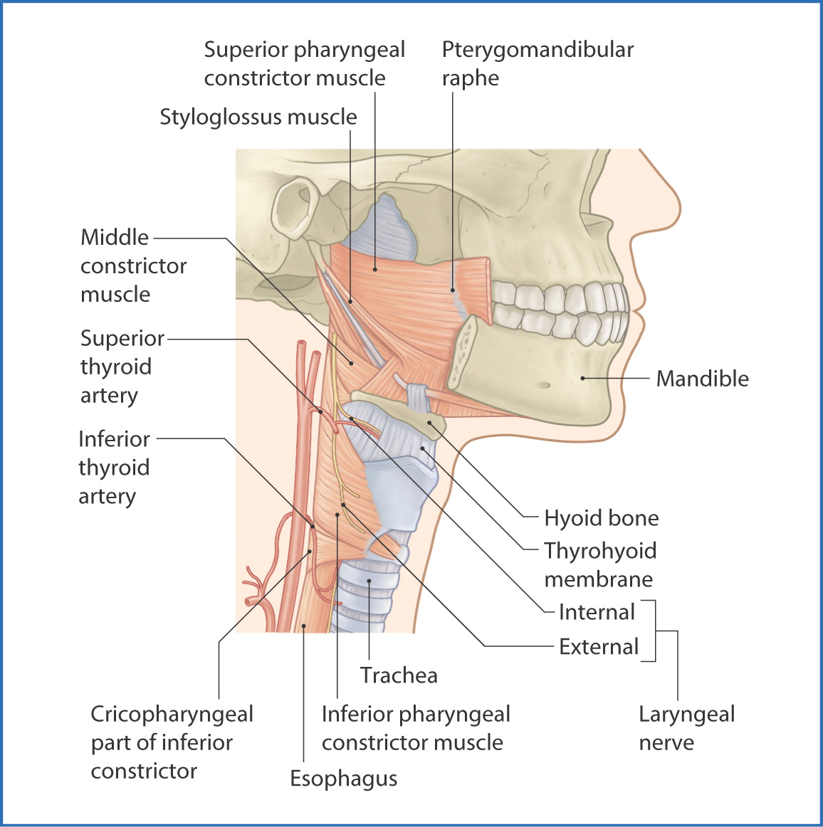
FIGURE 10.2 Pharyngeal constrictor muscles.
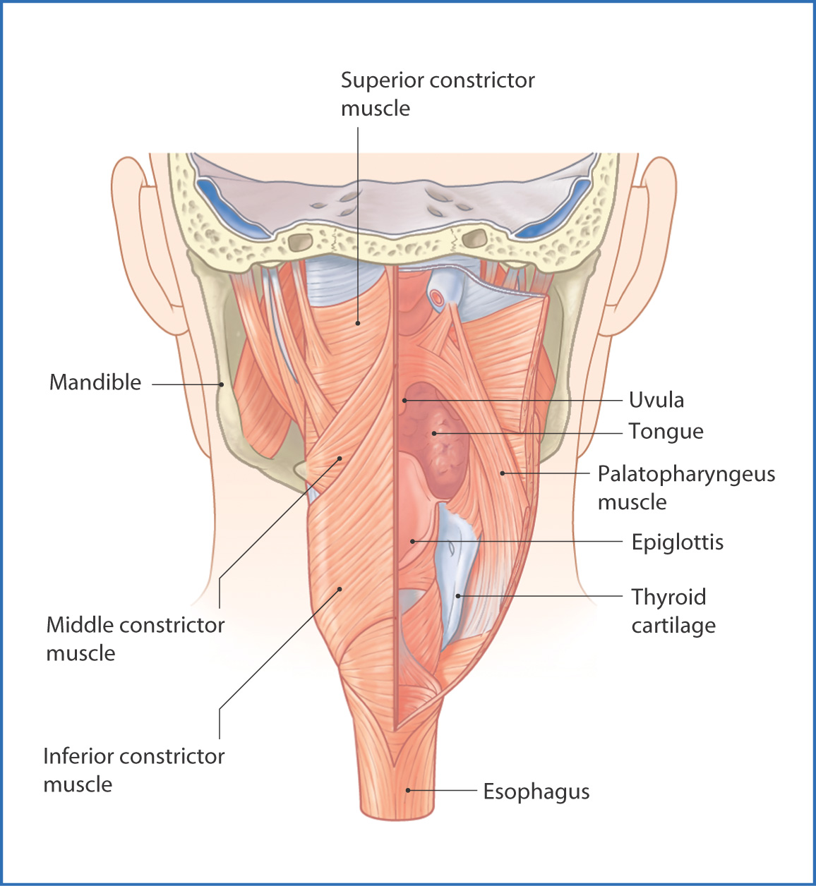
FIGURE 10.3 Posterior view of the pharynx.
NERVES
Sensory and motor innervation to the pharynx originates from the pharyngeal plexus on the posterior aspect of the pharyngeal constrictors. This plexus is formed by pharyngeal branches of the vagus [X] and glossopharyngeal [IX] nerves and by postganglionic sympathetic nerve fibers from the superior cervical ganglion.
Motor fibers in the plexus are derived from the cranial part of the accessory nerve [XI], which runs with the vagus nerve [X] and supplies all muscles of the pharynx, larynx, and soft palate except for the tensor veli palatini and stylopharyngeus muscles, which are innervated by the trigeminal [V] and glossopharyngeal [IX] nerves, respectively.
Sensory innervation of the pharynx is from the glossopharyngeal nerve [IX]. The maxillary [V2] and vagus [X] nerves also carry sensation from a small part of the pharynx.
ARTERIES
Blood supply to the pharynx is by branches of the ascending pharyngeal, superior thyroid, lingual, facial, and maxillary arteries.
VEINS AND LYMPHATICS
Venous drainage of the pharynx is through the pharyngeal plexus
Stay updated, free articles. Join our Telegram channel

Full access? Get Clinical Tree


