38
Perineum
The perineum is the area outlined by the diamond-shaped pelvic outlet. It contains the external genitalia and anus. The pelvic outlet is bounded
- anteriorly by the pubic symphysis
- posteriorly by the coccyx
- laterally by the medial margins of the inferior pubic rami (anteriorly) and the sacrotuberous ligaments (posteriorly)
- posteriorly by the coccyx
The perineum is further subdivided into urogenital and anal triangles by an imaginary line that joins the space between the anterior margins of the ischial tuberosities. Osseous support is provided by the pubic and ischial parts of the hip bones, the sacrum, and the coccyx (see Chapter 36).
The floor of the perineum is formed by the skin and structures of the external genitalia and the roof (the pelvic diaphragm) by a group of flat, sheet-like muscles (levator ani and coccygeus). These muscles extend across and cover the pelvic outlet to support the pelvic viscera. All the structures inferior or superficial to the pelvic diaphragm are constituents of the perineum (e.g., penis, vagina, and anus).
Muscles
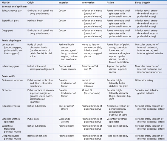
The muscles of the perineum (Table 38.1) can be divided into three distinct groups: the pelvic diaphragm and supporting muscles, the urogenital diaphragm, and the superficial perineal muscles. These three layers of muscle are separated by several spaces containing fascia, fat, and loose connective tissue. These perirectal and perineal spaces are all adjoining, and infection in one area can quickly spread to another.
TABLE 38.1 Muscles of the Perineum

PELVIC DIAPHRAGM
The pelvic diaphragm is a collection of flat muscles that create a supportive basket-like floor for the pelvic viscera. These muscles are adjacent to each other and comprise the levator ani (puborectalis, pubococcygeus, and iliococcygeus) and ischiococcygeus muscles. The three levator ani muscles span the space between the body of the pubis, ischial spine, and coccyx. The coccygeus muscle attaches to the ischial spine and pelvic surface of the sacrospinous ligament and to the coccyx and inferior surface of the sacrum. The coccygeus is superior and lateral to the levator ani muscles. These muscles support and elevate the pelvic floor when contracted.
The piriformis and obturator internus muscles provide additional support to the pelvic viscera. The piriformis muscle (see Chapter 42) is on the internal surface of the lateral aspect of the sacrum and covers the opening in the pelvic outlet between the coccygeus muscle and the lateral wall of the pelvis. The obturator internus muscle is lateral to the levator ani muscles, spans the obturator foramen on the lateral wall of the pelvis, and leaves the pelvis through the lesser sciatic foramen to insert onto the femur.
UROGENITAL DIAPHRAGM
The urogenital diaphragm is composed of two muscles: the deep transverse perineal muscle and the urethral sphincter. These two muscles are superficial or inferior to the pelvic diaphragm. The deep transverse perineal muscles arise from the ischial tuberosities on the pelvic girdle and extend toward the midline, where they meet and help form the perineal body. Muscle fibers from the external anal sphincter and puborectalis (from the levator ani) also insert on this midline collection of muscle and soft tissue fibers. The perineal body is between the root of the penis/vaginal fornix and the anus. Muscles of the superficial perineal group (superficial transverse perineal, bulbospongiosus, external anal sphincter) attach to it.
Just anterior to the deep transverse perineal muscles is the urethral sphincter muscle, which forms a circular ring around the urethra in both sexes; in females it also encircles the vagina. Small slips of muscle leave this ring laterally to insert onto the inferior pubic ramus. As a group, the urethral sphincter and deep transverse perineal muscles can be controlled voluntarily to delay micturition (urination). They are innervated by the perineal branch of the pudendal nerve.
SUPERFICIAL PERINEAL MUSCLES
The superficial perineal muscles are the bulbospongiosus, ischiocavernosus, and superficial transverse perineal muscles and the external anal sphincter (see Figs. 38.1 and 38.3). These muscles are superficial or inferior to the urogenital diaphragm. All are innervated by the pudendal nerve. The bulbospongiosus muscle originates from the perineal body and inserts into the corpus spongiosum of the penis, where it maintains erection by preventing venous outflow. In women, the bulbospongiosus compresses the erectile tissue in the vestibule of the vagina and helps constrict the vaginal opening. This muscle also aids in expelling urine from the urethra in males and females.
The ischiocavernosus arises from the ischial tuberosity and inserts onto the corpus cavernosum of the penis or clitoris. It maintains erection in males and constricts the vagina and clitoris in females.
Like the other superficial perineal muscles, the superficial transverse perineal muscle arises from the ischium and inserts onto the perineal body. Its action stabilizes the perineal body.
The external anal sphincter is a circular muscle that surrounds the anus and provides voluntary control of defecation through the pudendal nerve.
Male External Genitalia
The penis
Stay updated, free articles. Join our Telegram channel

Full access? Get Clinical Tree


