Elongated and anastomosing rete ridges


• Basaloid, warty, and mixed (warty/basaloid) PeIN and VIN (a.k.a. usual VIN, HPV-related)
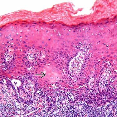
 Warty VIN/PeIN: Pleomorphic cells with koilocytic changes replace most to full thickness of epithelium
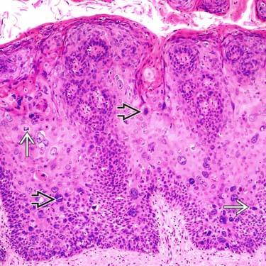
 Warty-basaloid VIN/PeIN: Pleomorphic cells with koilocytic changes seen on upper epithelium and basaloid cells replace lower epithelium
 Warty VIN/PeIN: Pleomorphic cells with koilocytic changes replace most to full thickness of epithelium Warty-basaloid VIN/PeIN: Pleomorphic cells with koilocytic changes seen on upper epithelium and basaloid cells replace lower epithelium
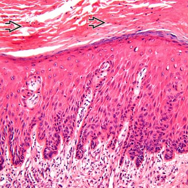
Acanthotic epithelium with subtle abnormal maturation and hyperchromatic-staining atypical basilar cells are features of differentiated penile intraepithelial neoplasia/vulvar intraepithelial neoplasia (PeIN/VIN). Prominent parakeratosis is seen on the surface
 .
.
Enlarged keratinocytes with abundant eosinophilic cytoplasm throughout most of the epithelium are seen in differentiated PeIN/VIN. Characteristic keratin pearl formation is present
 .
.
Warty PeIN and VIN show pleomorphic and hyperchromatic-staining nuclei
 , bi- and multinucleation, and multiple mitoses
  throughout the epithelium.
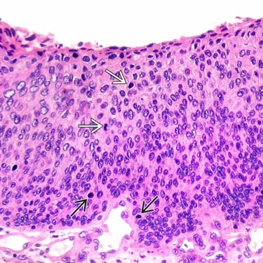
The epithelium is completely replaced by atypical cells with basaloid features. Numerous mitoses
 and apoptotic bodies
  are seen. The diagnosis of carcinoma in situ is easily achieved in high-grade basaloid PeIN and VIN.
TERMINOLOGY
Abbreviations


Definitions
• VIN and PeIN are considered intraepithelial (in situ) precursor lesions of invasive SCC
 Recommended terminology for HPV-associated squamous lesions of anogenital tract is LSIL and HSIL, which may be further classified by applicable -IN sub-categorization
• LAST (2012) recommends 2-tiered nomenclature system for HPV-related precursor lesions (applicable to entire anogenital region)

 Recommended terminology for HPV-associated squamous lesions of anogenital tract is LSIL and HSIL, which may be further classified by applicable -IN sub-categorization


CLINICAL ISSUES
Epidemiology


Presentation


• Warty, basaloid, and mixed PeIN and VIN (a.k.a. VIN of “usual type” in vulvar pathology)


Stay updated, free articles. Join our Telegram channel
Full access? Get Clinical Tree
