Autosomal dominant disease due to mutations in mismatch repair genes MLH1, MSH2, MSH6
Multiple sebaceous tumors including sebaceous adenomas, less likely carcinomas and sebaceomas
Clinical Issues
• Rare tumors; typically occur in adults
• Most common on face
• Usually single but may be multiple, especially in MTS patients
Microscopic
• Nodular, dermal-based adnexal tumor
• Well circumscribed, noninfiltrative
• Composed mostly of basaloid cells
• Minor population of clear cells with multivacuolated cytoplasm, consistent with mature sebaceous cells
Show nuclear hyperchromasia with indentations
• Basaloid cells may show mild cytologic atypia and increased mitotic figures
• Sebaceous cells do not show significant atypia or mitotic activity
Top Differential Diagnoses
• Basal cell carcinoma with sebaceous differentiation
Basal cells show greater atypia and peripheral palisading
Mucinous stroma and retraction artifact often present (lacking in sebaceoma)
• Sebaceous adenoma
> 50% clear cells
• Sebaceous carcinoma
• Trichoblastoma with sebaceous differentiation
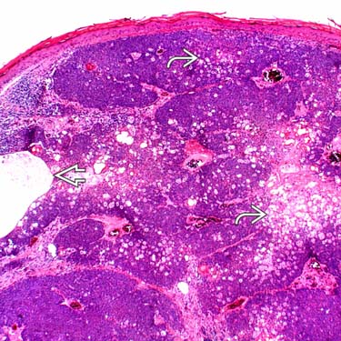
Sebaceoma at Low Magnification Low magnification of a sebaceoma shows a dermal-based basaloid to clear cell neoplasm with peripheral basaloid cells surrounding clear cells , sebaceous secretions, and a focal cystic space .
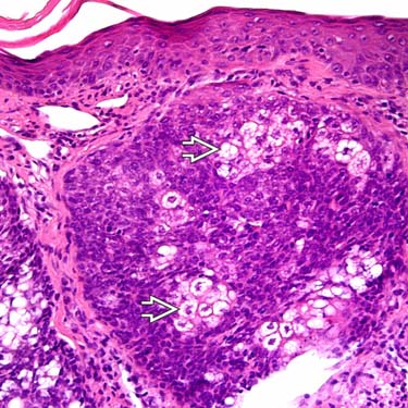
Sebaceoma at Higher Magnification Higher magnification of sebaceoma shows a superficial, well-circumscribed lobule of predominantly basaloid cells with a smaller population of bland-appearing, mature sebaceous cells .
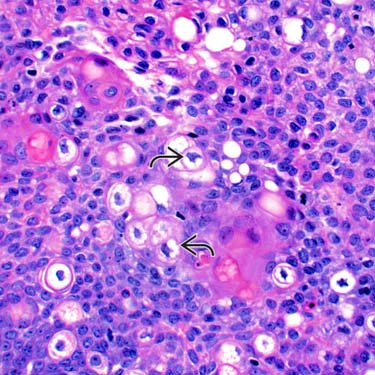
Sebaceoma at High Magnification High magnification of a sebaceoma shows a population of scattered, bland-appearing, multivacuolated clear cells demonstrating nuclear hyperchromasia and nuclear indentations , which are due to cytoplasmic lipids.
Cystic Sebaceoma This is an example of a sebaceoma with cystic areas, which shows a proliferation of predominantly basaloid and focal clear cells surrounding the cystic space .
TERMINOLOGY
Synonyms
• Sebaceous epithelioma (older term, should be discouraged)
Definitions
• Benign proliferation of mature sebaceous cells associated with predominant basaloid cell population
ETIOLOGY/PATHOGENESIS
Unknown in Most Cases
• Some cases are part of Muir-Torre syndrome (MTS)
Autosomal dominant disease due to mutations in mismatch repair genes MLH1, MSH2, MSH6
Patients present with multiple sebaceous tumors, including sebaceous adenomas (most common), sebaceomas, and sebaceous carcinomas
Also associated with internal malignancies including gastrointestinal carcinomas (most common), genitourinary, breast, and ovarian tumors
• Rare cases arise in nevus sebaceous of Jadassohn
CLINICAL ISSUES
Epidemiology
• Incidence
Rare tumors
• Age
Typically occur in adults
Site
• Often occur on face but may also present on trunk
Presentation
• Slow-growing papular to nodular lesion
Usually single, but may be multiple, especially in MTS
• Flesh-colored to yellowish
Treatment
• Surgical approaches
Complete conservative excision is curative
Prognosis
• Excellent; very low malignant potential
Possible transformation to sebaceous carcinoma in longstanding lesions
MACROSCOPIC
Size
• Usually measure between 0.5-3.0 cm in diameter
Only gold members can continue reading. Log In or Register to continue


 , sebaceous secretions, and a focal cystic space
  .
.
 .
.
 , which are due to cytoplasmic lipids.
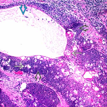
 and focal clear cells
  surrounding the cystic space
  .
.


