Includes angiomyolipoma (AML), clear cell sugar tumor of lung (CCST), lymphangioleiomyomatosis (LAM), clear cell myomelanocytic tumor of falciform ligament/ligamentum teres
Etiology/Pathogenesis
• Only AML, CCST, and LAM are associated with tuberous sclerosis, but not other types
Clinical Issues
• Most are benign, but rare malignant cases reported
• Rare tumors overall; very rare in skin
Microscopic
• PEC cell component consists of epithelioid to spindled cells arranged around vessels extending outward radially
• Clear to granular, lightly eosinophilic cytoplasm and round to oval nuclei with small nucleoli
• Myoid component with densely eosinophilic cytoplasm
• Adipose tissue component present in lesions termed AML
• Express smooth muscle markers and melanoma markers but lack S100 expression
• TFE3 rearrangements
Top Differential Diagnoses
• True smooth muscle tumors
• Renal cell carcinoma
• Clear cell sarcoma
• Melanoma
• Myoepithelioma
• Well-differentiated liposarcoma
• Metastatic gastrointestinal stromal tumor
• Metastatic hepatocellular carcinoma
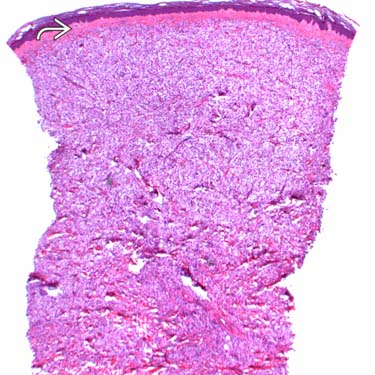
PEComa at Scanning Magnification This rare primary cutaneous PEComa shows a clear cell neoplasm diffusely involving the dermis and associated with many small blood vessels. There is a grenz zone separating the tumor from the overlying epidermis .
PEComa at High Magnification Higher magnification examination shows a clear cell tumor with numerous fibrovascular channels between the spindled and epithelioid clear cells.
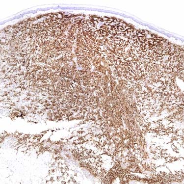
Cutaneous PEComa, SMA Labeling SMA shows strong and diffuse cytoplasmic expression in this rare primary cutaneous PEComa.
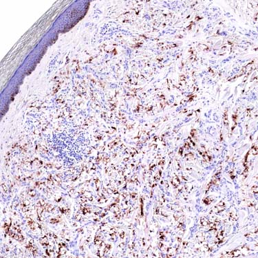
PEComa, HMB-45 Staining HMB-45 shows scattered, strong cytoplasmic expression in a primary cutaneous PEComa. These lesions also express other markers of melanocytic differentiation as well as CD117 and CD10, but typically lack S100 and keratin expression.
TERMINOLOGY
Abbreviations
• Perivascular epithelioid cell (PEC)
Thus, neoplasms are termed PEComa
Synonyms
• Perivascular epithelioid cell tumor
• Extrapulmonary sugar tumor
• Monotypic epithelioid angiomyolipoma
Definitions
• Mesenchymal neoplasms composed of distinctive perivascular epithelioid cells; category includes
Angiomyolipoma (AML)
Clear cell sugar tumor of lung (CCST)
Lymphangioleiomyomatosis (LAM)
Clear cell myomelanocytic tumor of falciform ligament/ligamentum teres (CCMMT)
• In many respects, PEComas are simply angiomyolipomas without fat
• Subset displays overt histologic features of malignancy and malignant clinical behavior
ETIOLOGY/PATHOGENESIS
Association With Tuberous Sclerosis
• Genetic alterations of tuberous sclerosis complex (TSC), losses of TSC1 (9q34) or TSC2 (16p13.3) genes
• Autosomal dominant inheritance
• Benign tumors of brain (most common), kidneys, heart, eyes, lungs, and skin
Name comes from characteristic tuber or potato-like nodules in brain, which calcify with age and become hard or sclerotic
• AML, CCST, and LAM are associated with tuberous sclerosis but not other types
CLINICAL ISSUES
Epidemiology
• Incidence
AML, CCST, LAM are rare
– Other PEComas extremely rare
• Age
CCMMT typically encountered in girls in late childhood
Most others seen in adults 50-60 yr old
AML detected in younger patients in setting of tuberous sclerosis
• Sex
Marked overall female predominance
Site
• Reported in multiple sites; rare in skin, but reported
Kidney, liver, falciform ligament, deep soft tissues of extremities, uterus, vulva, heart, gallbladder, gastrointestinal tract
Presentation
• CCMMT presents as painful abdominal mass
• Uterine examples manifest as uterine bleeding
• Most other categories of PEComas present as painless masses
• Brain tumors in patients with tuberous sclerosis present with seizures, developmental delay, behavioral problems
Treatment
• Surgical excision
Prognosis
• Most are benign
Rare documented examples of malignancy
– Usually not in AML, LAM, or CCST types
– Malignant examples behave as aggressive sarcomas
MICROSCOPIC
Histologic Features
• PEC cell component consists of epithelioid to spindled cells arranged around vessels extending outward radially
Clear to granular, lightly eosinophilic cytoplasm and round to oval nuclei with small nucleoli
Lesions are richly vascular
Small arching vessels divide tumor into packets (similar to pattern in renal cell carcinoma)
• Myoid component with densely eosinophilic cytoplasm
Nuclei less rounded than those of true smooth muscle
• Adipose tissue component present in lesions termed AML
• CCMMT is almost exclusively spindle cell lesion
Uniform moderate-sized cells set in elaborate lace-like vasculature
Rare Malignant Examples
• Criteria for malignancy
Infiltrative growth
Marked hypercellularity
Nuclear enlargement and hyperchromasia
Numerous &/or atypical mitotic figures
Only gold members can continue reading. Log In or Register to continue
 .
.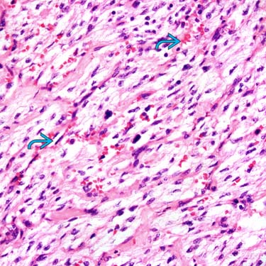
 between the spindled and epithelioid clear cells.