5
Parotid, Temporal, and Pterygopalatine Region
The temporal and infratemporal fossae are two anatomical areas on the lateral surface of the skull. The temporal fossa is the site of origin of the temporalis muscle, and the infratemporal fossa is the site of origin of the medial and lateral pterygoid muscles. These are all muscles of mastication. The masseter muscle, the fourth muscle of mastication, is in the vicinity of the parotid gland (Fig. 5.1) on the lateral aspect of the ramus of mandible (see Chapter 4 and later). The infratemporal fossa contains
- the mandibular division of the trigeminal nerve [V3]
- the parasympathetic otic ganglion, which sends postganglionic nerve fibers to the parotid gland via the auriculotemporal nerve (see Chapter 4)
- the maxillary artery
- the pterygoid venous plexus
- the chorda tympani [VII], which contains taste and preganglionic parasympathetic nerve fibers
- the parasympathetic otic ganglion, which sends postganglionic nerve fibers to the parotid gland via the auriculotemporal nerve (see Chapter 4)
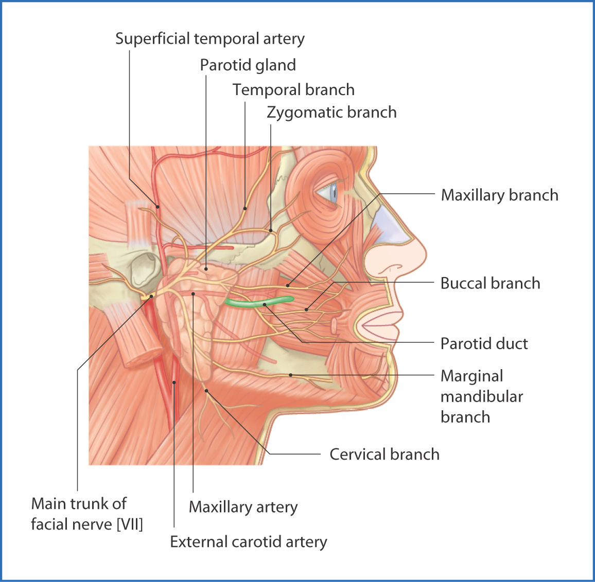
FIGURE 5.1 Temporal region: parotid gland and branches of the facial nerve [V].
The temporal fossa is bounded superiorly by the temporal lines, laterally by the zygomatic arch, and inferiorly by the infratemporal crest (Table 5.1). It is formed by the frontal bone, the parietal bone, the greater wing of the sphenoid, and the squamous part of the temporal bone. These bones unite to form an important clinical landmark—the pterion—which lies over the intracranial middle meningeal artery and vein and is the thinnest part of the skull; a skull fracture at this site can easily cause brain damage and intracranial bleeding.
TABLE 5.1 Boundaries of the Temporal, Infratemporal, and Pterygopalatine Fossae
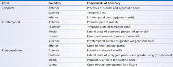
The infratemporal fossa is bounded anteriorly by the posterior part of the maxilla, posteriorly by the tympanic plate of the temporal bone, medially by the lateral plate of the pterygoid process of the sphenoid bone, and laterally by the ramus and coronoid process of the mandible (see Table 5.1). Its roof is formed by the infratemporal surface of the greater wing of the sphenoid; the floor of the fossa is open.
The pterygopalatine fossa is an irregularly shaped space posterior to the maxilla and inferior and deep to the zygomatic arch. It is medial to the infratemporal fossa, and its boundaries are the posterior surface of the maxilla anteriorly, the lateral plate of the pterygoid process and the greater wing of the sphenoid posteriorly, the perpendicular plate of the palatine bone medially, and the body of the sphenoid and orbital surface of the palatine bone superiorly. Laterally, the pterygopalatine fossa opens through the pterygomaxillary fissure (see Table 5.1).
Anteriorly, the pterygopalatine fossa is closely related to the orbit through the inferior orbital fissure; medially, it is related to the nasal cavity through the sphenopalatine foramen; inferiorly, it is related to the oral region through the greater and lesser palatine foramina; laterally, it is related to the infratemporal fossa through the pterygomaxillary fissure; and posterosuperiorly, it is related to the middle cranial fossa through the foramen rotundum and pterygoid canal. The pterygopalatine fossa contains the maxillary nerve [V2], the pterygopalatine ganglion, the nerve of the pterygoid canal, and the third or pterygopalatine part of the maxillary artery and its branches (see later). It is the contents and relationships of the pterygopalatine fossa that make understanding of it important.
The temporomandibular joint (TMJ) is a synovial joint with gliding and hinge functions, which are enhanced by the insertion of an articular disc between the head of the mandible and the mandibular fossa of the temporal bone. Major support for the TMJ is provided by the muscles of mastication (temporalis, medial pterygoid, lateral pterygoid, and masseter; see later). Additional support is provided by the lateral, stylomandibular, and sphenomandibular ligaments. The TMJ is innervated by the masseteric, auriculotemporal, and deep temporal nerves, which are all branches of the mandibular nerve [V3]. Blood supply to the TMJ arises from branches of the maxillary and superficial temporal arteries.
Muscles
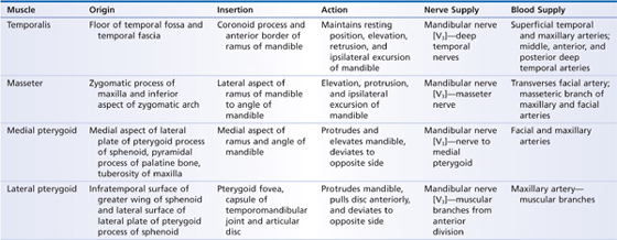
The muscles of mastication associated with the temporal and infratemporal fossae are as follows:
- The temporalis muscle is a large fan-like muscle originating from the temporal fossa, inserting inferiorly onto the coronoid process of the mandible, and acting to elevate the mandible.
- The masseter muscle originates from the zygomatic arch, inserts onto the lateral aspect of the ramus of the mandible, and elevates the mandible.
- The medial pterygoid muscle originates from the tuberosity of the maxilla and palatine bone, inserts onto the medial aspect of the mandible below the mandibular foramen, and elevates and protracts the mandible.
- The lateral pterygoid muscle joins the sphenoid bone to the neck of mandible and protrudes the mandible.
- The masseter muscle originates from the zygomatic arch, inserts onto the lateral aspect of the ramus of the mandible, and elevates the mandible.
These four muscles are innervated by branches of the mandibular nerve [V3] (Table 5.2).
TABLE 5.2 Muscles of Mastication
Nerves
The main nerves associated with the temporal, infratemporal, and pterygopalatine fossae are branches of the maxillary [V2] and mandibular [V3] nerves, which are divisions of the trigeminal nerve [V] (see Fig. 4.2).
The maxillary nerve [V2] leaves the middle cranial fossa through the foramen rotundum and enters the pterygopalatine fossa. It is a sensory nerve with no motor component, and its branches provide sensation to the midsection of the face (lower part of the orbit, nose, upper part of the mouth, and cheek).
Stay updated, free articles. Join our Telegram channel

Full access? Get Clinical Tree


