Paediatric disorders

16 Paediatric disorders


INTRODUCTION


Paediatric surgical disorders can present at any age. Paediatric in most centres is from birth to 16 years. Some surgical disorders in childhood are similar to the same condition in adults, and are discussed in those relevant chapters elsewhere in this book.


The aspects of paediatric surgery which will be covered in this chapter are those which arise from congenital anomalies, i.e. the child is born with a condition which has occurred during intrauterine development. Although many of these conditions present at or soon after birth – in the neonatal period, that is, the first 28 days after birth – some do not present until later in childhood, and some are never discovered throughout childhood or adult life, as they are completely compatible with normal life. About 3% of live-born infants have an obvious major malformation (e.g. diaphragmatic hernia, oesophageal atresia), which rises to 6% by the end of the first year as other major anomalies (e.g. congenital heart disease, renal anomalies) are discovered. Minor malformations (e.g. ear tags, simian crease) occur in about 14% of babies and are usually of no significance – although, if multiple, should alert the clinician to the possibility of an associated major malformation.


The causes of congenital anomalies are various – e.g. genetic, environmental – and are usually multifactorial. Pure genetic causes of anomalies which are surgically treated are uncommon but are seen in achondroplasia (limb lengthening is now offered), cystic fibrosis (with gastrointestinal problems) and haemophilia (venous access is often required).


Environmental factors include maternal illness with viruses such as rubella or cytomegalovirus and, depending on the timing of the infection, would lead to different effects on the fetus. Rubella in the first trimester, when the maximum amount of development and differentiation is occurring in the embryo, is often associated with congenital heart disease, deafness, cataracts and developmental delay. Exposure of the mother to radiation or other teratogens such as drugs (e.g. warfarin, anti-epileptics, thalidomide), again in the first trimester, can also produce congenital anomalies.


More commonly the cause of congenital anomalies is multifactorial, the combined effect of genetic and environmental interaction, where a genetic predisposition plus environmental effects are responsible for the anomaly – such as neural tube defects (spina bifida, etc.), congenital heart defects and cleft lip/palate.


The majority of anomalies do not have a known cause, although some are local effects – such as small bowel atresia due to a local vascular accident, or talipes (clubfoot) due to intrauterine pressure.


The reader needs to be fully conversant with normal development and anatomy in order to be able to recognise and understand congenital anomalies. The relevant information will be covered in each section of this chapter, but it will need to be put into the overall picture of normal embryological development to be fully understood. The implications of the various anomalies will also be mentioned, but only in the anatomical and clinical management sense.


The reader needs to remember the impact any anomaly will have on the patient and more importantly on the family. This particularly affects the mother who has an underlying feeling of failure at having produced a baby which is not perfect. Even the father may feel responsible, which is even less likely, but this must be recognised and dealt with when speaking to the parents about the anomaly – how and when it might have arisen and what is to be done about it. If there is a genetic implication, it is essential to refer the parents to seek expert genetic advice – for their own future children, for the siblings of the affected child, and for the children of this affected child.


EMBRYOLOGY


The reader is referred to any standard embryological text to obtain a full description of human embryology. A synopsis of the first eight weeks follows here in brief and will then be expanded in the appropriate later part of the chapter to form the basis of understanding of the congenital anomalies which lead to the fundamentals of paediatric surgery.


Fertilisation takes place between a male and female gamete, each containing 23 chromosomes, and their two nuclei coalesce to form a single nucleus containing the usual complement of 46 chromosomes, which then is called the zygote. A series of mitotic divisions then occurs which, through growth and differentiation, eventually leads to the formation of the embryo.


During the first week, the zygote divides and becomes the morula. Spaces develop within these cells and forms the blastocyst. This blastocyst continues to divide and develop, and undergoes implantation within the maternal uterine wall. As this occurs, there is differentiation into two distinct embryonic layers: the bilaminar embryonic disc. The outer layer of one side of this disc forms the amniotic and yolk sacs which connect the embryonic disc to the uterus – and will become the umbilical cord through which nutrients and oxygen are delivered to the developing embryo.


The next six weeks sees the most rapid period of development of this embryonic disc into the true embryo. The bilaminar disc is converted into a trilaminar disc within the third week by the primitive streak which develops within the embryonic disc and becomes the mesoderm.


The trilaminar disc has the three germ layers of ectoderm, mesoderm and endoderm – these three layers giving rise to the tissues and organs of the embryo. The embryonic ectoderm gives rise to the epidermis, nervous system, sensory epithelium of eye, ear and nose, and skin. The embryonic mesoderm becomes muscle, connective tissue, bone and blood vessels. The embryonic endoderm forms the linings of the digestive and respiratory tract.


From this primitive streak (or mesenchymal or mesodermal area) of the embryonic disc, cells migrate cranially and caudally as the notochord from the mouth to the cloaca. The notochord provides some rigidity, and the vertebral column develops. The embryonic ectoderm overlying this notochord thickens to form the neural plate which will subsequently develop into the brain, spinal cord and nerves and the neural crest.


As the notochord and neural tube form, the adjacent mesoderm forms longitudinal columns called paraxial mesoderm which divide into paired cuboidal bodies called somites. The first pair of somites develop at the cranial end, and subsequent pairs develop more caudally and develop into the vertebral column, ribs, sternum and skull and associated muscles. Lateral to this paraxial mesoderm is the mesoderm from which coelomic spaces will develop into the three body cavities: pericardial, pleural and peritoneal cavities.


The primitive streak continues to form mesoderm until the end of the fourth week, when it begins to shrink and is sited at the sacrococcygeal region and should degenerate and disappear, but it may persist and develop into a sacrococcygeal teratoma – a tumour of neonates which is initially benign, but will become malignant if not removed at birth.


The three germ layers continue to differentiate during the fourth to the eighth week, and all major internal and external structures and main organ systems appear, although the organ function is minimal. As this is such a crucial period of development, any disturbances during this time in pregnancy (for instance, from maternal teratogens) will give rise to congenital anomalies in the various systems.


The embryo folds and converts the flat trilaminar disc into a c-shaped cylindrical embryo (Fig. 16.1). The original endoderm has developed into the yolk sac, and part of this is incorporated into the embryo as the gut. As the cranial end of the embryo folds, it takes the mouth and heart ventrally, and incorporates the adjacent yolk sac as the foregut, and the most cranial part of the embryo is then the brain as it develops from the neural plate. As the caudal end of the embryo folds, the adjacent yolk sac is incorporated as the hindgut, and is carried ventrally as the cloaca, allantois and umbilical cord. The embryo also folds horizontally and incorporates part of the yolk sac as the midgut, which in these early stages is outside the embryo, within the umbilical cord. The rest of the yolk sac remains attached to the midgut as a stalk.
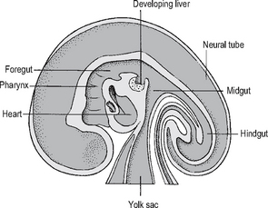


At the cranial end, six branchial arches develop in pairs, with the ears developing between the first (i.e. most cranial) and second arches. The limb buds are developing and grow into limbs with hands and feet, and the tail, which was prominent, has gone before the end of the eighth week.


And so, from an ovum and a spermatozoa, a miniature human being has developed. The various important surgical congenital anomalies which can affect the future baby will be discussed below.


PERSISTENCE OF THE BRANCHIAL ARCHES AND THEIR REMNANTS


During embryological development, the six branchial arches go through various stages of development and regression. They each originate from embryological mesenchyme and contain a core of cartilage, muscle, an artery and nerve supply from a cranial nerve.


The cartilage of the first arch develops into the malleus and incus (middle ear bones) and an associated ligament, the second into the stapes (third middle ear bone) and styloid and part of the hyoid bone, the third into the rest of the hyoid, and part of the fourth and sixth arch into the larynx, and most of the rest of the cartilage disappears. The muscles of the arches develop into facial muscles, each keeping their original nerve supply. The arteries become paired aortic arches, but the only ones which remain are the third (carotids), fourth (right subclavian on right and aortic arch on left) and sixth (right pulmonary artey on right and left pulmonary artery plus ductus arteriosus on left – which with its nerve supply from the vagus, the tenth cranial nerve, explains how the recurrent laryngeal nerve loops under the ductus arteriosus on the left and under the subclavian on the right).


Persistence or failure of complete regression of the branchial arches gives rise to many congenital abnormalities which are present at birth.


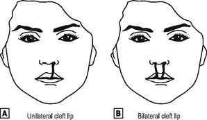
The first arch anomalies include cleft lip and palate (see p. 496). Upper preauricular sinuses and skin tags (which invariably contain cartilage) are usually superficial and can easily be excised, but a low preauricular sinus may have a deep internal connection to the first arch – a surprising finding for the unsuspecting surgeon who follows a track and ends up within the middle ear – and close to the facial nerve, with its consequent problems if damaged.


A branchial fistula arises from the second arch, from the anterior border of the bottom third of the sternomastoid muscle, and travelling up inside the neck to open in the tonsillar fossa in the pharynx. This fistula may present as a discharging dimple on the neck (as the fistula is lined by mucus-secreting glands), and it needs to be excised (usually requiring two separate neck incisions) in its entirety from the lower neck up to the pharynx to prevent continuous discharge, infection, or the rare possibility of subsequent malignant transformation. This second arch remnant may also present as a skin or cartilaginous tag at the site of the dimple.


A branchial cyst also originates from a remnant of the second arch without external connection, and contains the glairy fluid containing cholesterol crystals which typifies the mucus-secreting glands within the cyst. It should be excised before it gets infected.


Fistulae from the third or fourth arches are rare, but will also connect deeply internally, and can get infected and should be excised, again with the full awareness of the anatomy which may be involved.


LINGUAL THYROID AND THYROGLOSSAL CYSTS


The thyroid gland begins as an outgrowth from the midline of the tongue in the primitive pharynx, which moves caudally into the neck, looping around or through the hyoid bone and moving caudally further to its final site in the lower neck.


The thyroid may fail to descend, and remain as a small gland in the tongue at the site where it started its outgrowth (the foramen caecum, in the midline at the junction of the anterior two-thirds and posterior third of the tongue), or it may partially descend and be mistaken for a thyroglossal cyst – see below. This ‘ectopic’ or incompletely descended thyroid tissue is invariably hypoplastic, and should be removed, as it may suffer from all the pathological problems of a normally sited thyroid. The patient should be investigated to see if they have any normally sited thyroid, which most of them do not. The patient will usually become hypothyroid, as this gland is hypoplastic, and if it is their only thyroid tissue, and it is removed, the patient will certainly become hypothyroid, but without potential problems from the gland.


The thyroid may keep its connection to the tongue as the thyroglossal duct, and a cyst can develop anywhere along the line of this duct, the commonest site being at the level of the body of the hyoid bone. This thyroglossal cyst can present at any age, as a midline swelling in the upper neck, which moves upward when the tongue is protruded. Excision is advised, along with the whole thyroglossal tract (and consequently the midportion of the hyoid bone), in order to stop infection, following which excision is so much more difficult.


CONGENITAL DIAPHRAGMATIC HERNIA


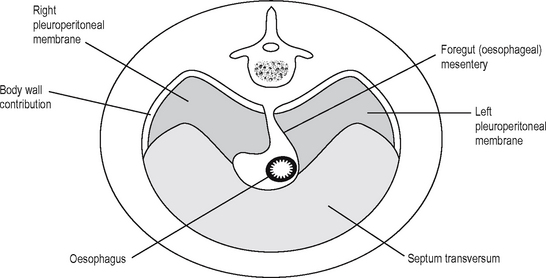
The diaphragm develops between the thoracic and abdominal cavity, and this is a complex process which is finished before the end of the eighth week (Fig. 16.4). As the embryo folds and carries the primitive heart and septum transversum caudally and ventrally, it carries part of the yolk sac dorsally to develop as the foregut. The lateral mesenchyme develops into the pericardioperitoneal canals, from which the pericardial cavity and the lungs develop, and is separated from the peritoneal cavity by the closure of the diaphragm. The motor nerve supply travels with the diaphragm as it descends, and so comes from a more cranial region than may be expected: C3–5. This explains the clinical observation that diaphragmatic inflammation/irritation, e.g. due to intraperitoneal blood, can demonstrate referred pain and can cause shoulder tip pain (which area is also supplied by C4).


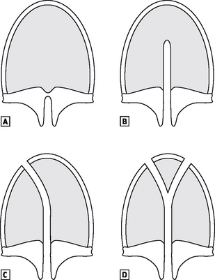
The diaphragm develops from the fusion of four parts:


There are different types of congenital diaphragmatic hernia, depending on which section has failed to close.


The most common defect is the posterolateral Bochdalek hernia (through the pleuroperitoneal canal) which is more common on the left, as that side closes last. Absence of the diaphragm can also occur, or absence of the central tendon. These three hernias tend to present early with respiratory distress soon after birth. The presence of the intestines within the pleural cavity antenatally prevents the normal development of the lung on the ipsilateral side, and mediastinal shift also prevents normal development of the contralateral lung. If the lung hypoplasia is severe, it is not compatible with life. Urgent supportive ventilation is required, and nasogastric aspiration of the gut, to decrease direct pressure on the lungs.


These diaphragmatic hernias must be dealt with surgically, after resuscitation of the patient (this is sometimes not possible in a neonate because of the severity of the lung hypoplasia) – up to 50% of babies born with congenital diaphragmatic hernias will die even today with all the modern management possibilities of oscillatory and jet ventilation, or extracorporeal membrane oxygenation (bypass).


Morgagni hernias are small defects in the anterior diaphragm close to the sternum, and are rarely associated with lung hypoplasia. They may be a coincidental finding on a chest x-ray taken for another reason. These hernias also require surgical repair.


GASTROINTESTINAL TRACT


The foregut develops from the yolk sac which folds in to the embryo at its cephalad end during the fourth week. From this foregut is derived the:


OESOPHAGEAL ATRESIA


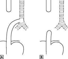
The foregut starts to divide into the oesophagus and the laryngotracheal tube during the fourth week. If it fails to do so correctly, there can be pure oesophageal atresia (in 8% of cases), or atresia associated with tracheo-oesophageal fistula – the commonest (in 80% of cases), being a fistula between the lower trachea and the distal oesophagus (Fig. 16.5).


The baby presents soon after birth, unable to swallow saliva, and an attempt to pass a tube into the stomach fails. An x-ray taken then will show the tube in the proximal oesophagus, and either no gas below the diaphragm (in pure oesophageal atresia) or gas below the diaphragm (in patients with oesophageal atresia and tracheo-oesophageal fistula). There is a high incidence (50% of babies) of associated anomalies described by the acronym VACTERL:


Management involves protection of the lungs from aspiration of saliva prior to surgical repair. This is usually by a right thoracotomy to ligate the fistula, freeing the distal oesophagus which is then anastomosed primarily to the upper oesophageal pouch. If primary repair is not possible, a feeding gastrostomy is performed to feed the baby until it has grown enough to perform a delayed primary anastomosis or oesophageal substitution, e.g. with stomach, colon, or small bowel.


INFANTILE HYPERTROPHIC PYLORIC STENOSIS


The stomach develops from a simple tubular part of the foregut by localised dilatation. The stomach rotates clockwise so that the vagus which followed the left side of the oesophagus supplies the anterior stomach. The mesentery which suspends the stomach from the posterior abdominal wall enlarges and becomes the greater omentum. The exit of the stomach into the duodenum is the pyloric canal.


All of the gastrointestinal tract has two layers of muscle: circular and longitudinal. The circular muscle only of the pylorus can become hypertrophied in some babies. This is often called ‘congenital’ hypertrophic pyloric stenosis, but does not actually exist at birth. The baby usually presents after 10–50 days (most commonly 3–5 weeks), as the pyloric canal is narrowed by the hypertrophied muscle, and milk is prevented from leaving the stomach. The stomach becomes full and peristalses vigorously to try to empty. This peristalsis may be visible on the baby’s abdomen. The baby will then vomit forcefully, which is described as projectile.


As the baby vomits fluid and gastric hydrochloric acid, the baby becomes dehydrated, hypochloraemic and alkalotic. This is reflected in the baby’s electrolytes and blood gases at presentation. The diagnosis is made by feeding the baby, to relax the baby. The visible peristalsis may be seen, and the abdomen is palpated to feel for the pylorus, which can be felt as a lump in the right upper quadrant, about the size and shape of an olive. After rehydration and correction of the acid-base balance, the baby is taken to theatre for a laparotomy, and the hypertrophied muscle is split, without opening the mucosa – a pyloromyotomy. This is a curative operation, and the baby will be fully fed within 24–36 h postoperatively and discharged home.

Stay updated, free articles. Join our Telegram channel

Dec 12, 2016 | Posted by in GENERAL & FAMILY MEDICINE | Comments Off on Paediatric disorders

Full access? Get Clinical Tree

Get Clinical Tree app for offline access