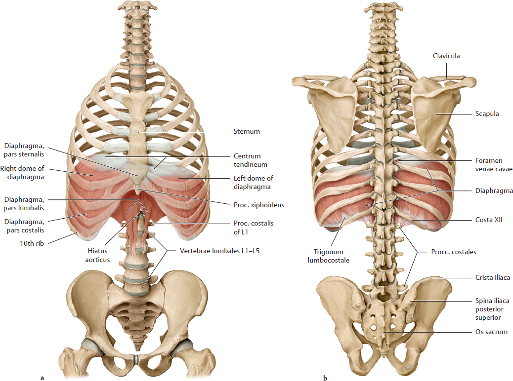
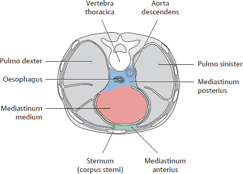
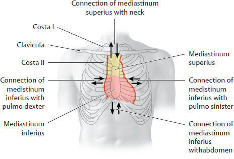
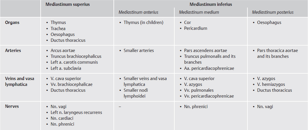
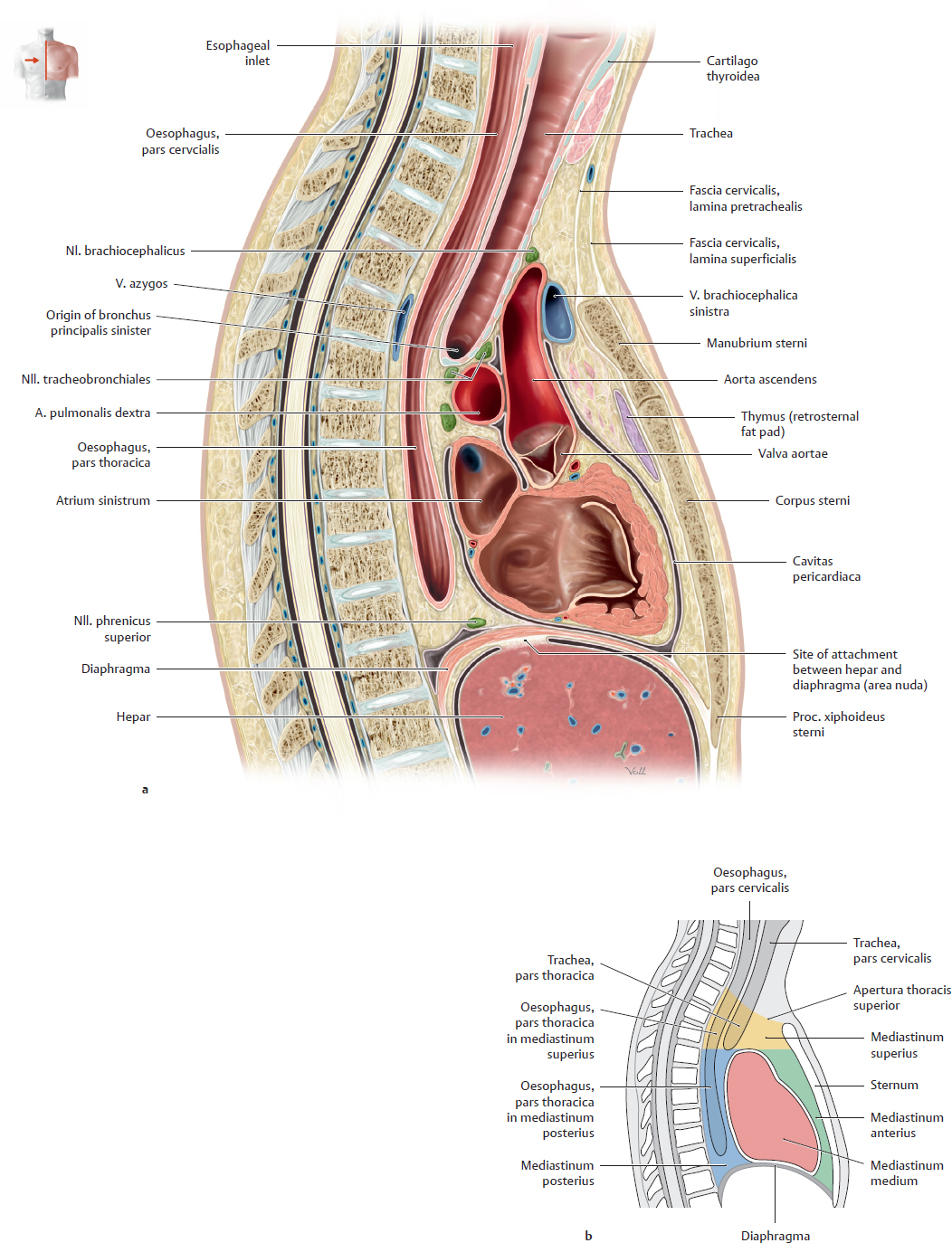
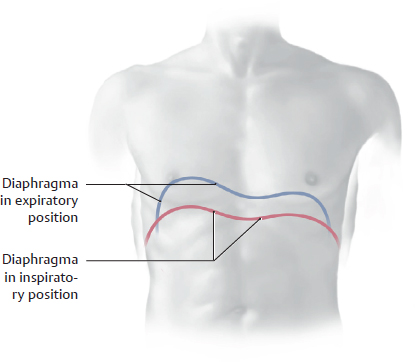
10. Overview and Diaphragma A Divisions of the cavitas thoracica and mediastinum Transverse section, superior view. The cavitas thoracica is divided into three large spaces: • The mediastinum, in the midline, is divided into an upper, smaller mediastinum superius and a lower, larger mediastinum inferius (see B). The mediastinum inferius is further subdivided, from front to back, into the mediastinum anterius, medium, and posterius. The mediastinum anterius is an extremely narrow space between the sternum and pericardium, containing only small vascular components (see table, C). • The paired cavitates pleurales on the left and right sides of the mediastinum are lined by tunica serosa (pleura parietalis) and contain the pulmones sinister and dexter. They are completely separated from each other by the mediastinum. The mediastinum extends further to the left than to the right owing to the asymmetrical position of the cor and pericardium. Because of this, the cavitas pleuralis (and pulmo) is smaller on the left side than on the right. The cavitates pleurales terminate blindly at their upper end, but the mediastinum is continuous with the connective tissue of the neck. B Principal neurovascular structures that enter and leave the mediastinum Mediastinum superius (borders the neck, yellow): • The nn. vagus and phrenicus, veins (tributaries of the v. cava superior), oesophagus, and trachea enter the mediastinum superius from the neck. • Arterial branches from the arcus aortae and the cervical part of the truncus sympathicus leave the mediastinum superius to enter the neck. Mediastinum inferius (borders the abdomen and cavitates pleurales, red): • The ductus thoracicus and ascending abdominal lumbar veins (the v. azygos on the right side, the v. hemiazygos on the left side) pass through the diaphragma to enter the mediastinum inferius. • The nn. vagus and phrenicus, portions of the pars sympathica of the nervous system, the aorta, and the oesophagus descend from the mediastinum inferius and pass through the diaphragma to enter the abdomen. Aa. and vv. pulmonales, vasa lymphatica, autonomic nerves (plexus pulmonalis), and the bronchi principales connect the mediastinum to the pulmones (and vice versa). C Contents of the mediastinum (for divisions see A) D Subdivisions of the mediastinum Midsagittal sections viewed from the right side. a Detailed view: simplified drawing of the pericardium, cor, trachea, and oesophagus in midsagittal section. This lateral view demonstrates how the atrium sinistrum of the cor narrows the mediastinum posterius and abuts the anterior wall of the oesophagus. Because of this proximity, abnormal enlargement of the atrium sinistrum may cause narrowing of the esophageal lumen that is detectable by radiographic examination with oral contrast medium. Radiologists call the area between the images of the cor and columna vertebralis the retrocardiac space. b Schematic view: subdivisions of the mediastinum (described in A, with contents listed in C). Note: Single diagrams cannot adequately show the components and configuration of the mediastinum, because of its asymmetry and extensions in all three axes. The anatomical relations in this space are best appreciated when viewed from multiple directions, at different planes (see also pp. 182 and 183). A Projection of the diaphragma onto the trunk Anterior view. The positions of the diaphragma in expiration (blue) and inspiration (red) are shown. The right hemidiaphragm rises as high as the fourth rib during expiration, and the diaphragma may fall almost to the level of the seventh rib at full inspiration. Note: • The exact position of the diaphragma depends on body type, sex, and age. • The left diaphragma leaflet is lower than the right due to the asymmetrical position of the cor. • Inspiration is marked by an overall depression of the diaphragma and also by a flattening of the diaphragm leaflets. • The diaphragma is higher in the supine position (pressure from the intra-abdominal organs) than in the standing position. • The degree of diaphragmatic movement during inspiration can be assessed by noting the movement of the hepatic border, which is easily palpated. • The diaphragma in a cadaver occupies a higher level than the expiratory position in vivo due to the loss of muscular tone.
10.1 Divisions of the Cavitas Thoracica and Mediastinum
10.2 Diaphragma: Location and Projection onto the Trunk
Basicmedical Key
Fastest Basicmedical Insight Engine