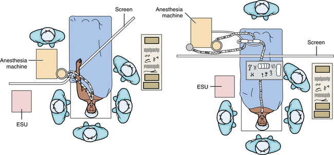
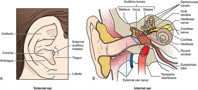
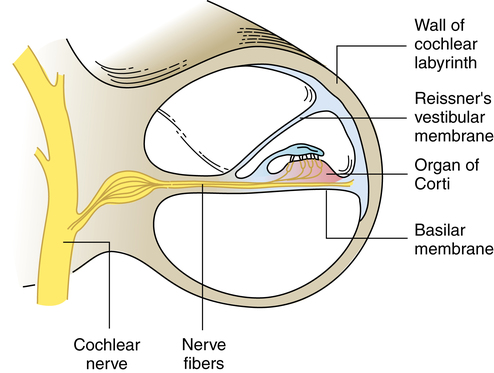
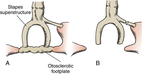
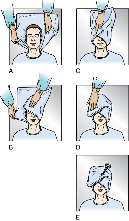
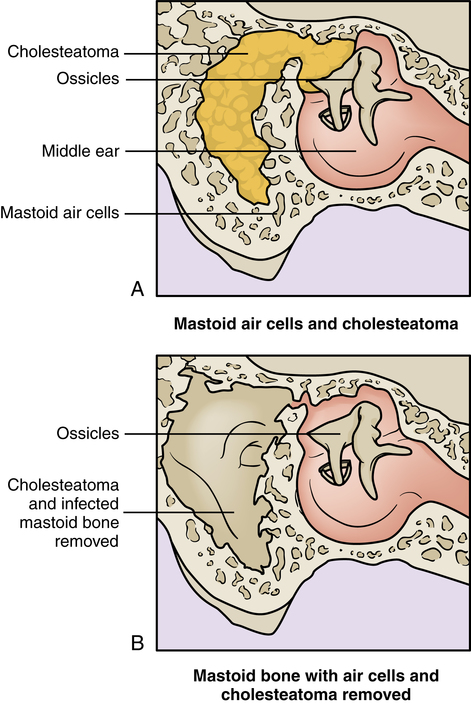
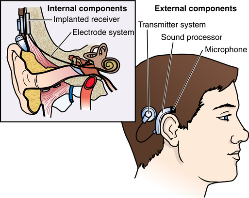
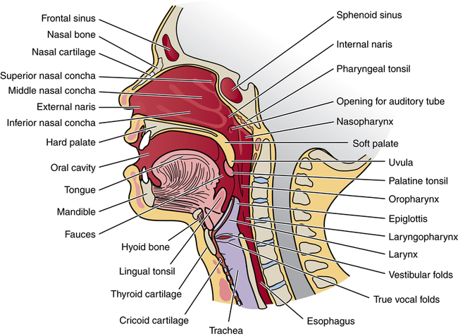
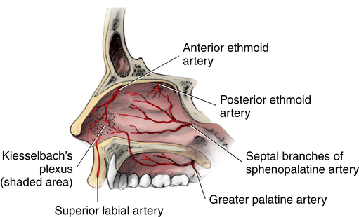
Otorhinolaryngologic and head and neck surgery

Chapter 41


Otorhinolaryngologic and head and neck surgery


General considerations in ear, nose, and throat procedures


Surgical procedures of the structures of the head and neck region are not within the province of any one surgical specialty. Subspecialists from many disciplines, most notably general surgeons, plastic surgeons, otolaryngologists, and dentists, limit their surgical practice to specific types of problems involving areas of the head and/or neck. Training in these subspecialties is included in specialty postgraduate programs.


Otorhinolaryngology has traditionally been concerned with research and surgical treatment of diseases of the ear (oto), nose (rhino), and throat (laryngo). Advances in scientific knowledge, diagnostic capabilities, and technology have broadened the scope of this field, which has led to subspecialization. General otorhinolaryngologists, commonly called ear, nose, and throat (ENT) surgeons, practice within the total scope of this specialty. Other surgeons confine their practice to one of the subspecialties: otology, facial surgery, or head and neck oncology. The certifying body for this specialty is the American Board of Otolaryngology, Head and Neck Surgery.


Dental and skeletal deformities of alignment and function coexist with aesthetic appearance. These deformities may be congenital, or they may be caused by trauma or disease. They may interfere with breathing, eating, swallowing, speaking, seeing, or hearing. A multidisciplinary team of surgical specialists is often required to reconstruct complex deformities. This team may be all-inclusive with a plastic surgeon, neurosurgeon, oral surgeon, orthodontist or prosthodontist, otolaryngologist, ophthalmologist, radiologist, and general surgeon; or it may be limited to two or three specialists. The team may also include a psychiatrist or psychologist, speech pathologist, and social worker. A team includes all of the specialties needed for complete preoperative assessment, intraoperative care, and postoperative rehabilitation of the individual patient.


Specific considerations include the following:


Anesthesia considerations


Anesthesia can be local or general. Use of local anesthesia minimizes bleeding and postoperative discomfort in addition to affording the surgeon observation of patient response. It allows patient cooperation. A qualified perioperative nurse may deliver moderate sedation and monitor vital signs, including cardiac function with electrocardiogram (ECG) and oxygenation with a pulse oximeter in the absence of an anesthesia provider. The nurse records the amount of local anesthesia administered and documents the patient’s intraoperative responses.


Monitored anesthesia care (MAC) may be used with an anesthesia provider in attendance for more complicated patients who need additional adjunctive medication and monitoring.


General anesthesia requires good control of the airway. The presence of an endotracheal tube in the mouth during general anesthesia can distort features during a surgical procedure in the oral cavity. Nasotracheal intubation may be necessary. In complex cases involving large areas of the head and neck, the patient may have a tracheostomy created and sutured in place for the duration of the procedure. The tracheostomy may be permanent or temporary depending on the procedure performed. Special endotracheal tubes are used if a laser is employed. The use of methylene blue dye in sterile saline for filling the endotracheal tube cuff is recommended. If the cuff is perforated, the dye acts as an indicator.


The anesthesia provider requires periodic access to the patient’s head and neck, as do the surgeon and sterile team. Positioning of the OR bed must provide space for the anesthesia machine and other equipment, such as the microscope. The anesthesia machine may be positioned near the patient’s side or feet. The endotracheal tube is taped to prevent becoming dislodged during the procedure; all connections must be tight, and tubes must be unkinked. Breathing circuits may need an extension hose if the anesthesia machine is at the foot of the OR bed. The anesthesia provider usually sits alongside the OR bed near the lower half of the patient on the opposite side of the surgeon (Fig. 41-1). Provisions are made for the anesthesia provider to have access to the intravenous (IV) line when the patient’s arms are tucked in at the sides. Extension IV tubing is used.


Pharmacologic considerations


Epinephrine in a local anesthetic, such as lidocaine, is frequently used as a vasoconstrictor in ENT procedures. Cocaine 4% provides local anesthesia and vasoconstriction when used topically on the surgical site. Cocaine can systemically block the effects of epinephrine and should be applied first and allowed to take effect. The epinephrine in the local anesthetic can cause cardiac dysrhythmias, and the anesthesia provider should be alerted when it is injected.7


Nitrous oxide diffuses into the middle ear, causing pressure. During ear procedures, such as tympanoplasty, the nitrous oxide is decreased or discontinued once the tympanic graft is placed to minimize inner ear pressure that could displace the graft.7


Neuromuscular blockade affects the nerves of the head and neck. The use of nerve conduction monitoring or nerve stimulators is adversely affected by strong skeletal relaxation drugs.


Positioning, prepping, and draping


Although the oral cavity is considered contaminated, sterile equipment and sterile technique are preferred to avoid introducing exogenous bloodborne microorganisms.


The patient is positioned supine. The arms are usually tucked in at the sides or secured over the abdomen. The patient’s head is positioned in a donut or horseshoe head rest to prevent movement during the procedure. Many procedures are done with the patient in a slight reverse Trendelenburg’s position or with a roll under the shoulders to hyperextend the head. Some surgeons prefer to have the patient in a modified beach-chair position. This helps anatomic positioning and controls bleeding.


The patient’s eyes should be protected by corneal shields and sterile ophthalmic lubricant. Care is taken not to lean on the patient’s draped face during the procedure. Pressure on the patient’s eye can cause eye damage and cause cardiac dysrhythmias.


Care with antiseptic solutions during a facial prep is necessary because they are painful and irritating if they are allowed to touch a perforated eardrum or get into the eyes. A cotton or rayon ball can be used to occlude the ear during facial prepping. Cotton fibers can stick inside the ear, causing itching and discomfort postoperatively, and a small nonradiopaque gauze (2 × 2) can be used instead. Some solutions, such as chlorhexidine or hexachlorophene, can cause permanent damage. Consult the manufacturer’s recommendations before use.


The awake patient under local anesthesia may experience anxiety with drapes over the head. Oxygen (6 to 8 L/min) administered during the procedure affords relief. A patient with chronic lung disease should be given oxygen at 3 L/min.


Care is taken not to permit the use of cautery in an oxygen-rich environment. Oxygen builds up under the drapes and could create a fire hazard. Oxygen is combustible.


A patient having a procedure longer than 2 hours should have a Foley catheter inserted to monitor urine output. The drainage bag should be positioned in the view of the anesthesia provider.


Some surgeons prefer a turban-style head drape. Figure 41-2 depicts the method used to create a turban from a drape. Patients undergoing local anesthesia may wear pajama-type bottoms or underpants to the OR in some facilities. Patients having general anesthesia should wear only a gown.


Equipment and instrumentation


Illumination is provided by the overhead spotlight, the operating microscope, the endoscope, or the surgeon’s fiberoptic headlight. Some surgeons want the room darkened during endoscopy. Various types of fiberoptic endoscopes with appropriate accessories are used. The diameter of aspirating tubes and forceps is small enough to pass through small lumens, and the length is long enough to extend beyond the end of the scope. The lighting mechanism and all accessories should be checked for working order before the patient is brought to the room.


Suction should be available at all times and should include several patent cannulas. The degree of suction should be variable and usually includes a thumb hole for intermittent release of the vacuum. Cannulas with very fine suction tips should be irrigated frequently to avoid blockage. Fine suction tips are equipped with stylets for clearing the lumens.


Suction irrigators are used with sinus endoscopes. Irrigation equipment and solution at body temperature should be available to remove bone dust, clean burrs, and rinse suction apparatus. Warm solutions would cause vasodilation, with the potential for obscured vision by venous bleeding.


Special care with electric appliances is indicated. Bipolar and monopolar electrosurgery are used to control bleeding. Many ENT surgeons use Coblation technology, which is a form of high-frequency bipolar energy in a conductive medium, usually saline. The target tissue is removed using a lower temperature than traditional electrosurgery devices.


Compressed air or nitrogen drills with a foot-pedal control are used on bone, such as the mastoid area. Only the operator should activate the foot pedal. A number of drills for extremely fine work, such as stapes sculpturing, are powered by small electric motors fitted into a handpiece. The tip of the drill is cooled by small drips of irrigation solution during use.


Instrumentation is varied to suit the area. It includes very delicate, small endoscopic and microsurgical implements in addition to various sized bone instruments because of the extensive involvement of cartilage and bone in the facial structures and skull. These areas have relatively little soft tissue. Many instruments are angled with small jaws to permit insertion without obscuring visualization.


Lasers may be used. Carbon dioxide (CO2) lasers are used to vaporize tissue. Argon lasers coagulate tissue by heat generation. Potassium titanyl phosphate (KTP) lasers coagulate to shrink or debulk tissues. Neodymium:yttrium-aluminum-garnet (Nd:YAG) lasers cut and coagulate to minimize bleeding. Appropriate ebonized or dulled instrumentation and attachments must be available for each type of laser.


Laser surgery is usually done in conjunction with the operating microscope or endoscope. Microsurgical techniques are used because they facilitate distinction between normal and diseased tissue and allow more accurate dissection. All safety precautions are taken to avoid ignition and injury when a laser is used. Team members must wear protective eyewear of the correct optical density for type of laser in use.


Cryosurgery may be used to destroy tissue in accessible areas, especially those that bleed profusely if incised.


Hemostasis and drugs on the sterile field


One or two drops of blood can obscure a microsurgical field. Hemostatic aids and sponges should be ready at all times. Gelfoam pledgets soaked in 1:1000 epinephrine are commonly used. Soaked pledgets are kept in a labeled Petri dish and passed only with forceps. Press the saturated pledget against the side of the dish to wring out some of the moisture before passing to the surgeon.


Various solutions and medications are on the sterile field, for example, cocaine, lidocaine, and epinephrine. Each container and delivery device should be clearly labeled for distinction between irrigation solutions and drugs. Unused drugs are discarded in the presence of another team member after the case is completed. The amount of unused cocaine should be documented before discarding.


Sponges and compressed patties are relatively small and are a distinct hazard when blood soaked because they can occlude an airway. All items used in the OR are counted or accounted for in their entirety at the end of the procedure.


Ear


Anatomy of the ear


The structures of the outer and inner ear (Fig. 41-3, A and B) are concerned with two functions:


Anatomically, the ear is divided into three parts: external, middle, and inner.


Middle ear


The middle ear consists of the tympanic cavity, a closed chamber that lies between the tympanic membrane and the inner ear. Within this cavity are the three smallest bones in the body—an ossicular chain comprising the malleus, incus, and stapes. They resemble a hammer, anvil, and stirrups, respectively. The malleus, which is attached to the eardrum, joins the incus, the extremity of which articulates with the stapes, the innermost bone. The footplate of the stapes fits in the oval (vestibular) window, an opening in the wall of the inner ear.


The bones of the ossicular chain must be able to move mechanically to conduct sound from the eardrum to the inner ear. The round (cochlear) window, also between the middle and the inner ear, equalizes pressure that enters through the oval window.


The middle ear opens into the nasopharynx by way of the eustachian tube. Normally closed during swallowing or yawning, the eustachian tube aerates the middle ear cavity. This mechanism is essential for adequate hearing.


Posteriorly the middle ear exits to the mastoid process. This inferior projection of the temporal bone is a honeycomb of air cells lined with mucous membrane. Because the antrum of the mastoid process connects with it, middle ear infection may produce mastoiditis. The middle ear is situated in the tympanic portion of the temporal bone; the inner ear is situated in the petrous portion, which integrates with the base of the skull. The tympanic portion also forms part of the ear canal.


Inner ear


The end organs of hearing and equilibrium are situated in the inner ear. The two main sections—cochlear and vestibular—have distinct, although coordinated, functions. The cochlea, a bony spiral, relates to hearing. The vestibular labyrinth, which is composed of three semicircular canals, relates to equilibrium. These structures house two separate fluids—endolymph and perilymph—which nourish and protect the hearing receptors.


The neuroepithelium of the organ of Corti, the end organ of hearing, holds thousands of minute hair cells, which respond to sound waves that enter the cochlea via the oval window. The neuroepithelium of the vestibular portion also contains hair cells. Rapid head motion produces current in the endolymph that may result in nausea or vertigo. The eighth cranial (vestibulocochlear) nerve governs reflexes to muscles to maintain equilibrium and controls hearing.


Physiology of hearing


Sound or pressure waves enter the auricle. They pass along the ear canal to the tympanic membrane. The vibration of the waves is transmitted across the middle ear sequentially by the ossicles. Amplification of sound is enhanced to some extent by mechanical action of the ossicles but mainly by aerial ratio. A large volume of sound wave pressure from the tympanic membrane funnels to a small reactive area, the stapedial footplate, intensifying sound. At the footplate of the stapes, sound pressure is transferred to the inner ear via the oval window. The hair cells of the organ of Corti are set in motion by disturbance of the inner ear fluids as sound wave pressure moves from the oval to the round window. Mechanical energy is converted to electrical potential, which is delivered to the brain along the eighth cranial nerve. The brain interprets the impulse (sound) as hearing.


Pathology of hearing


Hearing affects the quality and quantity of interpersonal interactions. It is a major sense for communicating within one’s environment.5 Loss of hearing therefore affects social relationships. The type of deafness or hearing loss in varying degrees may result from the following:


1. Disease, such as otosclerosis, in which changes in the bony capsule of the labyrinth occur. Otosclerotic bone invades the stapedial footplate, resulting in its fixation and ultimate inability to vibrate in the oval window. Hearing loss is gradual but progressive. This type of deficiency can be surgically corrected when auditory nerve endings are not destroyed.


2. Trauma, such as a perforated eardrum, requiring repair to restore function and aerial ratio.


3. Infection, usually controlled by antibiotics. Although it is more common in children, infection may also occur in adults. It may cause accumulation of fluid in the middle ear. Mastoiditis results from extension of otitis media.


a. Serous otitis media may result from obstruction of the pharyngeal orifice of the eustachian tube. If blocked, for example, by hypertrophied adenoid tissue, infection, or allergic swelling, the eustachian tube is unable to equalize pressure because air cannot enter the middle ear from the pharynx. The vacuum or negative pressure thus created causes serum to be drawn into the tympanic cavity from blood vessels in the middle ear mucosa. Recurrent otitis media may require drainage of purulent exudate if conservative treatment fails.


b. Acute otitis media may require drainage of purulent exudate if conservative treatment fails.


c. Chronic otitis media with or without mastoiditis may follow recurrent otitis media with tympanic membrane perforation. It can produce a chronically draining ear.


Differential diagnosis


Measurements that compare bone conduction with air conduction are important in differential diagnosis. Bone conduction refers to hearing as transmitted through the skull; air conduction refers to transmission of sound waves from the tympanic membrane to the inner ear via air. Hearing loss caused by a defect in the external or middle ear, referred to as conductive loss, is a mechanical obstruction of air conduction that usually can be helped by surgical intervention. When the decrement is in the inner ear, referred to as perceptive or sensorineural loss, damage to nerve tissue and/or sensory paths to the brain is not benefited by a surgical procedure. Cochlear nerve endings are the main component of sensorineural hearing (Fig. 41-4).


Auditory acuity and function are measured by various tests. The audiogram is one measurement tool. Computer-averaged tomography is used to measure and analyze electrical impulses, known as auditory brainstem responses, from the brain and cortical auditory pathway. An acoustic reflex latency test of the stapedius reflex provides information about hearing sensitivity. Auditory brainstem evoked potentials assess the patient’s hearing threshold.


Surgical procedures of the ear


Current techniques, instrumentation, lasers, and the operating microscope have enhanced the capability of otologists.


General considerations


1. Local anesthesia may be used for a minor procedure on the external ear, but general anesthesia generally is used to avoid patient movement while the surgeon is manipulating delicate structures in the middle or inner ear.


2. The patient’s head is turned with the affected side up and stabilized in a donut. The pinna on the unaffected side should be protected from pressure.


3. Lint-free drapes are preferred. It is mandatory that gloves be free of powder and lint. The formation of a powder granuloma in the oval window can cause irreversible sensorineural hearing loss.


4. The operating microscope is used for many otologic procedures.


5. Compressed absorbent patties (cottonoids) moistened with normal saline solution, rather than gauze sponges, are frequently used. They must be counted.


6. Hemostasis may be achieved with epinephrine, absorbable hemostatic sponges or oxidized cellulose, laser, and bone wax.


7. Prosthetic devices should be available in an assortment of types and sizes. Tissue allografts may be used.


8. A nerve stimulator may be used to identify facial, acoustic, cochlear, and/or vestibular nerve branches. Evoked potential audiometry also may be used to monitor the seventh and eighth cranial nerves.


9. Bone instruments, including powered drills, are used for opening the temporal bone.


10. CO2, Nd:YAG, argon, and KTP lasers are used during otologic procedures to control bleeding, divide nerves, and/or vaporize tissues.


11. Pressure dressings are usually applied. Some surgeons place 1 drop of phenylephrine (Neo-Synephrine) in the ear canal postoperatively.


External ear procedures


Middle ear procedures


Mastoidectomy.

Mastoidectomy, the eradication of mastoid air cells, may be indicated to relieve complications of acute or chronic mastoiditis. Mastoidectomy is more commonly performed in conjunction with a reconstructive procedure (see “Tympanomastoid Reconstruction”). A cholesteatoma can form after repeated infections (Fig. 41-5).


Tympanoplasty.

Tympanoplasty, as a general term, refers to any procedure performed to repair defects in the eardrum and/or middle ear structures for the purpose of reconstructing sound conduction paths. The degree of hearing improvement after tympanoplasty is related to the degree of damage. Preferably, the ear should be uninfected at the time of the surgical procedure. If not, infected tissue is debrided. As microsurgical procedures, tympanoplasties are classified into five types:


1. Type I (myringoplasty) is closure of a perforation in the tympanic membrane caused by infection or trauma. The ossicular chain is normal. Autologous fascia or a vein is used to repair the perforation. A vein graft is taken from the patient’s forearm or hand. Fascia, more commonly used as a patch over a perforation, is obtained from the temporalis muscle.


2. Type II is closure of a perforated tympanic membrane with erosion of the malleus. The graft is placed against the incus or remains of the malleus.


3. Type III replaces the tympanic membrane to provide protection for the stapes and round window. The tympanic membrane, malleus, and incus have been destroyed by disease. The stapes is intact and mobile. A homograft of tympanic membrane with attached malleus and incus is placed in contact with the normal stapes, permitting transmission of sound.


4. Type IV is similar to type III except that the head, neck, and crura of the stapes are missing. The mobile footplate may be left exposed with the graft placed around it. The air pocket between the graft and the round window provides sound protection for the round window. To conserve the middle ear hearing mechanism, homograft transplantation of tympanic membrane and ossicles may be used to rebuild the chain.


5. Type V is similar to type IV except that the stapedial footplate is fixed because of otosclerosis (osteospongiosis). A fenestra (small opening) is made in the horizontal semicircular canal. The homograft seals off the middle ear to provide sound protection for the round window.


Reconstruction of the middle ear may be done with a synthetic bioinert material such as high-density polyethylene sponge (Plastipore). Fibrin glue also is used. Partial and total ossicular replacement prostheses have been developed.


Tympanomastoid reconstruction.

Tympanoplasty may be combined with either simple or radical mastoidectomy. The mastoid is drained and cleaned before reconstruction of the eardrum or middle ear ossicles. After incision behind the auricle, the tympanic membrane and tympanic cavity are inspected. The mastoid antrum is entered by drilling through mastoid bone.


Sometimes in chronic otitis media, the mucous membrane of the tympanic cavity is replaced by epithelium from the ear canal as it grows through a perforation in the eardrum. Desquamated skin cells that cannot escape form a ball or cyst, known as a cholesteatoma. If present in the middle ear or mastoid, a cholesteatoma is removed during mastoidectomy and/or tympanoplasty. If left intact, a cholesteatoma can cause permanent hearing loss, balance disturbance, infection, and facial nerve paralysis.


Stapedectomy and stapedotomy.

Conductive hearing loss can result from fixation of the stapes, most often caused by otosclerosis. The surgeon aims to restore vibration from the incus to the mobile oval window membrane to transmit sound. A stapedectomy involves partial or total removal of the stapes.


A partial stapedectomy removes only the fixed footplate (Fig. 41-6); a total stapedectomy removes the entire stapes, including the footplate. A stapedotomy (small opening into the footplate) may be the preferred procedure. The opening is made with a handheld perforator, microsurgical power drill, or laser. An argon, CO2, or KTP laser may be used for this purpose. A low-wattage lasing beam avoids thermal damage to the perilymph and inner ear structures. After partial or total stapedectomy or stapedotomy, the remaining superstructure is used to reconstruct the sound-conducting mechanism.


An incision is made deep in the canal near but not in the eardrum. The eardrum is folded over, giving access to the middle ear. The stapes is disconnected from the incus, fractured by fine microinstruments, and removed. The oval window is sealed by a graft of vein, perichondrium, fascia, fat, or an absorbable hemostatic sponge over the oval window. A prosthesis is inserted and connected to the incus and to the graft, thus restoring sound conduction. Prostheses are made of various inert materials such as polyethylene, stainless steel, or tantalum.


By performing the microsurgical procedure with the patient under local anesthesia, the surgeon can reposition the eardrum and use his or her voice to test whether the patient’s hearing is improved. Otosclerosis usually involves both ears, but stapedectomy is performed on only one ear at a time.


Stapes mobilization.

The stapes is manipulated at the footplate to restore normal function. A break through an otosclerotic lesion is achieved by means of transcrural pressure or direct application of chisels and picks to the footplate. A mobile, unaffected portion of a functioning stapes remains. Various techniques are used, with or without the use of prosthetic devices. The advantage of the procedure is that the preserved stapedial footplate provides natural protection for the inner ear. The disadvantage is that frequently a continuing otosclerotic process causes the footplate to become refixed. Therefore stapedectomy is more popular because it produces long-lasting results.


Stapes procedures are performed under direct vision with the operating microscope. The procedures do not disturb the integrity or position of the eardrum.


Inner ear procedures


Removal of an acoustic neuroma.

Acoustic neuroma resection may be performed by an otologist and/or a neurosurgeon, depending on its location and the extent of neurologic involvement. An acoustic neuroma is a slow-growing, encapsulated, benign tumor of the eighth cranial (acoustic) nerve. It originates in the neural sheath in the internal auditory canal but grows to involve nerve fibers in the posterior fossa. Initially the patient experiences unilateral hearing loss and disturbances, especially tinnitus, and equilibrium problems such as mild vertigo.


The syndrome may resemble Ménière’s disease or an expanding intracranial tumor. Early differential diagnosis is enhanced by brainstem evoked response audiometry, vertebral angiography, and small-volume air-contrast computed tomography (CT).


A small neuroma, confined to the internal auditory canal, may be resected by the otologist using a microsurgical technique through a middle fossa approach to preserve hearing. If a translabyrinthine approach is used to gain access to the internal auditory canal posterior to the inner ear structures, the patient will have total hearing loss after the surgical procedure. Acoustic neuromas extending into the cranial cavity are resected by the neurosurgeon. The CO2 laser may be used to excise acoustic neuromas through a transmastoid or craniotomy approach. Cerebrospinal fluid leak and meningitis are complications of the craniotomy approach.


For select patients, stereotactic radiosurgery can be performed on an outpatient basis with local anesthesia. Hearing is preserved in 50% of these patients.


Implantation of a cochlear prosthesis.

Cochlear implants have been used for adults since the 1980s, but it was not until the 1990s that they were used for children. Children as young as 1 year old can be candidates for implantation. A cochlear implant can restore perception of sound to patients who have profound sensorineural deafness not responsive to external amplification of a hearing aid. They are indicated for use in patients who have intact eighth cranial nerve function.


The implant is an electronic device that converts sound waves into electrical signals to stimulate cochlear nerve fibers in the absence of functioning hair cells. Several devices are available. All of them have external and internal components (Fig. 41-7). The external part, which is attached behind the ear, has a microphone/transmitter and a speech processor/receiver. The internal part has a receiver/stimulator and electrode/channel that is threaded through the cochlea.


The internal electrode attaches to wires permanently implanted in the cochlea. Through a postauricular incision, a simple mastoidectomy is performed. Under the operating microscope, the facial recess between the posterior canal and the facial nerve is enlarged to expose the chorda tympani nerve and the middle ear. The electrode is securely seated in the mastoid cavity and placed through a recess into the middle ear. It is directed through an opening made in the round window into the scala tympani until it meets resistance. Placement is critical. A plug of fascia from the temporalis muscle is placed around the electrode at the round window to prevent perilymph leakage.


The internal electrode stimulates the auditory nerve to interpret sound. The single-channel model stimulates the nerve randomly so that the patient can discern environmental sounds but not speech. A multichannel electrode enables the patient to distinguish environmental sounds and some speech by differentiating the frequency, volume, and pitch of sound waves. With a single-channel electrode, a ground wire is placed under the temporalis muscle in the mastoid or middle ear. A multichannel electrode does not have a ground wire. The internal receiver is sutured in position over the temporal bone behind the ear.


Electromagnetic components and/or a titanium enclosure for the receiver may act as an electrical ground device. Only bipolar electrosurgery is used if additional surgery is needed after placement of the receiver. Patients with cochlear implants should avoid MRIs because of metal components within the implanted device.


The external microphone/transmitter component of the implant activates the internal receiver/stimulator. Transmission may be percutaneous or transcutaneous. In the percutaneous model, a direct wire to a receiver implanted behind the ear connects the transmitter. In the transcutaneous model, the transmitter converts electrical energy into magnetic currents that pass through intact skin to the receiver. In both models, sound enters the microphone and is transmitted to a speech processor, worn outside the body, where it is encoded into electrical signals or energy and amplified. The coded signals return to the transmitter and are passed to the internal receiver. The patient can adjust amplification of environmental sounds.


Nose


Anatomy of the nose


The supporting structures of the nose consist of two nasal bones and the nasal processes of the maxillary bones superiorly, the lateral cartilages and connective tissue inferiorly, and the septum. The septum, composed of bone posteriorly and cartilage anteriorly, divides the nose into two chambers lined by mucous membrane. The anterior portion, or vestibule, holds the nasal hairs. The external anterior orifices are called nares.


The internal portion of the nose, the nasal cavity (Fig. 41-8) extends to the nasopharynx, the space behind the choanae (funnel-like posterior nasal orifices). The nose communicates with the ear via the eustachian tube. The hard and soft palates divide the nasal and oral cavities. The ethmoid bone separates the nasal and cranial cavities.


The paranasal sinuses are the frontal, maxillary, ethmoid, and sphenoid. Ostia (openings from the sinuses and nasolacrimal ducts) are located in the nasal lateral walls. The ostia provide a drainage system for the sinuses, as well as aerate them. Three turbinate bones (superior, middle, and inferior) are also situated in the lateral walls. These bones are covered with a vascular mucosa. Beneath each turbinate is a corresponding meatus. Tears drain into the nose through the nasolacrimal duct that enters the inferior meatus. Drainage from the paranasal sinuses is passed to the nose through the middle and superior meatus.


External and internal carotid arteries and their branches supply blood to the nasal region. Because of the extensive vascularity, lymphatic supply, and proximity to the brain, infections on or about the face are potentially very dangerous. Microorganisms may readily be carried to the cavernous sinus, or thrombi may form in the cavernous sinus. The sense of smell is derived from the first cranial (olfactory) nerve. The sensory nerve supply of the nasal area is associated with the trigeminal or fifth cranial nerve.


Surgical procedures of the nose


Nasal procedures are concerned with two factors: adequate ventilation to accessory spaces and adequate drainage from them. Abnormalities in structure, congenital or traumatic, and disease processes hinder function. Corrective procedures are done to relieve obstruction, to ensure drainage, to resect tumors, or to control bleeding (epistaxis).


General considerations


1. CT and rhinoscopy are often performed preoperatively to diagnose the nature and extent of pathologic conditions, especially in the paranasal sinuses.


2. Many nasal procedures are performed with the patient under local anesthesia with or without IV sedation.


3. Paranasal sinuses and tissues underlying the mucosa are considered sterile. Therefore, instrumentation should be sterile, although the nasal cavity is considered a contaminated area because the instrumentation is entering a vascular bed. Venous drainage could easily carry microorganisms to the cavernous sinus, causing an intracranial infection.


4. CO2, Nd:YAG, and KTP lasers may be used. Endoscopes also are used. All equipment and accessories should be checked for working order.


5. Nasal packing is inserted at the end of most procedures except endoscopic sinus and laser procedures. A mustache dressing may be placed under the nose to act as a drip pad. Postoperatively the patient should be positioned in a head-up position to minimize bleeding. Ice is placed over the nasal bridge for the first 48 hours. The ice should be used in 20-minute intervals only.


Nasal cavity procedures


The supporting structures surrounding nasal air passages can be injured or displaced. Acute or chronic disease processes can cause dysfunction or obstruction.


Epistaxis.

Most nosebleeds are caused by trauma, usually at Kiesselbach’s plexus of arteries and veins (also known as Little’s area) in the anterior part of the nasal septum (Fig. 41-9). Dehumidified air may cause changes in the mucosa and splitting of tiny vessels. Bleeding may be spontaneous, as in patients with arteriosclerosis, hypertension, or blood dyscrasia. Epistaxis is usually anterior and unilateral. In people with systemic disease, such as leukemia, or with severe fracture, the bleeding is frequently posterior and more severe. Management involves locating the precise bleeding site and promptly instituting appropriate therapy. Severe hemorrhage places the patient in a precarious condition. Hypovolemia should be corrected preoperatively.


Posterior pack.

A posterior pack may be necessary for constant pressure when bleeding is severe in the posterior part of the nose. The pack consists of rolled gauze securely tied to the middle of a length of narrow tape or strong string; commercial packs are available. To control infection and odor, gauze is lubricated with antibiotic ointment before insertion.


After a catheter is passed into the mouth via the nose, one end of the tape is tied to the oral end of the catheter. The catheter and attached tape are then drawn back through the mouth and out one nostril, thereby pulling the pack up into the nasopharynx. Thus one end of the tape comes out the nose, the other end out the mouth. These ends are secured to the patient’s cheek with adhesive tape. The nasal end is taped to prevent the pack from slipping into the throat; the oral end facilitates removal of the pack. Some oozing may persist despite packing. The pack is left in place until bleeding is arrested, usually for at least 48 hours, but prolonged use can lead to otitis media or paranasal sinusitis.


Patients with postnasal packs are often apprehensive and uncomfortable. They must breathe through the mouth. Posterior packs tend to reduce arterial oxygen tension. All patients, especially geriatric patients and those with marginal pulmonary function, should be observed carefully for respiratory problems that may result from the pack dropping into the hypopharynx.


Artery ligation.

A microsurgical procedure is performed to control persistent nasal hemorrhage by reducing the blood supply to the posterior portion of the nose. Some surgeons prefer artery ligation to packing, or it may be performed with a pack in place. Through an incision in the oral mucosa, removal of the posterior wall exposes the maxillary sinus for transantral ligation. Terminal branches of the internal maxillary artery, a branch of the external carotid artery, are exposed, identified, and ligated with metallic clips. Electrocoagulation is employed to control intraoperative bleeding, but if bleeding is excessive, creation of a nasoantral window establishes drainage. The replaced posterior mucosal flap is covered with an absorbable gelatin sponge, and the incision is closed.


Through an incision along the left side of the nose, ligation of the anterior and posterior ethmoidal arteries is helpful in controlling bleeding in the superior aspect of the nose.


The argon or KTP laser may be used to control severe epistaxis. If a laser is used to control bleeding, nasal packing usually is unnecessary.


Nasal obstruction.

Surgical intervention can provide relief for certain types of nasal obstructions. For example, after the mucosa is shrunk with a vasoconstrictor and secretions are suctioned, a foreign body is removed with a forceps. An abscess or hematoma may need to be drained to relieve pressure. Accumulation of pus or blood separates the perichondrium, the connective tissue, from underlying cartilage. This may cause necrosis of cartilage with resultant deformity. Infection must be eradicated to avoid extension to the brain.


Nasal deformity.

Surgical intervention can restore contour and/or improve function after an injury. A deformity in nasal structure also may be corrected to improve cosmetic appearance.


Stay updated, free articles. Join our Telegram channel

Apr 6, 2017 | Posted by in GENERAL SURGERY | Comments Off on Otorhinolaryngologic and head and neck surgery

Full access? Get Clinical Tree

Get Clinical Tree app for offline access