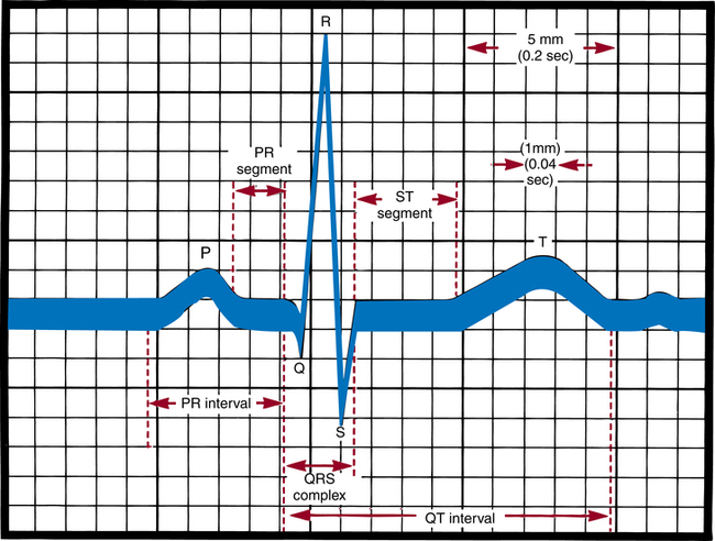
Physiologic maintenance and monitoring of the perioperative patient

Chapter 27


Physiologic maintenance and monitoring of the perioperative patient


The development of successful controllable anesthesia has made modern surgery possible.5,10,14 Because anesthesia is an adjunct to most surgical procedures, familiarity with various anesthetic agents, their interaction with certain drugs, and their potential hazards is a necessity. The perioperative nurse responsible for patient monitoring may detect the onset of complications and help avert an undesired outcome.


Working with anesthesia providers in the perioperative environment gives the learner an unparalleled opportunity to master immediate resuscitative measures and their effectiveness, and an understanding of the care of unconscious and critically ill patients. For example, the learner daily observes endotracheal intubation, ventilatory control, insertion of arterial and venous cannulas, fluid replacement, and sophisticated hemodynamic monitoring.14


No surgical procedure is minor. Surgery and anesthesia impose on the patient certain inescapable risks, even in supposedly ideal circumstances. Overall, however, the anesthetic-related surgical mortality rate is relatively low. Preexisting patient factors, such as age and medical condition, and those related to the circumstances of the surgical procedure, such as type, duration, and elective versus emergency procedure, are more significant in determining surgical mortality.


The American Society of Anesthesiologists (ASA) has established standards for basic perioperative monitoring. Specialization in monitoring equipment and the use of computers to record monitoring data are facets of contemporary anesthesia practice.


Monitoring physiologic functions


Preoperative patient assessment establishes a baseline and provides valuable information with which intraoperative and postoperative patient care is planned and evaluated. Establishing a preoperative baseline enables the caregiver to be alerted to changes in the patient’s physiologic condition that may require prompt attention.


Surgical and anesthesia techniques have become increasingly complex, which allows many critically ill patients of all ages to undergo surgical procedures. The anesthesia provider carefully monitors the patient’s condition through the entire perioperative process and keeps the surgeon informed of important changes.


Monitoring implies keeping track of vital functions. During extensive surgery with the patient under anesthesia, the body is subjected to physiologic stress. Bleeding, tissue trauma, potent drugs, large extravascular fluid shifts, multiple transfusions, and a surgical position that may inhibit breathing and circulation all contribute to altered physiology. These factors can induce significant cardiopulmonary dysfunction.


Evaluation of the patient’s responses to these stressors includes observation, auscultation, and palpation. The assessment is enhanced with the use of electronic or mechanical devices that reveal physiologic trends and subtle changes and indicate responses to therapy. Most of this equipment is expensive and is incorporated into the anesthesia machine. Compact precise devices with miniaturized circuitry, better visibility, and easier maintenance are incorporated into portable models that allow for continued monitoring after the patient leaves the OR.


Computers improve and expedite analysis of data. They are sophisticated data collection and management tools that assist in physiologic assessment, diagnosis, and therapy. Clinical computers vary from single-function devices to complex, multifunction, real-time systems that acquire, store, and display data; organize information; display trends; and perform calculations.


Personnel who use electronic and computerized monitoring equipment must understand its use and function, be experienced in its interpretation, and be able to determine equipment malfunction easily. Instrumentation should augment, not replace, careful observation of the patient. Monitoring equipment can be inaccurate. Information from monitors should be compared with physical assessment data.


Perioperative nurses involved with patient monitoring should remain current in the knowledge of physical assessment and the use of the equipment. Periodic competency testing should be performed. Written policies, procedures, and guidelines should be available for reference.


The spectrum of monitoring devices is broad. It ranges from noninvasive to invasive. Noninvasive monitors do not penetrate the body or a body orifice. Conversely, invasive monitors penetrate skin or mucosa, or they enter a body cavity. Some parameters can be measured with both noninvasive and invasive methods. Perioperative nursing responsibilities may include assisting with sophisticated hemodynamic monitoring to evaluate the interrelationship of blood pressure, blood flow, vascular volumes, and physical properties of blood, heart rate, and ventricular function.


Detection of early changes in hemodynamics allows prompt action to maintain cardiac function and adequate cardiac output. Monitoring facilitates rapid, accurate determination of decreased perfusion. It reflects immediate response to therapeutic measures and stress.


Invasive hemodynamic monitoring


Hemodynamics is the study of the movement of blood. Measurements of cardiac output and intracardiac pressures provide information related to functions of the heart and other major organ systems.


Invasive hemodynamic monitoring uses basic physiologic principles to detect and treat a wide variety of abnormalities. Its purpose is to avoid problems in high-risk patients and to accurately diagnose and treat patients with established life-threatening disorders.14 Hemodynamic monitoring involves direct intravascular measurements and assessments by means of indwelling catheters connected to transducers and monitors.


Pressures and forces within arteries and veins are converted to electrical signals by the transducer, a device that transfers energy from one system to another. These electrical signals are then processed and amplified by the monitor into a continuous waveform displayed on an oscilloscope or monitoring screen that reproduces images received via the transducer; or the monitor may digitally display the values.


These measurements yield specific information that is otherwise not usually attainable or as accurate. Although these measurements may be pertinent in guiding patient care, they present additional risks because obtaining them requires invasion of the great vessels or heart. The benefits of invasive monitoring must be balanced against the risks.


Various types of equipment, monitors, and catheters are in use. Everyone caring for patients with invasive monitors must have knowledge of anatomy and physiology and an understanding of the entire monitoring circuit. Every precaution must be taken to ensure patient safety. Strict adherence to policies and procedures, manufacturer instructions for use, and sterile technique is absolutely essential to minimize complications and misinterpretation of data that could lead to errors in therapy.


Indwelling arterial, venous, and intracardiac catheters permit rapid accurate assessment of physiologic alterations in high-risk patients. Intravascular access is justified because of the high yield of information with minimal discomfort to patients. But hemodynamic monitoring techniques must not be abused.


Cardiac dysrhythmias, thrombosis, embolism, and infection are serious, sometimes fatal, complications of intravascular cannulation. Some facilities require the patient to sign a consent form before insertion of an invasive catheter.


Intravascular cannulation


Intravascular catheters usually are inserted before induction of anesthesia by the anesthesia provider. They may be inserted percutaneously or by cutdown, depending on the type of catheter, intended purpose, and location of the vessel to be cannulated. Intracardiac catheters may be placed under fluoroscopic control, or their position may be verified on a chest x-ray after insertion. In addition to their use in hemodynamic monitoring, intravenous (IV) catheters can be used to administer blood, drugs, and nutrients. Catheters may be inserted into the right atrium or pulmonary artery via the vena cava through a subclavian, jugular, brachial, or femoral vein.


Intraarterial catheters are inserted for direct pressure measurements and to obtain blood for arterial blood gas (ABG) analyses. Potential sites for cannulation include the radial, ulnar, axillary, brachial, femoral, and dorsalis pedis arteries. The radial artery is most commonly used if ulnar circulation to the hand is adequate. A Doppler ultrasound device may be used to determine a dominant artery and to locate a weakly palpable one. When radial artery dominance exists, the ulnar, brachial, or other artery is used.


As a precaution, adequacy of perfusion to the extremity below the catheter should be established before insertion, in case thrombosis or occlusion occurs. A radial artery distal to a brachial artery previously used for cardiac catheterization is avoided because of the possibility of distorted pressures or occlusion. Some physicians cannulate the femoral artery if the catheter is to remain in place for more than 24 hours. The incidence of thrombosis is lower when a large vessel is used.


Thrombosis may result from irritation of the vessel wall or hypercoagulation or inadequate flushing of the catheter and line. The larger the catheter in relation to the arterial lumen, the greater the incidence of thrombosis. Other complications of arterial cannulation include embolic phenomena, blood loss from a dislodged catheter or disconnected line, bruise or hematoma formation, arteriovenous fistula or aneurysm formation, systemic infection, and ischemic fingers from arterial spasm.


Intravascular catheters


Most catheters are radiopaque and have centimeter calibrations. They are flexible. They may be made of silicone, polyethylene, polyvinyl chloride, polytetrafluoroethylene (Teflon), or polyurethane. Those with soft pliable tips are safer than are stiff catheters. Shearing of a vessel with extravascular migration of the catheter has occurred from stiffness and sharpness of the catheter and movement of the patient. Soft catheters are introduced over a guidewire or with flow-directed balloons. The catheter and related introducer, guidewire, and caps may be supplied as a prepackaged sterile kit.


Many catheters have a heparin coating to prevent clot formation. Polyurethane or other uncoated catheters are available for the patient who is allergic to heparin. The catheter is kept open with a slow continuous infusion. Routine flushing of the catheter is necessary. Normal saline solution may be used if heparin is unnecessary or contraindicated. Continuous-flush devices with fast-flush valves release small amounts of solution. Limited pressure diminishes the possibility of ejecting a large clot. The catheter usually is fast-flushed both hourly and after blood samples are withdrawn. Air bubbles in the line must be avoided. After flushing, the drip rate in the drip chamber is checked.


A catheter may have a single lumen or two or three lumens. The catheters discussed are used for hemodynamic monitoring of the following:


Catheter insertion.

Catheter insertion is a sterile procedure. The necessary sterile supplies should be collected before the patient arrives. Although catheters are different, the technique for insertion is basically the same for all types. Insertion is a team effort. The circulating nurse’s responsibilities may vary but usually include the following:


1. Explain the procedure and reassure the patient. If the patient will be awake, sedation may be ordered.


2. Document the patient’s vital signs and pulse distal to the selected insertion site. If the pulse weakens after cannulation, circulation may be inadequate in an extremity and the catheter may need to be removed.


3. Position the patient as appropriate.


4. Prepare the skin per routine procedure. Wearing sterile gloves, the anesthesia provider then drapes the area. Warn patients if their face will be covered.


5. Inform patients, if awake, that they may have a burning sensation for a few seconds when the local anesthetic is injected before the area becomes numb. Explain that pressure, but not pain, may be felt during insertion. The skin and subcutaneous tissues are infiltrated with a local anesthetic because the skin is incised to facilitate entrance of the catheter. A cutdown, or opening of the skin and tissues to access a vein may be necessary.


6. Assist the anesthesia provider as appropriate. Be familiar with and follow manufacturer directions for the brand of catheter and monitoring equipment used.


7. Make sure the connections between the catheter and infusion line are secure after the catheter has been inserted and properly placed. The catheter is sutured in place with a synthetic monofilament suture to prevent inadvertent advancement or removal and is taped to the skin. Lumens on the three-way stopcock and catheter may be capped to prevent fibrin deposits and retrograde contamination.


8. Connect the catheter line to the transducer or monitor, and take baseline pressure readings.


9. Dress the puncture site. An antibacterial ointment may be put around the site. Tape must not apply pressure directly over the insertion site or catheter. A transparent dressing is preferable. The catheter beneath it must not be bent or curled.


10. Take the patient’s vital signs. Use a sphygmomanometer with the blood pressure cuff on the opposite arm from the insertion site, and check the blood pressure to compare with the monitor’s pressure reading to verify the monitor’s accuracy. The monitor may read higher systolic and lower diastolic pressures than the blood pressure cuff readings.


11. Document the procedure and initial readings. Include the insertion site; type and gauge of catheter; type of infusion solution and amount of heparin, if added; flow rate and pressure; pulse before and after insertion; tolerance of the procedure; color, sensation, and warmth of the area distal to the insertion site; time of insertion; and names of insertion team members.


Frequent checks of circuitry and calibrations are necessary to validate the recorded data. Conscientious attention to every detail is mandatory during catheter insertion and monitoring.


Drawing blood samples.

When the arterial or venous catheters are in place, the perioperative nurse may be asked to collect blood samples for analysis or to take measurements, although this is not universal practice. These procedures require special training, skill, and knowledge of equipment and hazards involved.


Samples for ABG measurements are sometimes drawn from an indwelling catheter line kept open with a continuously running infusion. The tubing incorporates a plastic three-way stopcock, usually close to the catheter insertion site. One lumen of the stopcock goes to the infusion solution, one to the cannulated vessel, and one to outside air. The last-mentioned one is normally closed or covered with a sterile cap, or a sterile syringe is kept inserted in the lumen to prevent bacteria and air from entering. With a three-way stopcock, two of the three lumens are always open.


In drawing blood samples from an indwelling catheter, always use strict sterile technique. Blood may be drawn through a stopcock on a single-lumen catheter or from one lumen of a multilumen catheter. A sterile ABG monitoring kit with administration tubing and pressure transducers may be used for intraarterial pressure monitoring. Manufacturer instructions should be followed for turning the stopcock to draw blood samples and to flush lines. Drawing blood from a multilumen catheter is simplified when an injection port can be used. The basic procedure is similar to the following, using a stopcock (always wipe the stopcock or end of the catheter with alcohol before entering the system):


1. Wear sterile gloves. A sterile heparinized syringe is used to prevent the blood samples from clotting. To heparinize, draw 1 mL of aqueous heparin 1:1000 into a 10-mL syringe. While rotating the barrel, pull the plunger back beyond the 7-mL calibration. With the syringe in an upright position, slowly eject the heparin and air bubbles while rotating the barrel.


2. Attach a sterile 5-mL syringe to the stopcock lumen going to outside air. Turn off (close) the infusion lumen. This automatically opens the line between the patient and the syringe. Aspirate to clear the line of fluid, and close the lumen to the patient. Discard this diluted sample.


3. Quickly attach the sterile heparinized syringe to a lumen to outside air, and open the lumen to the patient. This closes the lumen to the infusion, permitting aspiration of undiluted blood for analysis. Arterial pressure forces blood into the syringe. Withdraw 3 to 5 mL of blood. Hold the barrel, and the plunger, of the syringe to avoid their separation. Cap the syringe for placement in a properly labeled specimen bag.


4. Close the lumen to the patient, and flush the line and stopcock by letting the infusion solution run through them to prevent clot formation inside the catheter wall or stopcock, which could result in arterial embolization.


5. Close and recap the lumen to outside air (being careful not to contaminate the cap), thereby restarting the infusion to the patient. Regulate the infusion rate with the clamp on the infusion tubing.


6. If air bubbles are in the syringe, remove them. Send the samples immediately to the laboratory. If more than 10 minutes elapses between blood drawing and analysis, the analysis cannot be considered accurate. In the event of delay, the syringe with blood should be immersed in ice immediately and refrigerated at near-freezing temperature. Iced specimen bags may be used.


7. Attach the appropriate laboratory slips that include information such as the patient’s name and location, the time and date, and whether the patient is receiving oxygen supplement or breathing room air.


Physiologic parameters monitored


Noninvasive methods can be used to monitor some cardiopulmonary and neural functions and to determine body temperature and urinary output (Box 27-1). Both noninvasive and invasive techniques are used for monitoring hemodynamic parameters to show minute-to-minute changes in physiologic variables. Normal ranges of hemodynamic parameters are given in Table 27-1.


BOX 27-1   Noninvasive Methods of Monitoring Vital Functions


Cardiopulmonary functions


Blood pressure (BP): Measurement of pressure exerted against arterial vessel walls to force blood through circulation.


Capnometry: Measurement of end-tidal concentration of carbon dioxide, by exposing expired air to infrared light.


Cardiac index (CI): Measurement of cardiac output in relation to body surface, with use of ultrasound.


Chest x-ray study: Determination via radiology of the position of intravascular catheters and endotracheal or chest tubes.


Echocardiogram: Assessment of intraventricular blood volume by observing two-dimensional color images of the beating heart produced by an ultrasonic probe placed in the esophagus.


Electrocardiogram (ECG): Recording of electrical forces produced by the heart to evaluate changes in rhythm, rate, or conduction.


Near-infrared reflectance: Determination of the amount of oxygen in hemoglobin being delivered to the brain, with use of a niroscope (NIRS).


Pulse oximetry: Determination of arterial hemoglobin oxygen saturation with measurement of the optical density of light passing through tissues.


Respiratory tidal volume (VT): With use of a respirometer, measurement of the volume of air moved with each respiration.


Stethoscopy: Detection of cardiac rate and rhythm and pulmonary sounds, with auscultation.


Total blood volume (TBV): Measurement of plasma and red blood cell volumes, with use of an electronic device.


Electrocardiogram


Every heartbeat depends on the electrical process of polarization. Muscles in the heart wall are alternately stimulated and relaxed. An ECG is a recording of electrical forces produced by the heart and translated as waveforms (Fig. 27-1). It shows changes in rhythm, rate, and conduction, such as dysrhythmias, appearance of premature beats, and block of impulses. An ECG does not provide an index of cardiac output (CO). Cardiac monitoring has become standard procedure in the OR and postanesthesia care unit (PACU).2,3


Cardiac monitoring systems generally consist of a monitor screen; a cathode ray oscilloscope, on which the ECG is continuously visualized; and a printout system, which transcribes the rhythm strip to paper to permit comparison of tracings and provide a permanent record. The printout may be controlled or automatic. A heart rate meter may be set to print out a rhythm strip and sound an alarm if the rate goes above or below a preset figure. Lights and beepers may provide appropriate visual and audible signals of the heart rate.


Monitor leads or electrodes are attached to the chest or extremities. These electrodes detect electrical impulses that the heart generates. Connecting lead wires and cables transmit them to the cardiac monitor. A complete cardiogram includes 12 different leads, but usually only two or three electrodes are used. Careful placement of leads is important to show waves and complexes on the ECG rhythm strip. Leads to the anterior, lateral, or inferior cardiac surfaces, where ischemia most often occurs, provide myocardial ischemia monitoring. Use of multiple leads allows better definition of dysrhythmia and ischemia—the main reason for cardiac monitoring in the OR. The choice of leads is made by the anesthesia provider or by the surgeon in unattended local anesthesia.


When placing disc electrodes, the underlying skin must be clean and dry for adequate adherence. The sites are shaved, if necessary, because hair can interfere with adherence. The skin is abraded slightly with a gauze pad or rough material to facilitate conduction. The paper backing is peeled off the disc. As much as possible, avoid touching the adhesive. The conductive gel within the gauze pad at the center of the disc is checked. If it is not moist, another is used. The electrode is placed on the desired site, adhesive side down, and secured tightly by applying pressure. Begin at the center and move outward to avoid expressing gel from beneath the electrode. Placing gel over a bony area is avoided because bone interferes with conduction.


One ECG tracing is taken as a baseline before induction of anesthesia. An ECG is especially valuable during induction and intubation, when dysrhythmias are prone to occur. Early detection and rapid identification of abnormal rhythms and irregularities of the heart’s actions permit treatment to be more specific. Tracings may show changes related to the anesthetic itself or to oxygenation, coronary blood flow, hypercapnia (increased Paco2), or alterations in electrolyte balance or body temperature.


The ECG tracing becomes a flat line when heart action ceases, but preceding tracings may define the type of cardiac arrest, which is of value in treatment. It is beyond the scope of this text to describe normal and abnormal cardiac rhythms interpreted by the ECG. However, perioperative nurses who monitor patients under local anesthesia should become familiar with them. Box 27-2 shows the characteristics of sinus rhythm, and Figure 27-2 shows an example of sinus rhythm in each of 12 leads.


Apr 6, 2017 | Posted by in GENERAL SURGERY | Comments Off on Physiologic maintenance and monitoring of the perioperative patient

Full access? Get Clinical Tree

Get Clinical Tree app for offline access