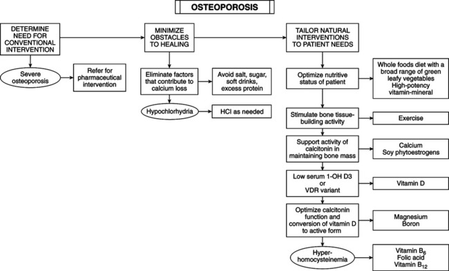
• Osteoporosis (OP) is the most common bone disease in human beings and is a health threat for PM women. Features include low bone mass, deterioration of microarchitecture of bone tissue, fragility of bones, and increased risk of fracture. Approximately 1.5 million OP fractures occur each year in the United States, of which 250,000 are hip fractures. Twenty percent of women with hip fractures die of complications within a year; an additional 25% require long-term nursing care. Half the women who have a hip fracture are unable to walk without assistance. • Vertebral fractures of the thoracic and/or lumbar region cause pain, height loss, and exaggerated kyphosis or deformity of thoracic spine, with restricted range of motion, changes in posture, restricted lung function, and digestive problems. • Depression, anxiety, low self-esteem, and tooth loss are also caused by OP. • Bone remodeling is process of bone resorption (breakdown) and bone formation. Osteoclasts induce enzymatic dissolution of minerals and protein for bone resorption. Osteoblasts create protein matrix of collagen for remineralization and bone formation. Bone remodeling is normally a balance of resorption and formation. Imbalance between removal and replacement causes bone loss and risk of fracture. • Bone mass rapidly increases in childhood, slows in the late teens, but continues to increase during 20s. In women, the building process is complete by age 17 years. Peak bone mass is at 28 years, then bone mass is slowly lost at a rate of 0.4% per year in the femoral neck. After menopause, the loss accelerates to 2% per year during the first 5 to 10 years. Loss continues in women older than 70 years at much slower rate. • Genetic factors: level of peak bone mass is attributable to genetic factors. Young daughters of women with OP fractures and first-degree relatives of women with OP have subnormal bone mass. Black women have greater bone mass compared with white women. Genomic testing is now available to assess the genetic predisposition to a defect in the vitamin D receptor sites (VDR). Women with the VDR defect require substantially greater levels of vitamin D, preferable as 1-OH D3. • Lifestyle: lifestyle, hormonal factors, calcium (Ca) and vitamin D intake, exercise, age of menarche, menstrual regularity, and alcohol and tobacco use have less impact than genetics. Several lifestyle factors affect OP risk after menopause: physical activity, animal protein intake, acid-base homeostasis, Ca and vitamin D intake, heavy smoking and alcohol intake. Requirements for peak bone mass: balanced diet, adequate calories, protein and Ca throughout life. Ca and vitamin D are critical in older women. Ca requirements change with age; menopause increases Ca needs. After age 65 years, women absorb 50% less Ca than do young women. Renal enzymatic activity that produces active vitamin D decreases. • Dietary protein: high intake of animal protein, but not plant protein, is linked to increased risk of forearm fracture. Red meat is acid producing, inducing release of salts from bone to balance acid and maintain acid-base homeostasis. Diets high in fruits and vegetables and plant proteins are alkaline forming. • Smoking: women smokers lose bone more rapidly, have lower bone mass, and have higher fracture rates. Smokers reach menopause up to 2 years earlier. Smoking may interfere with estrogen by an unknown metabolism. • Alcohol: ≥7 oz/week of alcohol increases the risk of falls and hip fractures. Moderate alcohol intake lowers risk of hip fractures in older women; it inhibits bone resorption by increasing estradiol levels and calcitonin excretion. • Physical activity: highly active persons have higher bone mass; prolonged bed rest or confinement to a wheelchair causes rapid, dramatic bone loss. Exercise reduces OP risk by stimulating osteoblasts. • Menopause: all women lose bone, but this loss is accelerated in the first 5 years of menopause. Estrogen decline increases rate of bone resorption. The earlier that occurs before the average age of menopause (51 years), the sooner the bones lose the protective effect of endogenous estrogen. Premature menopause (before 40 years), late onset of menarche in adolescence, and periods of amenorrhea or infrequent menses increase risk of OP. Women who missed up to half of their expected menstrual periods have 12% less vertebral bone mass; those who missed more than half had 31% less bone mass than healthy control subjects. • Ca blood concentration is strictly held within narrow limits. Ca decline triggers increased secretion of parathyroid hormone (PTH) and decreased secretion of calcitonin from the thyroid and parathyroids. Ca excess triggers decreased secretion of PTH and increased secretion of calcitonin. • PTH increases serum Ca by increasing osteoclast catabolism of bone, decreases excretion of Ca by kidneys, increases absorption of Ca in intestines, and increases kidney conversion of 25-(OH)D3 to 1,25-(OH)2D3. • Estrogen deficiency makes osteoclasts more sensitive to PTH, increasing bone breakdown and raising blood Ca. Elevated blood Ca decreases PTH, diminishing active vitamin D and increasing Ca excretion. — Early menopause without hormone replacement therapy or oral contraceptive use — History of amenorrhea (from anorexia nervosa, hyperprolactinemia, or exercise-induced amenorrhea), infrequent menses, late menstrual onset, anovulation — Kidney disease and kidney stones, rheumatoid arthritis, multiple myeloma, chronic obstructive pulmonary disease, scoliosis, anorexia nervosa, diabetes mellitus, Cushing’s disease, hyperthyroidism, primary hyperparathyroidism, hypercalciuria, vitamin D deficiency — Gallbladder disease, primary biliary cirrhosis, fat malabsorption, hypochlorhydria, lactose intolerance — History of chronic low back pain for more than 15 years — Prolonged bed rest, paralysis, or time in wheelchair — History of other fractures after age 45 years — Dental conditions: bone loss in the jaw, dentures before age 60 years, increased tooth loss — Nulliparous (never had a full-term pregnancy, that is, no periods of sustained high estrogens) • Environmental and lifestyle factors: — Inadequate Ca and vitamin D intake during pregnancies and nursing — Dietary factors: excessive caffeine, high animal protein, high sodium, high phosphate, low dietary Ca — Lifestyle factors: sedentary, moderate (or more) alcohol intake, cigarette smoking — Isoniazid, furosemide, heparin, tetracycline, anticonvulsants, gonadotropin-releasing hormone, cortisone or prednisone, and aluminum-containing antacids, lithium • Assess all PM women for risk factors of OP—history, physical examination, diagnostic tests. Goals of evaluation: identify women at risk for OP or fracture; diagnose OP and/or determine severity of OP; rule out secondary causes of bone loss; identify risk factors for falls or injuries. • Focus history and physical examination on identifying risk factors. Physical signs of OP: loss of height >1.5 inches (measure height annually), excess kyphosis of thoracic spine, dowager’s hump, dental caries, tooth loss, receding gums, back pain. • Radiologic tests of bone mineral density (BMD): BMD testing is optimal to diagnose OP. Gold standard is dual energy x-ray absorptiometry (DEXA). Other methods include computerized tomography (CT) scans, ultrasound of heel, radiographs; none of these tests is optimal for diagnosis and follow-up. The following tests measure BMD and are compared by accuracy. Adapted from Jergas M, Genant HK: Current methods and recent advances in the diagnosis of osteoporosis, Arthritis Rheum 36:1649-1662, 1993.
Osteoporosis
GENERAL CONSIDERATIONS
Pathophysiology

Risk Factors
Hormonal Factors
Additional Factors
DIAGNOSTIC CONSIDERATIONS
Method
Site
Accuracy
Dual-energy x-ray absorptiometry (DEXA)
Hip, spine, total body
90%-99%
Peripheral dual-energy x-ray absorptiometry (PDXA)
Forearm, finger, heel
90%-99%
Single-energy x-ray absorptiometry (SXA)
Heel
98%-99%
Quantitative ultrasound (QUS)
Heel, shin
Not applicable
Quantitated computed tomography (QCT)
Spine
95%-97%
Peripheral quantitative computed tomography (PQTC)
Forearm
92%-98%
![]()
Stay updated, free articles. Join our Telegram channel

Full access? Get Clinical Tree


Basicmedical Key
Fastest Basicmedical Insight Engine
