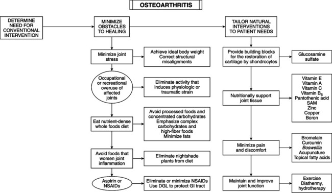
• Symptoms: mild early morning stiffness, stiffness after periods of rest, pain that worsens on joint use, and loss of joint function. • Signs: local tenderness, soft tissue swelling, joint crepitus, bony swelling, restricted mobility, swelling of Heberden’s (proximal interphalangeal joints) and/or less common Bouchard’s (distal interphalangeal joints) nodes, and other signs of degenerative loss of articular cartilage. • Radiographic findings: narrowed joint space, osteophytes, in-creased density of subchondral bone, subchondral sclerosis, bony cysts, soft tissue swelling, and periarticular swelling. • Diseases thought to be OA of specific joints: hands (Heberden’s and Bouchard’s nodes), hips (malum coxae senilis), temporomandibular (Costen’s syndrome), knees (chondromalacia patellae), and spine (ankylosing hyperostosis, interstitial skeletal hyperostosis). • Weight-bearing joints and peripheral and axial articulations are principally affected. Hyaline cartilage destruction is followed by hardening and formation of large bone spurs (calcified osteophytes) in joint margins, causing pain, deformity, and limited joint motion. Inflammation usually is minimal. • Two categories of OA: (1) primary OA arises from wear and tear after the fifth and sixth decades, with no predisposing abnormalities. The cumulative effects of decades of use stress collagen matrix. Damage releases enzymes that destroy collagen components. With aging, the ability to synthesize restorative collagen decreases. (2) Secondary OA entails predisposing factors for degeneration. Factors include congenital abnormalities in joint structure or function (e.g., hypermobility and abnormally shaped joint surfaces), trauma (obesity, fractures along joint surfaces, surgery), crystal deposition, presence of abnormal cartilage, previous inflammatory disease of joint (rheumatoid arthritis [RA], gout, septic arthritis). Source of pain is not OA, but: • Pathologic changes of adjacent bone (tumor, osteomyelitis, metabolic bone disease) • Referred pain of neuritis, neuropathy, or radiculopathy Source of pain is OA but not at joint suspected: • OA of hip, pain localized to knee • OA of cervical spine, causing pain in shoulder • OA of lumbar spine, causing pain in hip, knee, or ankle Source of pain is caused by secondary soft tissue alterations of OA: • Tendonitis or ligamentitis (especially of knee) • Enthesopathy, tendinopathy from joint contracture • BursitisMisinterpretation of deformity: • Pseudohypertrophic osteoarthropathy • Flexion contracture of joints • Calcium pyrophosphate dihydrate crystal deposition disease Misinterpretation of x-ray films: • Arthritis with previous OA changes • Initial stage of OA, may have normal radiograph • Flexion contracture can cause virtual loss of joint space width • Neurogenic and metabolic arthropathies • Pain: severity of OA (by radiograph) does not correlate with degree of pain. Forty percent of patients with the worst radiographic classification for OA are pain free. The cause of pain in OA is still not well defined. Depression and anxiety increase OA pain. • Aspirin (ASA) and nonsteroidal antiinflammatory drugs (NSAIDs): ASA is effective in relieving pain and inflammation and is inexpensive. Therapeutic dose is high (2-4 g q.d.) and toxicity is frequent, including tinnitus and gastric irritation. Side effects of other NSAIDs are gastrointestinal upset, headache, and dizziness. They are only recommended for short periods. Unexpected side effects may increase the rate of cartilage degeneration; they inhibit collagen matrix synthesis and accelerate cartilage destruction. NSAID use is associated with acceleration of OA. NSAIDs suppress symptoms but accelerate OA progression. Main drug categories and individual drugs used to treat OA: • NSAIDs: salicylates (aspirin), propionic acids (ibuprofen [Motrin], naproxen [Anaprox], oxaprozin [Daypro], flurbiprofen [Ansaid], ketoprofen [Orudis]), acetic acids (diclofenac [Voltaren], tolmetin [Tolectin], etodolac [Lodine], sulindac [Clinoril], indomethacin [Indocin]), and oxicams (piroxicam [Feldene]) • Irritants and counterirritants: capsaicin 0.025% cream • Hyaluronic acids: hylan G-F 20 (Synvisc), sodium hyaluronate (Hyalogen) • Estrogen: higher prevalence of OA in women suggests estrogen involvement. Estradiol worsens OA. The antiestrogen drug tamoxifen improves experimental OA by decreasing erosive lesions; this suggests a therapeutic role for estrogen blockade. Botanicals (e.g., Glycyrrhiza glabra and Medicago sativa) used for OA contain phytoestrogens that bind to estrogen receptors, acting as estrogen antagonists. Food sources of phytoestrogens include soy, fennel, celery, parsley, nuts, whole grains, and apples. Insulin, growth hormone (GH), and somatomedin (SMM): diabetics have greater incidence and more severe OA than non-diabetics. DM features causing OA: insulin insensitivity or deficiency, increased GH, and decreased SMMs (insulinlike growth factors secreted by liver in response to GH). Insulin stimulates chondrocytes to increase synthesis and assembly of proteoglycans. The most prominent early changes in articular cartilage are decreased proteoglycans and state of aggregation; thus insulin insensitivity or deficiency predisposes to OA. Excessive GH is detrimental to bone and joint structures; and increased incidence of OA is found in acromegaly. Women with primary osteoporosis have higher basal GH levels than do control subjects. GH is detrimental to chondrocytes. SMMs mediate normal chondrocyte activity. Impaired hepatic function, diabetes, and malnutrition suppress liver secretion of SMMs in response to GH, increasing risk of OA. • Thyroid: hypothyroidism increases risk of OA compared with age- and sex-matched population samples.
Osteoarthritis
DIAGNOSTIC SUMMARY
GENERAL CONSIDERATIONS

DIAGNOSIS
Causes of Misdiagnosis of Osteoarthritis
THERAPEUTIC CONSIDERATIONS
Conventional Pharmacologic Treatment
Hormonal Considerations
![]()
Stay updated, free articles. Join our Telegram channel

Full access? Get Clinical Tree


Osteoarthritis
