Diagnostic Checklist

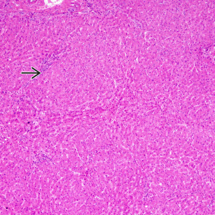
Nodular regenerative hyperplasia (NRH) is characterized by diffuse replacement of liver by small nodules
 . Nodules are typically 1-3 mm in size but can be as large as 1 cm.
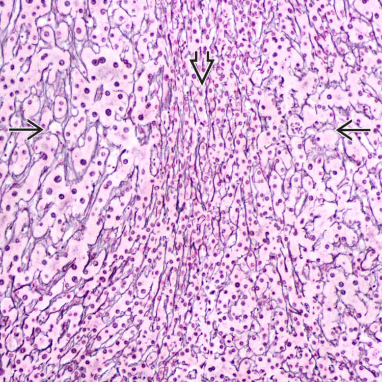
Reticulin stain highlights the nodules
 . The reticulin network is compressed in the parenchyma between the nodules
  . Reticulin stain is very useful for the diagnosis as the nodularity in NRH can be subtle.
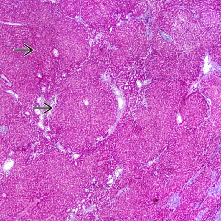
Trichrome stain highlights the nodules
 . By definition, there are no fibrous septa between the nodules in nodular regenerative hyperplasia.
TERMINOLOGY
Abbreviations
Definitions
• Pattern of liver injury, associated with many underlying causes, that does not represent specific entity


Stay updated, free articles. Join our Telegram channel

Full access? Get Clinical Tree


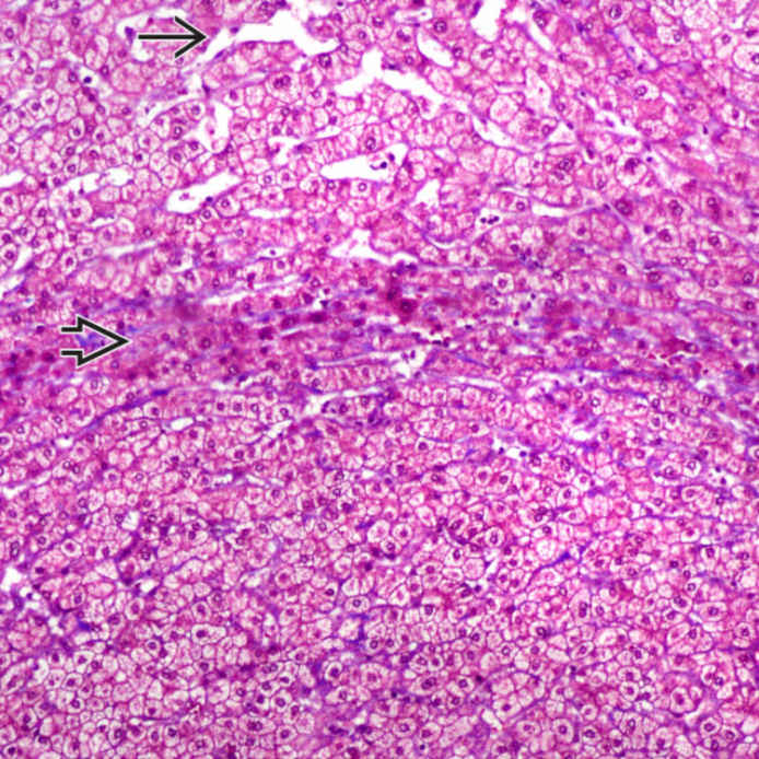
 . The hepatocytes between the nodules are compressed and atrophic
  .
.