After studying this chapter, the student is expected to: 1. Relate the focal effect of a lesion to the specific area of damage in the brain. 3. Explain the effects of herniation. 4. Compare the effects of brain tumors in different areas of the brain. 5. Compare transient ischemic attacks (TIAs) to cerebrovascular accidents (CVAs). 6. Explain how cerebral aneurysms develop, their effects, and possible complications. 7. Describe the cause, pathophysiology, and manifestations of bacterial meningitis. 8. Explain how a brain abscess may cause focal and general effects. 9. Differentiate the types of hematomas and describe the effect of a hematoma on the brain. 10. Explain how seizures may be related to infection or injury. 11. Describe how various types of spinal cord injury may occur. 12. Explain how the effects of spinal cord injury depend on the location of the damage. 13. Compare the signs of spinal shock with the permanent effects of spinal cord injury. 15. Describe the signs of increasing intracranial pressure in the neonate. 16. Describe the major types of spina bifida and the effect on a child who has the defect. 17. Describe the types of cerebral palsy and signs of each. 18. Differentiate the types of seizures. 19. Describe the pathophysiology, course, and effects of multiple sclerosis. 20. Relate the pathophysiology to the signs of Parkinson’s disease. 22. Describe the pathophysiology of myasthenia gravis and its effects on the body. 23. Describe the inheritance of Huntington’s disease and the onset and early signs. 24. Describe the changes in the brain as Alzheimer’s disease develops and the effects on function. afferent amnesia anencephaly anomalies aphasia athetoid atresia aura bifurcation choreiform clonic cognitive coma contralateral disorientation efferent fissure flaccid flaccid foramina fulminant ganglion gyri hyperreflexia hyperreflexia infratentorial ipsilateral labile nuchal rigidity paralysis paresis paresthesia photophobia postictal precursor pressoreceptors prodromal ptosis retina scotoma spastic stupor sulcus, sulci supratentorial sutures tetraplegia tonic transillumination The nervous system consists of the central and peripheral nervous system. The brain is the communication and control center of the body. It receives, processes, and evaluates many kinds of input; decides on the response or action to be taken; and then initiates the response. Responses include both involuntary activity that is required to maintain homeostasis in the body (regulated by the autonomic nervous system) and voluntary actions (controlled by the somatic nervous system). With both reflex and voluntary activities, the individual is often not aware of the amount and diversity of input received or the integration or assessment of that input, but knows only of the response (Fig. 14-1). The brain is protected by the rigid bone of the skull, the three membranes or meninges, and the cerebrospinal fluid (CSF). The cranial and facial bones are connected by sutures, which are relatively immovable joints consisting of fibrous tissue. If pressure inside the skull increases in infants before the sutures fuse or ossify, the cranial bones may separate, causing the head to enlarge. The skull contains a number of cavities, or fossae, as well as foramina (openings) and canals through which nerves and blood vessels pass. The largest opening, the foramen magnum, is located in the occipital bone at the base of the skull, where the spinal cord emerges. The meninges consist of three continuous connective tissue membranes covering the brain and spinal cord. The meninges and the contents of the spaces between the layers are as follows: • The arachnoid (arachnoid mater), a loose, weblike covering, is the middle layer. • The pia mater, a delicate connective tissue that adheres closely to all convolutions on the surface of the brain, is the inner layer. Many small blood vessels are found in the pia mater (Fig. 14-2). The cerebrospinal fluid (CSF) provides a cushion for the brain and spinal cord. Similar to plasma in appearance, it is a clear, almost colorless liquid, but it differs from plasma in the concentrations of electrolytes, glucose, and protein (Table 14-1), which remain relatively constant. A change in the characteristics of the CSF is a useful diagnostic tool (see Fig. 14-11). For example, the presence of significant numbers of erythrocytes in CSF indicates bleeding in the central nervous system. TABLE 14-1 Characteristics of Normal Cerebrospinal Fluid Cerebrospinal fluid is formed constantly in the choroid plexuses in the ventricles and then flows into the subarachnoid space, where it circulates around the brain and spinal cord and eventually passes through the arachnoid villi, returning into the venous blood. To maintain a relatively constant pressure within the skull (intracranial pressure), it is important for equal amounts of CSF to be produced and reabsorbed at the same rate. The blood-brain barrier is a protective mechanism provided primarily by relatively impermeable capillaries in the brain. The endothelial cells of the capillaries are tightly joined together by tight junctions rather than possessing pores. This barrier limits the passage of potentially damaging materials into the brain and controls the delicate but essential balance of electrolytes, glucose, and proteins in the brain. There is a similar blood-CSF barrier at the choroid plexus to control the constituents of CSF. The blood-brain barrier is poorly developed in neonates, and therefore substances such as bilirubin (see Chapter 22) or other toxic materials can pass easily into the infant’s brain, causing damage. When fully developed, the blood-brain barrier can be a disadvantage, because it does not allow the passage of many essential drugs into the brain (i.e., certain antibiotics and anticancer drugs). Lipid-soluble substances, including alcohol, pass freely into the brain. The cerebral hemispheres make up the largest and most obvious portions of the brain. The outer surface is covered by elevations, or gyri (sing., gyrus), that are separated by grooves, or sulci (sing., sulcus). The longitudinal fissure separates the left and right hemispheres. The corpus callosum consists of nerve fibers connecting the left and right hemisphere for the purpose of communication between the two hemispheres. Each hemisphere is divided into four major lobes, each of which has specific functions (Fig. 14-3). Complex functions, such as language and memory, involve many areas of the brain. Each hemisphere is concerned with voluntary movement and sensory function in the opposite (contralateral) side of the body, and these areas of the cortex have been well mapped. In Figure 14-3, note the large number of nerve cells required to innervate the face compared with the amount of cortex allocated to the trunk. The cells of the motor cortex of the frontal lobe initiate specific voluntary movements, and these cells are often referred to as upper motor neurons (UMNs). Their axons form the corticospinal tracts in the spinal cord. Because the crossover of most of these tracts occurs in the medulla, damage to the motor cortex in the left frontal lobe adjacent to the longitudinal fissure (on top of the head) results in paralysis or paresis of the muscles of the right leg. Each special sensory area of the cortex has an association area surrounding the primary cortex, in which the sensory input is recognized and interpreted. For example, the occipital lobe contains the primary visual cortex, which receives the stimuli from the eye, and the surrounding association cortex identifies the object seen. If the primary cortex is damaged, the person is blind, but if the association area is damaged, the person can see an object but cannot comprehend its significance. The right and left hemispheres are generally similar in structure but not necessarily in function (Table 14-2). The term dominant hemisphere refers to the side of the brain that controls language, which in most people is the left hemisphere. There are two special areas involved in language skills. Broca’s area is considered the motor or expressive speech area, in which the output of words, both verbal and written, is coordinated in an appropriate and understandable way. This area is located at the base of the premotor area of the left frontal lobe. Wernicke’s area is the integration center that comprehends language received, both spoken and written. This area is located in the posterior temporal lobe and has connecting fibers to the prefrontal, visual, and auditory areas. The left hemisphere also appears to be responsible for mathematical, problem-solving, and logical reasoning abilities. The right hemisphere has greater influence on artistic abilities, creativity, spatial relationships, and emotional and behavioral characteristics. TABLE 14-2 Major Functional Areas of the Brain The prefrontal cortex lies anterior to the motor and premotor cortex and recent research indicates that it functions in coordinating complex cognitive behavior as well as providing components for expression of personality. It plays a very significant role in social relationships and impulse control. The basal nuclei (previously called the basal ganglia) are clusters of cell bodies or gray matter located deep among the tracts of the cerebral hemispheres. These are part of the extrapyramidal system (EPS) of motor control, which controls and coordinates skeletal muscle activity, preventing excessive movements and initiating accessory and often involuntary actions, such as arm-swinging when walking. Two additional nuclei located in the midbrain, the substantia nigra and the red nucleus, are also connected to the basal nuclei and the EPS. The limbic system consists of many nuclei and connecting fibers in the cerebral hemispheres that encircle the superior part of the brain stem. The limbic system is responsible for emotional reactions or feelings, and for this purpose, it has many connections to all areas of the brain. Part of the hypothalamus is involved with the limbic system. The hypothalamus provides the link for the autonomic responses, such as altered blood pressure or nausea, which occur when one experiences fear, excitement, or an unpleasant sight or odor. Any cognitive (intellectual) decision arising from the higher cortical centers may be accompanied by an emotional aspect mediated through the limbic system. The diencephalon is the central portion of the brain. It is surrounded by the hemispheres and contains the thalamus, the hypothalamus, and the epithalamus. The thalamus consists of many nerve cell bodies, the major function of which is to serve as a sorting and relay station for incoming sensory impulses. From the thalamus, connecting fibers transmit impulses to the cerebral cortex and other appropriate areas of the brain. The hypothalamus has a key role in maintaining homeostasis in the body, controlling the autonomic nervous system and much of the endocrine system through the hypophysis, or pituitary gland. It is responsible for the regulation of body temperature, intake of food and fluid, and the regulation of sleep cycles. The hypothalamus is also the key to the stress response and plays major roles in emotional responses through the limbic system and in biologic behaviors, such as the sex drive (libido). The inferior portion of the brain, called the brain stem, is the connecting link to the spinal cord. The pons is composed of bundles of afferent (incoming) and efferent (outgoing) fibers. Several nuclei of cranial nerves are also located in the pons. The medulla oblongata contains the vital control centers that regulate respiratory and cardiovascular function and the coordinating centers that govern the cough reflex, swallowing, and vomiting. The medulla is the location of the nuclei of several cranial nerves. It is distinguished by two longitudinal ridges on the ventral surface, termed the pyramids, marking the site of crossover (decussation) of the majority of fibers of the corticospinal (pyramidal) tracts, which results in the contralateral control of muscle function. The reticular formation is a network of nuclei and neurons scattered throughout the brain stem that has connections to many parts of the brain. The reticular-activating system (RAS) is part of this formation and determines the degree of arousal or awareness of the cerebral cortex. In other words, these neurons decide which of the incoming sensory impulses the brain ignores and which it notices. Many drugs can affect the activity of the RAS, thus increasing or decreasing the input to the brain. The cerebellum is located dorsal to the pons and medulla, below the occipital lobe. It functions to coordinate movement and maintain posture and equilibrium by continuously assessing and adjusting to input from the pyramidal system, the proprioceptors in joints and muscles, the visual pathways, and the vestibular pathways from the inner ear. Blood is supplied to the brain by the internal carotid arteries and the vertebral arteries. Each internal carotid artery is a branch of a common carotid artery (right or left) and includes the carotid sinus, which is the location of the pressoreceptors, or baroreceptors, that signal changes in blood pressure and the chemoreceptors that monitor variations in blood pH and oxygen levels. At the base of the brain, each internal carotid artery divides into an anterior and middle cerebral artery (see Fig. 14-14). Posteriorly, the vertebral arteries join to form: At the base of the brain, the basilar artery divides into: • The right and left posterior cerebral arteries, which supply blood to the occipital lobes. • The anterior, middle, and posterior cerebral arteries follow a course over the surface of each hemisphere, with many branches penetrating into the brain substance (see Fig. 14-13A). Anastomoses between these major arteries at the base of the brain are provided by the: • Anterior communicating artery between the anterior cerebral arteries • Posterior communicating arteries between the middle cerebral and posterior cerebral arteries Blood flow in the cerebral arteries is relatively constant because the brain cells constantly use oxygen and glucose (essential nutrients for neurons) and have little storage capacity. Autoregulation is a mechanism by which increased carbon dioxide levels or decreased pH in the blood, or decreased blood pressure, in an area of the brain results in immediate local vasodilation. The pressoreceptors (baroreceptors) and chemoreceptors protect the brain from damage related to abnormal blood pressure or pH levels in the systemic flow. As mentioned, venous blood from the brain collects in the dural sinuses and then drains into the right and left internal jugular veins, to be returned to the heart. There are 12 pairs of cranial nerves. They originate from the brain stem and pass through the foramina in the skull to serve structures in the head and neck, including the eyes and ears. The vagus nerve (cranial nerve X) serves a more extensive area, branching to innervate many of the viscera. A cranial nerve may consist of motor fibers only (with associated sensory fibers from proprioceptors in the skeletal muscles) or of sensory fibers only, or it may be a mixed nerve, containing both motor and sensory fibers (Table 14-3). Four cranial nerves (III, VII, IX, X) include parasympathetic fibers. 14-3 Think about TABLE 14-3 Major Components of Cranial Nerves The spinal cord is protected by the bony vertebral column, the meninges, and the CSF. The cord is continuous with the medulla oblongata and ends at the level of the first lumbar vertebra. Beyond this extends a bundle of nerve roots known as the cauda equina. This arrangement is significant because there is little risk of damaging the cord when a needle is inserted into the subarachnoid space below the first lumbar level (usually in the space between L3 and L4) to obtain a sample of CSF (see Fig. 14-11). The spinal cord consists of the white matter (outer region) containing the spinal cord tracts, composed of myelinated fibers, and the gray matter (inner region) containing the nerve cell bodies, dendrites, and nonmyelinated axons. Within the gray matter three distinct areas can be identified:
Nervous System Disorders
Learning Objectives
Key Terms
Review of Nervous System Anatomy and Physiology
Brain
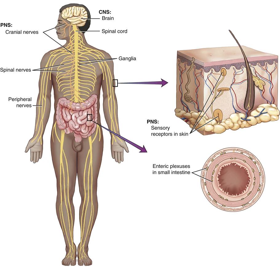
The nervous system is divided into two components: the central nervous system composed of the brain and the spinal cord, and the peripheral nervous system containing the cranial and spinal nerves, ganglia, and sensory receptors. (From VanMeter K, Hubert R: Microbiology for the Healthcare Professional, St. Louis, 2010, Elsevier.)
Protection for the Brain
Meninges
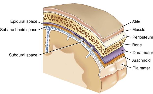
The brain and the spinal cord are covered by three protective membranes, collectively called meninges. The outermost membrane is the dura mater, the middle layer is the arachnoid, and the innermost membrane is the pia mater. (From VanMeter K, Hubert R: Microbiology for the Healthcare Professional, St. Louis, 2010, Elsevier.)
Cerebrospinal Fluid
Appearance
Clear and colorless
Pressure
9-14 mmHg or 150 mm H2O
Red blood cells
None
White blood cells
Occasional
Protein
15-45 mg/dL
Glucose
45-75 mg/dL
Sodium
140 mEq/L
Potassium
3 mEq/L
Specific gravity
1.007
pH
7.32-7.35
Volume in the system at one time
125-150 mL
Volume formed in 24 hours
500-800 mL
Blood-Brain Barrier and Blood-Cerebrospinal Fluid Barrier
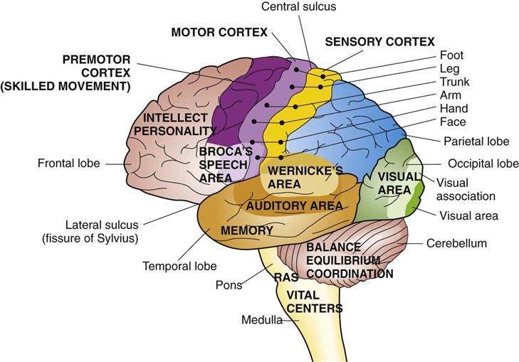
Functional Areas
Cerebral Hemispheres
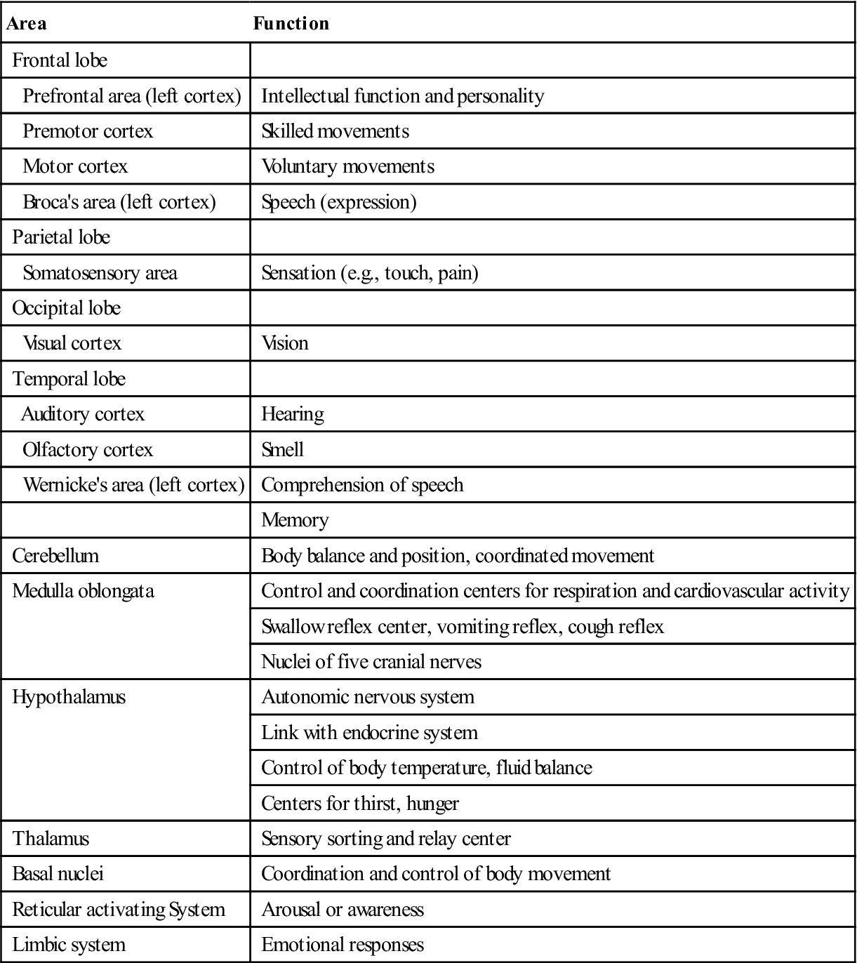
Area
Function
Frontal lobe
Prefrontal area (left cortex)
Intellectual function and personality
Premotor cortex
Skilled movements
Motor cortex
Voluntary movements
Broca’s area (left cortex)
Speech (expression)
Parietal lobe
Somatosensory area
Sensation (e.g., touch, pain)
Occipital lobe
Visual cortex
Vision
Temporal lobe
Auditory cortex
Hearing
Olfactory cortex
Smell
Wernicke’s area (left cortex)
Comprehension of speech
Memory
Cerebellum
Body balance and position, coordinated movement
Medulla oblongata
Control and coordination centers for respiration and cardiovascular activity
Swallow reflex center, vomiting reflex, cough reflex
Nuclei of five cranial nerves
Hypothalamus
Autonomic nervous system
Link with endocrine system
Control of body temperature, fluid balance
Centers for thirst, hunger
Thalamus
Sensory sorting and relay center
Basal nuclei
Coordination and control of body movement
Reticular activating System
Arousal or awareness
Limbic system
Emotional responses
Diencephalon
Brain Stem
Cerebellum
Blood Supply to the Brain
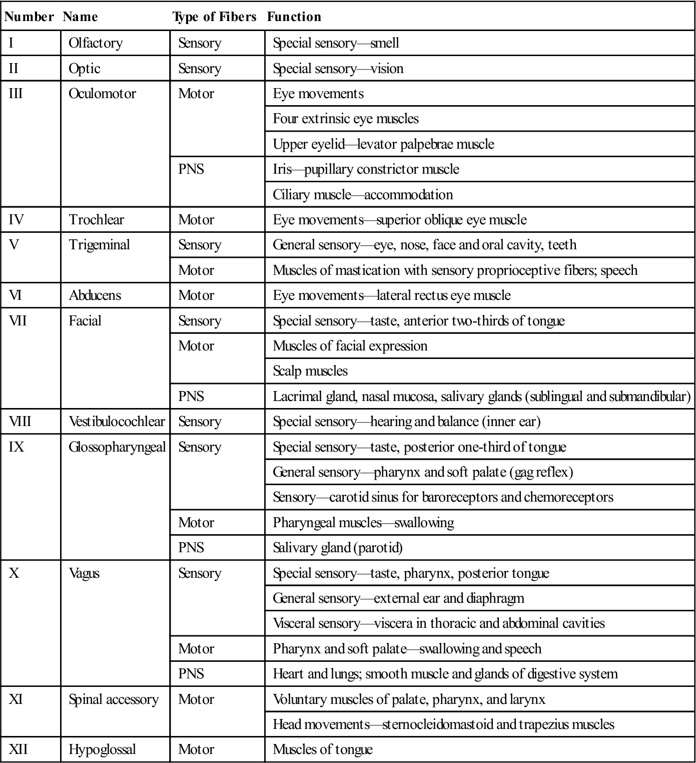
Cranial Nerves

Number
Name
Type of Fibers
Function
I
Olfactory
Sensory
Special sensory—smell
II
Optic
Sensory
Special sensory—vision
III
Oculomotor
Motor
Eye movements
Four extrinsic eye muscles
Upper eyelid—levator palpebrae muscle
PNS
Iris—pupillary constrictor muscle
Ciliary muscle—accommodation
IV
Trochlear
Motor
Eye movements—superior oblique eye muscle
V
Trigeminal
Sensory
General sensory—eye, nose, face and oral cavity, teeth
Motor
Muscles of mastication with sensory proprioceptive fibers; speech
VI
Abducens
Motor
Eye movements—lateral rectus eye muscle
VII
Facial
Sensory
Special sensory—taste, anterior two-thirds of tongue
Motor
Muscles of facial expression
Scalp muscles
PNS
Lacrimal gland, nasal mucosa, salivary glands (sublingual and submandibular)
VIII
Vestibulocochlear
Sensory
Special sensory—hearing and balance (inner ear)
IX
Glossopharyngeal
Sensory
Special sensory—taste, posterior one-third of tongue
General sensory—pharynx and soft palate (gag reflex)
Sensory—carotid sinus for baroreceptors and chemoreceptors
Motor
Pharyngeal muscles—swallowing
PNS
Salivary gland (parotid)
X
Vagus
Sensory
Special sensory—taste, pharynx, posterior tongue
General sensory—external ear and diaphragm
Visceral sensory—viscera in thoracic and abdominal cavities
Motor
Pharynx and soft palate—swallowing and speech
PNS
Heart and lungs; smooth muscle and glands of digestive system
XI
Spinal accessory
Motor
Voluntary muscles of palate, pharynx, and larynx
Head movements—sternocleidomastoid and trapezius muscles
XII
Hypoglossal
Motor
Muscles of tongue
Spinal Cord
Spinal Cord
![]()
Stay updated, free articles. Join our Telegram channel

Full access? Get Clinical Tree


Basicmedical Key
Fastest Basicmedical Insight Engine