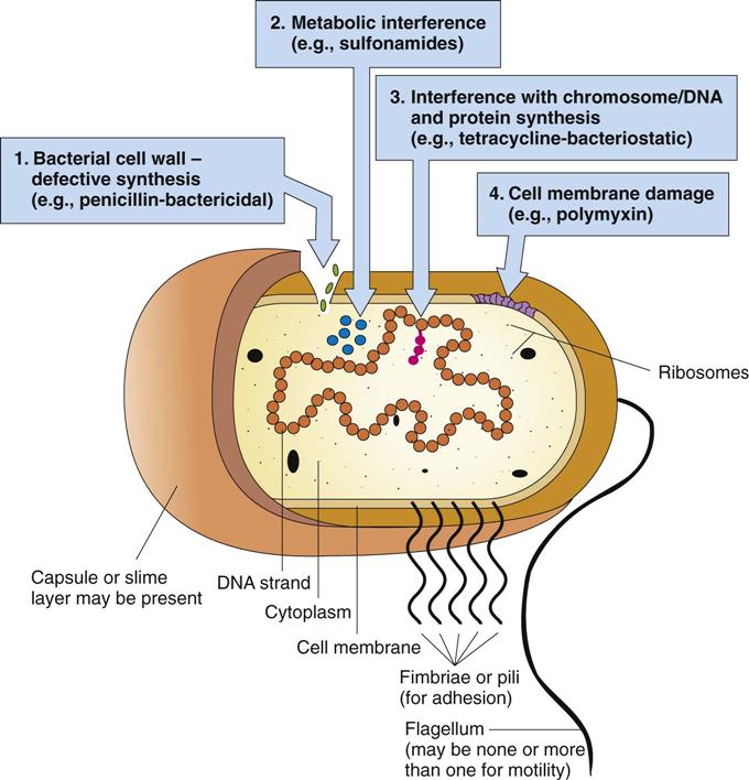
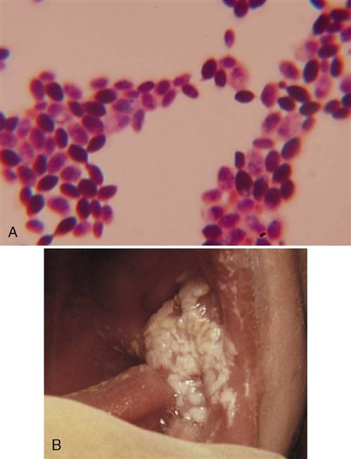
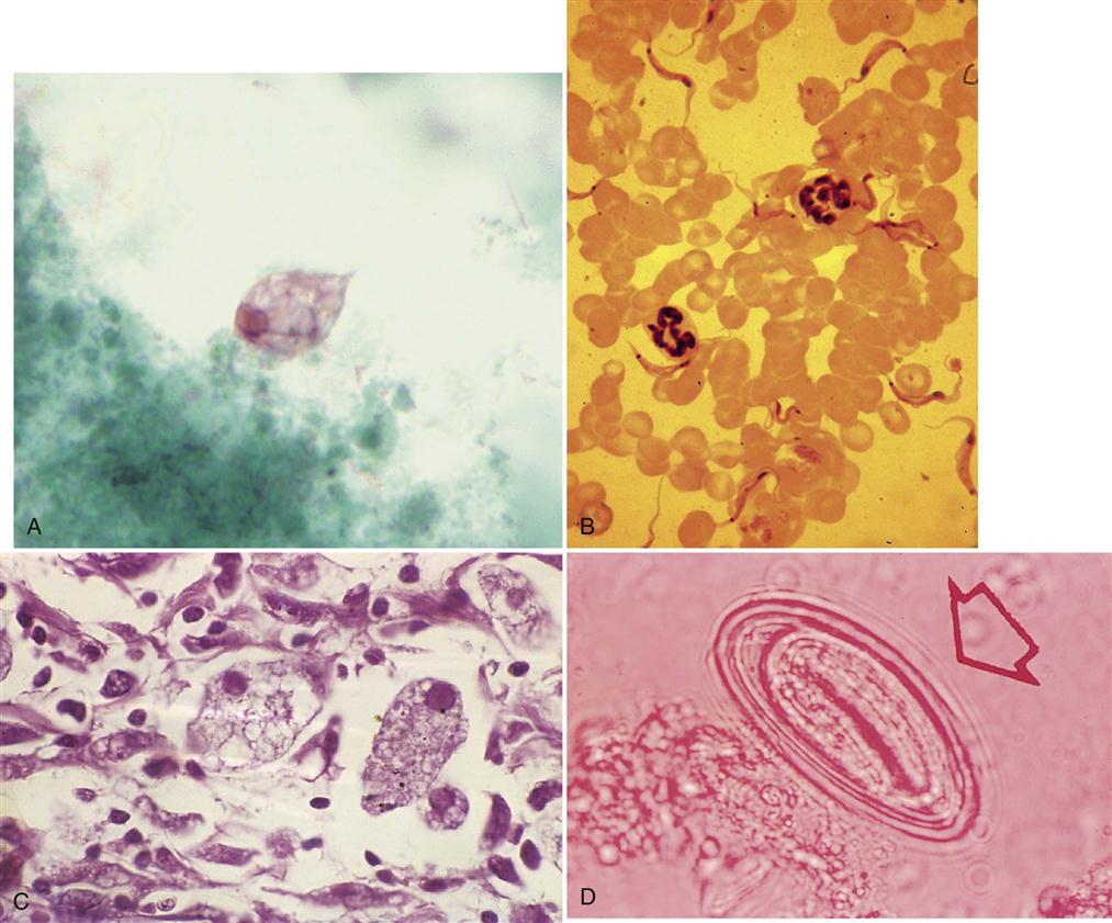
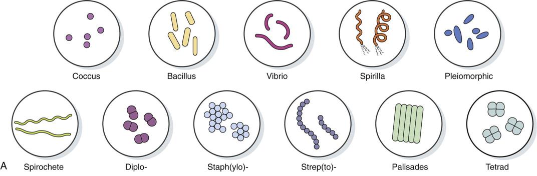
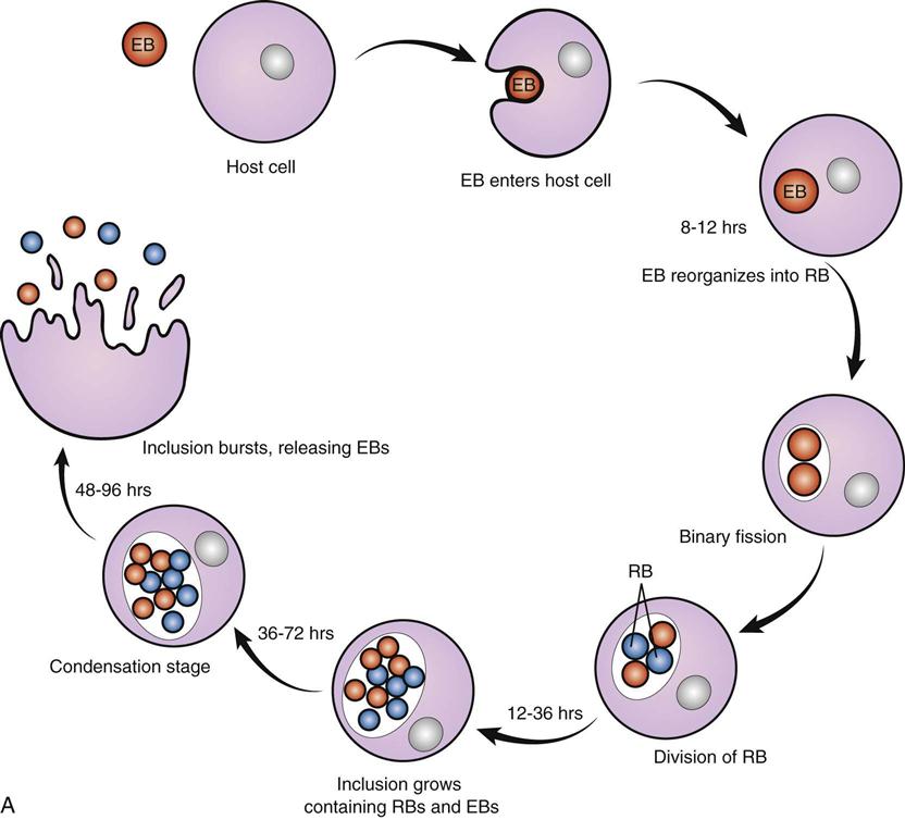
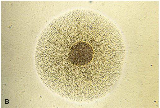
After studying this chapter, the student is expected to: 2. Discuss the locations, advantages, and disadvantages of resident (normal) flora. 3. Describe the modes of transmission of microbes. 4. Describe the factors determining host resistance. 5. Explain the factors contributing to pathogenicity and virulence of microbes. 6. Discuss methods of preventing and controlling infection. 7. Describe the stages in the development and course of an infection. 8. Describe typical, local, and systemic signs of infection. 9. State the common diagnostic tests for infection and the purpose of each. 10. Describe the mechanisms of action of common antimicrobial drugs. 11. Explain the basic guidelines for use of antimicrobial drugs. antiseptics autoclaving culture disinfectants endemic epidemics fimbriae infection leukocytosis leukopenia lymphadenopathy monocytosis mutation neutropenia nosocomial obligate parasite pathogens pili prions prosthetic seizures septicemia sterilization toxins unicellular Microbiology refers to the study of microorganisms or microbes, very small living forms that are visible only with a microscope. Microorganisms include bacteria, fungi, protozoa, and viruses (Fig. 6-1). Detailed classifications of organisms with their proper names are available in microbiology references (e.g., Bergey’s Manual). Selected examples of microorganisms are examined briefly here. Bacteria are classified as prokaryotic cells because they are very simple in structure—lacking even a nuclear membrane—but they function metabolically and reproduce. They also have a complex cell wall structure. By comparison, eukaryotic cells are nucleated cells found in higher plants and animals, including humans. They lack cell walls (except in plants) but their DNA is enclosed in a nuclear membrane and the cell membrane has a complex structure. Many microorganisms are classified as nonpathogenic because they do not usually cause disease; in fact, they are often beneficial. Pathogens are the disease-causing microbes often referred to as “germs.” Infectious diseases result from invasion of the body by microbes and multiplication of these microbes, followed by damage to the body. These agents and their ability to cause disease vary widely. In the 18th and 19th centuries, scientists experimented on fermentation and spoilage of foods. This resulted in the concept of the “germ theory of disease” as well as explanations of how wine and other foods became unfit for consumption. The transmission of pathogens and infection through hands, surfaces, water, and the air was documented, and the practices of asepsis were begun. Microorganisms vary widely in their growth needs, and their specific requirements often form the basis for identification tests. Many microbes can be grown in a laboratory using an appropriate environment and a suitable culture medium in a Petri dish (Fig. 6-2A) or a test tube. The culture medium provides the required nutrients for specific microbial groups. The culture base may be synthetic or a broth base with additives. The need for oxygen, carbohydrates, a specific pH or temperature, or a living host depends on the needs of the particular microbe. Those microbes that require living cells in which to survive are particularly difficult to identify without specialized laboratory techniques such as cell culture, immunoassays, or electron microscopy. The specific growth factors play a role in determining the site of infection in the human body. For example, the organism causing tetanus is an anaerobic bacterium that thrives in the absence of oxygen and therefore can easily cause infection deep in the tissue. Bacteria are unicellular (single cell) organisms that do not require living tissue to survive. They vary in size, shape, and arrangement and are classified and named accordingly (see Fig. 6-1). These obvious characteristics may assist in rapid identification of microbes. The major groups of bacteria based on cellular shape are: Bacterial cells can further be categorized by their characteristic groupings or arrangement. • Diplo- prefix, indicating pairs • Strep(to)- prefix, indicating chains • Staph(ylo)- prefix, indicating irregular, grapelike clusters • Tetrads- refers to cells grouped in a packet or square of four cells • Palisade- refers to cells lying together with the long sides parallel The basic structure of bacteria includes: 1. An outer rigid cell wall protects the microbe and provides a specific shape (Fig. 6-3). A bacterium has one of two types of cell walls, gram-positive or gram-negative, which differ in their chemical composition. This difference can be quickly determined in the laboratory using a Gram stain and provides a means of identification and classification for bacteria. This classification is useful in selecting appropriate antimicrobial therapy; for instance, penicillin acts on the cell wall of gram-positive bacteria. Targeting cell wall structure and function is important because human cells do not have cell walls. A drug such as penicillin thus does not damage human cells but is effective against gram-positive bacteria. 4. One or more rotating flagellae attached to the cell wall provide motility for some species. 7. Some bacteria secrete toxic substances, toxins, and enzymes. Toxins consist of two types, exotoxins and endotoxins. • Exotoxins are usually produced by gram-positive bacteria and diffuse through body fluids. They have a variety of effects, often interfering with nerve conduction, such as the neurotoxin from the tetanus bacillus. Other toxins termed enterotoxins may stimulate the vomiting center and cause gastrointestinal distress. Exotoxins stimulate antibody or antitoxin production, which after being processed to reduce the toxic effect, can be used as toxoids to induce an immune response (see Chapter 7). • Enzymes are produced by some bacteria and are a source of damage to the host tissues or cells. For example, hemolysin is produced by bacteria called “hemolytic streptococcus.” This enzyme destroys red blood cells, as seen on a culture medium containing red blood cells (see Fig. 6-2B). Other enzymes assist the bacteria to invade tissue by breaking down components. For example, collagenase breaks down collagen, and streptokinase dissolves blood clots. 8. Several species can form spores, a latent form of the bacterium with a coating that is highly resistant to heat and other adverse conditions (Fig. 6-4). These bacteria can survive long periods in the spore state, but they cannot reproduce when in spore form. Later, when conditions improve, the bacteria resume a vegetative state and reproduce. Tetanus and botulism are two examples of dangerous infections caused by spores in the soil entering the body, where they return to the vegetative state and reproduce. Bacteria duplicate by a simple process called binary fission (see Fig. 6-4), a division of the cell to produce two daughter cells identical to the parent bacterium. The generation time or rate of replication varies from a few minutes to many hours, depending on the particular microbe. If binary fission occurs rapidly, a large colony of bacteria can develop very quickly, and this leads to the rapid onset of infection. The limiting factors to bacterial growth include insufficient nutrients and oxygen, the effects of increased metabolic wastes in the area, and changes in pH or temperature, all of which cause microbes to die faster than they can divide. Thus the colony eventually self-destructs, but pathogenic bacteria may cause life-threatening damage to tissues before self-destruction. There are several types of viruses, many of which include numerous subtypes. Table 6-1 lists some types of viruses and common pathogens causing disease in humans. A virus is a very small obligate intracellular parasite that requires a living host cell for replication. The need of viruses for living tissue complicates any laboratory procedure to grow or test viruses. When it is extracellular, a virus particle is called a virion. It consists of a protein coat, or capsid, and a core of either DNA or RNA (see Fig. 6-1B). The protein coat comes in many shapes and sizes and undergoes change relatively quickly in the evolution of the virions. The nucleic acid content and its form provide methods of classification of viruses. A retrovirus such as HIV, the human immunodeficiency virus, contains RNA only, plus an enzyme to convert RNA into DNA, a process activated when the virus enters the host cell. Most viruses contain DNA. Some viruses have an additional outer protective envelope. TABLE 6-1 When a virus infects a person, it attaches to a host cell, and the viral genetic material enters the cell. Viral DNA or RNA takes over control of the host cell, using the host’s capacity for cell metabolism to synthesize protein, producing many new viral components (Fig. 6-5). The new viruses are assembled, then released by lysis of the host cell or by budding from the host cell membrane (see Fig. 7-14 and Fig. 6-5D)—usually with destruction of the host cell—and the new viruses in turn infect nearby cells. Some viruses remain in a latent stage; they enter host cells and replicate very slowly or not at all until sometime later. Viruses can also insert their capsid proteins into the cell membrane of the host cells; these cells are then recognized as viral invaders and are attacked by the body’s immune system. Frequently one type of virus exists in many similar forms or strains, and viruses tend to mutate, or change slightly, during replication (e.g., the cold or influenza viruses). Some viruses such as the influenza virus are composed of nucleic acids from differing viral strains in animals and humans. Influenza H1N1 has components from both swine influenza and human influenza; these mixtures can change rapidly, leading to new combinations. These factors make it difficult for a host to develop adequate immunity to a virus, either by effective antibodies or vaccines. Because of their unique characteristics, viruses are difficult to control. They can hide inside human cells, and they lack their own metabolic processes or structures that might be attacked by drugs. Certain intracellular viruses may also alter host cell chromosomes, thus leading to the development of malignant cells or cancer. Several strains of the human papillomavirus (HPV) have been shown to be a major cause of cervical cancer. A vaccine is now available for this common cancer and is approved for use in females entering puberty to prevent later cancer. These three groups of microorganisms have some similarities to both bacteria and viruses (Table 6-2). They replicate by binary fission, but they lack some basic component; therefore they require the presence of living cells for reproduction. • Chlamydiae are considered very primitive forms related to bacteria that lack many enzymes for metabolic processes. They exist in two forms. One, the elementary body (EB) is infectious, possessing a cell wall and the ability to bind to epithelial cells. The other form, the reticulate body (RB) is noninfectious, but uses the host cell to make ATP and reproduce as an obligate intracellular organism (Fig. 6-6A). After large numbers of new microbes are produced inside the host cells, the new RBs change into EBs, rupturing the host cells’ membranes and dispersing to infect more cells. Chlamydial infection is a common sexually transmitted disease that causes pelvic inflammatory disease and sterility in women. Infants born to infected mothers may develop eye infections or pneumonia. • Mycoplasmal infection is a common cause of pneumonia (Fig. 6-6B) (see Chapter 13). These microbes lack cell walls—therefore are not affected by many antimicrobial drugs—and they can appear in many shapes. They are the smallest cellular microbe. TABLE 6-2 Comparison of Common Microorganisms Fungi are found everywhere, on animals, plants, humans, and foods. Growth of various types of fungi can be observed easily on cheese, fruit, or bread. They are often found on dead organic material such as plants. Fungal or mycotic infection results from single-celled yeasts or multicellular molds. These organisms are classified as eukaryotic and consist of single cells or chains of cells, which can form a variety of structures (see Fig. 6-1). Fungal growth is promoted by warmth and moisture. Fungi are frequently considered beneficial because they are important in the production of yogurt, beer, and other foods, as well as serving as a source of antibiotic drugs. The long filaments or strands of a fungus are hyphae, which intertwine to form a mass called the mycelium, the visible mass. Fungi reproduce by budding, extension of the hyphae, or producing various types of spores. Spores can spread easily through the air and are resistant to temperature change and chemicals. Inhaled spores can stimulate an allergic reaction in humans. Only a few fungi are pathogenic, causing infection on the skin or mucous membranes. Infections such as tinea pedis (athlete’s foot) result from the fungus invading the superficial layers of the skin. Tinea pedis infection is often transmitted in public pools, showers, or gymnasiums. Candida is a harmless fungus normally present on the skin (Fig. 6-7). However, it may cause infection in the oral cavity (see Fig. 17-5B), sometimes called thrush in infants, and is a common cause of vaginal infection. In immunodeficient individuals, Candida frequently becomes opportunistic, causing extensive chronic infection (see Fig. 7-17C) and perhaps spreading to cause serious systemic infection. Histoplasma is a fungus causing a lung infection that may become disseminated through the body in immunosuppressed patients. Histoplasmosis is transmitted by inhaling contaminated dust or soil particles. It is not always easy to clearly classify microorganisms because microbes may demonstrate characteristics of more than one group. For example, Pneumocystis carinii, an opportunist causing pneumonia, has some characteristics of fungi and some of protozoa. It was once considered a fungus, then a protozoon, but now may be classified as a fungus again (see Fig. 7-17A). Protozoa are eukaryotic or more complex organisms. They are unicellular, motile, and lack a cell wall, but occur in a number of shapes, sometimes within the life cycle of a single type. Many live independently, some live on dead organic matter, and others are parasites living in or on another living host. As in other microbial classifications, protozoa are divided into a number of subcategories. The pathogens are usually parasites. Some diseases caused by protozoan infection include trichomoniasis, malaria, and amebic dysentery. Trichomonas vaginalis is distinguished by its flagella (Fig. 6-8A). It causes a sexually transmitted infection of the reproductive tracts of men and women, attaching to the mucous membranes and causing inflammation (see Chapter 19). The causative agents for malaria, the Plasmodium species, belong to a group of nonmotile protozoa called sporozoa. Plasmodium vivax is found in temperate climates such as the southern United States (Fig. 6-8B). Clinically these microbes are found in the red blood cells, where they undergo several stages in their life cycle. The red blood cells become large and eventually rupture and release new microbes and toxins into the blood, causing acute illness. The microbe is transmitted by a blood-sucking insect, the female Anopheles mosquito. One form of malarial parasite, Plasmodium falciparum, is extremely virulent and has become resistant to almost all antimalarial drugs. It is expected that global warming will put more of the world’s population at risk of malaria in the future as the Anopheles mosquito extends its range and infects nonimmune individuals. The amebas are a motile group of protozoa, moving by extending part of their cytoplasm and flowing forward (ameboid movement). They engulf food in the same manner. The important pathogen in this group is Entamoeba histolytica, a parasite in the large intestine that causes amebic dysentery, a severe form of diarrhea, and liver abscesses if it penetrates into the portal circulation (Fig. 6-8C). These organisms exist in two forms. One form is actively pathogenic and is termed the trophozoite. Trophozoites secrete proteolytic enzymes, which break down the intestinal mucosa, causing flask-shaped ulcers. Trophozoites may invade blood vessels and spread to other organs, such as the liver. The organism also forms cysts, which are resistant to environmental conditions and are excreted in feces. Entamoeba histolytica infection is spread by the fecal-oral route. Although the infection is more common in less developed areas of the world, people may become infected and transmit the infection to family members and associates if proper handwashing is not employed. Simple treatment of water with chlorine or other halogens does not destroy the cysts; filtration and/or boiling of water are necessary to prevent infection when water has been fecally contaminated.
Infection
Learning Objectives
Key Terms
Review of Microbiology
Microorganisms
C, Fungus.

Types of Microorganisms
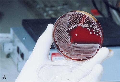
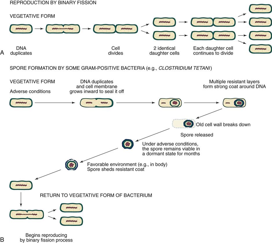
Bacteria
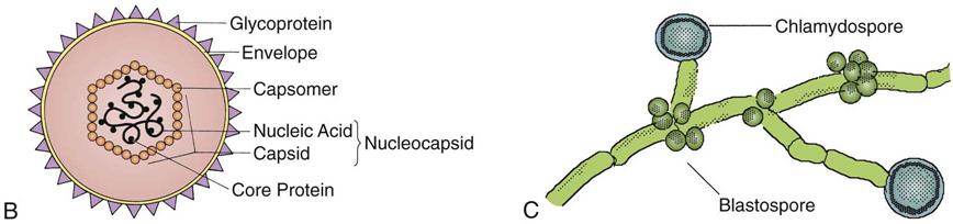
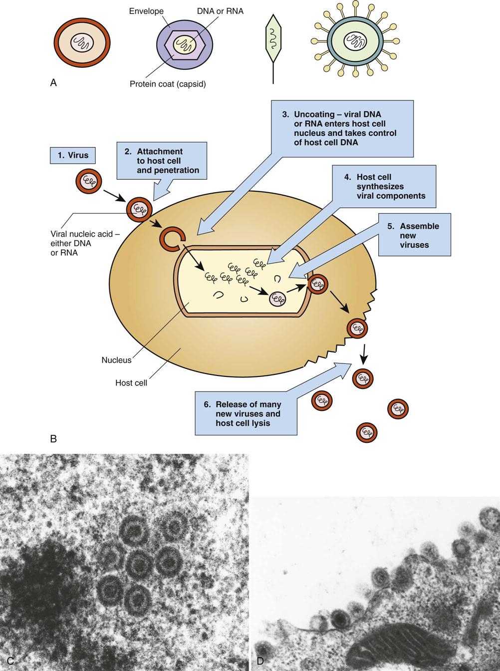
Viruses
Type of Virus
RNA or DNA
Example of Disease
Orthomyxoviruses
RNA
Influenza A, B, and C
Paramyxoviruses
RNA
Mumps, measles
Togavirus
RNA
Rubella virus (German measles), Hepatitis C virus
Herpesvirus
DNA
Herpes simplex, Infectious mononucleosis, Varicella (chickenpox)
Flaviviruses
RNA
West Nile virus, Encephalitis
Picornaviruses
RNA
Poliovirus, Hepatitis A virus
Hepadnaviruses
DNA
Hepatitis B virus
Papovaviruses
DNA
Warts, cancer (human papillomavirus/HPV)
Retrovirus
RNA
Human immunodeficiency viruses
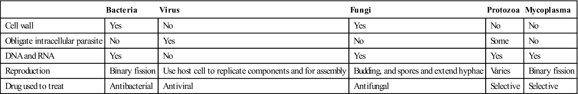
Chlamydiae, Rickettsiae, and Mycoplasmas
Bacteria
Virus
Fungi
Protozoa
Mycoplasma
Cell wall
Yes
No
Yes
No
No
Obligate intracellular parasite
No
Yes
No
Some
No
DNA and RNA
Yes
No
Yes
Yes
Yes
Reproduction
Binary fission
Use host cell to replicate components and for assembly
Budding, and spores and extend hyphae
Varies
Binary fission
Drug used to treat
Antibacterial
Antiviral
Antifungal
Selective
Selective
Fungi
Protozoa
Infection