5 Neoplasia
A neoplasm (‘new growth’) is a lesion that results from abnormal growth of a tissue, which is partly or completely autonomous of normal growth controls and persists after the initiating stimulus has been removed.
Neoplasms usually manifest themselves as tumours (abnormal swellings). However, some neoplasms, most notably those derived from haemopoeitic cells do not form tumours, and clinically tumourous lesions can be caused by non-neoplastic disease (e.g. tuberculosis).
This chapter will describe how neoplasms form, how they are classified and how they behave.
CARCINOGENESIS
Carcinogenesis is the process by which normal cells are converted into cells capable of forming neoplasms. There is no single cause of neoplasia, and it is generally accepted that most neoplasms require several events to occur in a single cell (the multistep hypothesis) before a sustainable neoplasm can form. This accounts for the relative rarity of neoplasms when compared with the number of cells in the body, all of which, theoret-ically, have the potential to form neoplasms.
Another factor that protects most cells from neoplasia is that, in order to form a neoplasm, a cell must divide. Thus cells which are postmitotic, such as nerve cells and skeletal muscle cells, rarely form neoplasms, whereas cells such as the epithelium of the gut and the epidermis of the skin, which continually divide, form neoplasms more frequently.
This section will describe the factors that predispose to the formation of neoplasms. Some examples of carcinogens are given in Table 5.1
Table 5.1 Examples of carcinogens
CARCINOGENS
Carcinogens are agents which cause the formation of neoplasms from cells exposed to them. The nature of carcinogens is diverse, but they all have the ability, directly or indirectly, to cause an inheritable change in the genes that control the growth and survival of the target cell.
Many carcinogens are now well known to the public as well as the medical profession: for example, tobacco smoke and asbestos. These have largely been identified by studies of the epidemiology of the neoplasms that they cause. Individual carcinogens can often cause neoplasms in more than one target tissue (e.g. tobacco derivatives can cause neoplasms of bronchial, laryngeal, oral, renal and bladder epithelium), and individual types of neoplasm can be caused by more than one carcinogen (e.g. bronchogenic carcinoma can be caused by tobacco derivatives, asbestos, nickel, or radon gas). However, for many types of neoplasm, the carcinogenic stimulus is not known.
Chemical
A variety of chemicals have been identified as carcinogens in man, and others are suspected to be on the basis of their carcinogenic effects in experimental animals. There is no common structural link between the different types of chemical carcinogen, but they appear to have in common the ability to modify the structure of DNA: for example, by forming adducts or by adding alkyl groups.
Many chemical carcinogens are procarcinogens which require metabolic conversion to their active form by enzymes. If the enzyme required is present in all cell types, the carcinogenic effect is likely to occur at the site of exposure. However, some carcinogens require metabolism in another tissue, which influences where they exert their carcinogenic effects. This is well illustrated by the aromatic amine β-naphthylamine, which requires metabolism by the liver before being active, and as a result causes neoplasms of the bladder where it is concentrated during excretion.
The major classes of chemical carcinogens currently known are as follows.
Polycyclic aromatic hydrocarbons
The first example of an occupation-related neoplasm was the description by Percival Pott in 1777 of scrotal carcinomas in adults who had been employed as chimney sweeps during childhood. It has subsequently been shown that this was due to exposure to polycyclic aromatic hydrocarbons. This class of compounds was found to be the carcinogenic component of tar, which can cause skin neoplasms if applied experimentally to the skin of rabbits, and was probably responsible for the high incidence of skin cancers in oil shale miners in West Lothian in Scotland during the nineteenth century. Of greater importance today is the carcinogenic effect of polycyclic aromatic hydrocarbons present in tobacco smoke, most notably 3,4-benzpyrene. Polycyclic aromatic hydrocarbons are procarcinogens which require the action of hydroxylating enzymes such as aryl carbo-hydrate hydroxylase to become active carcinogens. These enzymes are ubiquitous, so polycyclic aromatic hydrocarbons can be carcinogenic at their site of contact, but as they can be absorbed into the blood stream, they are also carcinogenic at distant sites such as the kidney and bladder. This accounts for the fact that, although smoking tobacco is most strongly associated with carcinogenesis in tissues directly exposed such as the bronchus and larynx, smokers have a slightly increased risk of neoplasia in many other tissues.
Aromatic amines
Epidemiological studies have shown an increased risk of bladder neoplasms in workers in the rubber industry. This has been found to be due to the aromatic amine, β-naphthylamine, which is converted into the active carcinogen 1-hydroxy-2-naphthylamine in the liver. Glucuronidation of this compound occurs in the liver, protecting the cells of the liver and other tissues from its carcinogenic effects. However, in the urinary tract, glucuronidase unconjugates the molecule, thus exposing the bladder urothelium to its carcinogenic effects.
Alkylating agents
The polycyclic aromatic hydrocarbons can act by adding alkyl groups to DNA, so one would expect that the alkylating agents such as cyclophosphamide that are used as chemotherapeutic agents might also be carcinogenic. While this risk is not sufficiently strong to contraindicate their use, there is certainly evidence that patients treated with these compounds for conditions such as Hodgkin’s disease have an increased risk of developing a different type of neoplasm later in life.
Azo dyes
These are an example of a class of compounds where recognition of their carcinogenic activity in laboratory studies has fortunately restricted their industrial use. For example, the dye dimethylaminoazobenzene causes liver cancer in rats.
Nitrosamines
This is another class of compounds that are strongly carcinogenic in laboratory animals. It is not known to what extent they are carcinogenic in humans, but it is possible that generation of nitrosamines by fungi in poorly stored food could be responsible for some gastrointestinal neoplasms.
Radiation
Electromagnetic radiation of wavelengths shorter than the visible spectrum can cause damage to DNA that can result in neoplasia. Ultraviolet light is a significant carcinogen because of the high levels of exposure that can occur during daily life, whereas ionising radi-ation (such as x-rays and gamma-rays) is significantly carcinogenic because of the high levels of energy it possesses.
Ultraviolet light
The relationship between exposure to ultraviolet light and skin neoplasms is now well established. Neoplasms of the epidermis (basal cell carcinoma and squamous cell carcinoma), and the related precancerous condition solar/actinic keratosis, usually occur on sun-exposed sites and become more frequent with greater sun exposure. Similarly, malignant melanoma, a malignant neoplasm of melanocytes, is most common in fair-skinned individuals living in environments with high levels of sunlight exposure, such as white Australians. Malignant melanoma is uncommon in individuals of Afro-Caribbean origin because the greater density of melanin in their skin reduces the amount of ultraviolet light that reaches the melanocytes, which reside along the basal (deepest) layer of the epidermis. The pattern of ultraviolet exposure is important in determining which cells are most affected: long-term chronic expos-ure is associated with an increased risk of the development of basal cell carcinoma or squamous carcinoma, whereas melanoma is more strongly associated with episodes of ultraviolet exposure of sufficient intensity to cause sunburn.
Ionising radiation
The first indication of the carcinogenic potential of ionising radiation came from the frequency with which early x-ray workers developed skin cancers on their hands. Further evidence subsequently accumulated from the development of neoplasms, particularly leukaemia, in the survivors of the World War II atomic bombs. Ionising radiation can cause neoplasms in a wide variety of tissues: for example, therapeutic irradiation can result in the development of bone and soft tissue sarcomas, and the Chernobyl disaster has caused a large increase in thyroid cancers in the Ukraine because of the release of radioactive iodine which resulted; this element is, of course, concentrated and stored in the thyroid gland. One of the great dangers of radioactive substances is, depending on their half-life, the persistence of their effect within the body. A good example of this is the persistence of the thorium dioxide from the radiological agent thorotrast within the liver. This has caused the development of hepatic angiosarcomas in some patients many years after exposure. Localised radiotherapy used to treat cancers is also associated with an increased risk of second malignancies developing in subsequent years, particularly sarcomas.
Viruses
A growing number of viruses have been implicated in the development of neoplasms. There are manyexamples of virally induced neoplasms in animals, study of which has done much to promote our understanding of the molecular genetics of neoplasia. Virally induced neoplasms in humans are (as far as we are aware) rather less common. The most ubiquitous oncogenic viruses are the human papilloma viruses; other viruses with well-established carcinogenic effects are the Epstein-Barr virus and the hepatitis B virus (see Table 5.2).
Virus | Neoplasm caused |
---|---|
Human papilloma virus | |
Epstein-Barr virus | |
Mechanisms of viral carcinogenesis
DNA viruses can be carcinogenic either through integration into the host genome in such a way that interferes with the function of growth-controlling genes, or through their ability to produce proteins that interfere with growth-regulating factors. For example, human papilloma viruses produce proteins that inhibit the function of the p53 and Rb1 gene products (see section on genetics). The best-known examples of oncogenic DNA viruses are the Epstein-Barr virus, which is strongly associated with Burkitt’s lymphoma and nasopharyngeal carcinoma, the hepatitis B virus, which is associated with hepatocellular (liver) carcinoma, and the human papilloma virus (HPV). HPV is associated with neoplasia of a number of different surface epithelia. It is responsible for the common viral wart of the skin and is also the main cause of carcinoma of the cervix, its precursor condition cer-vical intraepithelial neoplasia (CIN), and other forms of analogous intraepithelial neoplasia such as anal intraepithelial neoplasia (AIN). There are many different types of HPV. Individual types have preferred target tissues and have differing oncogenic potential. For example, many different HPV types infect the cervix, but only a small number of types (particularly types 16 and 18) are associated with the development of cervical carcinoma.
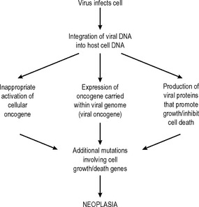
Oncogenic RNA viruses are retroviruses which integrate their genetic material into the host genome using the enzyme reverse transcriptase. Although there are many examples of oncogenic retroviruses causingneoplasms in animals, this is rare in man. The best-known examples are Human T-Lymphotrophic Virus-1 (HTLV-1) which causes a form of lymphoma/leukaemia which is endemic in Japan and the Caribbean, and the human immunodeficiency virus (HIV). However, HIV probably does not have a direct carcinogenic effect; the neoplasms that are associated with HIV infection probably arise as a consequence of immunosuppression and may actually be caused by other types of virus. Thus, HIV infection may act as a cofactor for oncogenesis by other viruses. There are other examples of this phenomenon, such as the Epstein-Barr virus requiring malaria infection as a cofactor in the development of Burkitt’s lymphoma.
Other associations between viruses and neoplasms are being described – for example, herpes virus 8 and Kaposi’s sarcoma and myeloma – and it seems likely that further causative associations will be established in the future, particularly in neoplasms of the lymphoreticular system. The sequence of events by which viruses can cause neoplasia is outlined in Fig. 5.1.
Other non-biological factors
Asbestos
The association between asbestos and malignant mesothelioma (a neoplasm of the pleural, pericardial, or peritoneal mesothelial lining) is so strong that this disease is almost unknown in individuals who have not been exposed to asbestos. Asbestos was a widely used building material because of its fire resistance, before the health risks of asbestos exposure were known. As a result of this, the incidence of mesothelioma con-tinues to rise despite the restrictions now placed on the use of asbestos. There is also a strong link between asbestos exposure and carcinoma of the bronchus. The mechanism responsible for the carcinogenic effect of asbestos is not known.
Metals
Industrial exposure to nickel is associated with an increased risk of nasal and bronchogenic carcinoma. In the setting of haemochromatosis, iron could be said to be an indirect carcinogen in the liver; however, the development of cirrhosis is required before the increased risk of hepatocellular carcinoma in this condition can be realised.
Other biological factors
Helicobacter pylori infestation
Helicobacter pylori infestation is a common cause of gastritis and peptic ulceration. Chronic Helicobacter pylori gastritis sometimes leads to intestinal metaplasia of the gastric mucosa. This results in the normal secretory epithelium of the gastric antrum being replaced by an epithelium with intestinal characteristics. Sometimes this epithelium is well differentiated with a mixture of absorptive and goblet cells identical to those seen in the small intestine. In other cases the epithelium is less well differentiated, being identifiable as intestinal rather than gastric by the type of mucin that it produces. In the latter case, there is a small risk of the development of dysplasia (see below, under ‘premalignant conditions’) and ultimately gastric carcinoma. However, the association between Helicobacter pylori infestation and gastric carcinoma appears to be weak and presumably requires multiple cofactors. Nonetheless, this causative link has recently been confirmed in experimental animals infected with Helicobacter pylori.
There is a more direct link between Helicobacter infestation and a far less common neoplasm of the stomach, the so-called mucosa-associated lymphoid tissue (MALT) lymphoma. It has been shown that, despite having characteristics of a malignant neoplasm, such as clonality and invasiveness, MALT lymph-omas sometimes regress when patients are treated with Helicobacter-eradicating antibiotics. However, it is more likely that Helicobacter infestation represents a growth-sustaining stimulus, rather than a conventional carcinogen. These observations have led to some debate about whether MALT lymphomas are true neoplasms or not.
Parasitic infestations
Schistosomiasis is associated with an increased risk of carcinoma of the bladder. Interestingly, Schistosomiasis-associated bladder carcinomas are squamous carcinomas, rather than transitional cell carcinomas which are the usual type of malignant neoplasm of the bladder. Clonorchis sinensis, the Chinese liver fluke, is also capable of inducing neoplasia of the bile ducts in which it dwells.
Hormones
Some neoplasms such as carcinomas of the breast and prostate may require the presence of hormones to maintain or promote their growth, as will be discussed below. There are also examples of abnormal exposure to some hormones being carcinogenic. For example, anabolic and androgenic steroids can cause the development of hepatocellular carcinoma, and oestrogens are associated with hepatocellular adenomas. Certain rare tumours of the female genital tract, such as clear cell carcinoma of the vagina, are very strongly associated with in-utero exposure to diethylstilboestrol, which was used therapeutically during pregnancy in the past.
Mycotoxins
It is likely that there are many toxins produced by fungi that are carcinogenic. To date the best-established carcinogenic effect is that of the aflatoxins produced by Aspergillus flavus. These toxins occur as dietary contaminants and are linked to the high incidence of hepatocellular carcinoma in some parts of central Africa.
HOST FACTORS
Age
Neoplastic disease is primarily a disease of old age. Although neoplasms can occur at any age, even in utero, neoplasms of almost all types become far more common after the age of 50. This presumably reflects the cumulative effects of exposure to carcinogens over an individual’s lifespan. A major reason for the continuing increase in the incidence of neoplastic disease is the increasing life expectancy of most populations.
Individual types of neoplasm have their own typical age distribution. For example, fibroadenoma of the breast usually occurs in women in their second, third and fourth decades, whereas carcinoma of the breast becomes more common after the menopause. Other types of neoplasm, for example, neuroblastoma of the adrenal, are restricted to children and are almost unknown in adults.
It is a general rule that familial neoplasms – that is, those occurring in individuals who have a genetic predisposition to them (see below under genetic factors) –occur at a younger age than sporadic neoplasms.
Race
Different races are subject to different profiles of neoplastic disease. This is almost entirely due to differences in lifestyle. For example, the commonest fatal neoplasm in the UK and the USA is carcinoma of the bronchus, which is caused largely by tobacco smoking. The commonest fatal neoplasm worldwide is hepatocellular carcinoma, which in Africa and South-East Asia is related to exposure to dietary carcinogens and viral hepatitis. Immigrant groups tend to eventually assume the disease profile of their adopted countries. There are, however, occasional examples of genetically determined racial differences, such as a high frequency of familial breast cancer in Ashkenazi Jews.
Endocrine status
Gender influences the risk of developing many types of neoplasm. This is generally related to differences in hormonal status, although lifestyle differences can play a part. For example, the far higher incidence of neoplasms of the breast in females than males is probably mainly due to endocrine influences, whereas, in the past, bronchogenic carcinoma was more common in men than women because of differences in the frequency of tobacco smoking between the sexes. There are, however, many examples where the influence of gender is not understood: for example, the higher frequency of osteosarcoma in males.
Diet
The risks of developing neoplasia as a result of dietary contaminants are well illustrated by the example of aflatoxin-induced hepatocellular carcinoma. Other dietary factors may also modify the risk of developing certain neoplasms, for example, there is a link between high levels of dietary fat and breast carcinoma. The risk of colorectal carcinoma seems to be associated with diet, but is probably multi-factorial. Dietary fiber, fruit and vegetable consumption seem to be protective and red meat consumption seems to be deleterious, but it has been difficult to consistently show an independent effect for any of these factors. There is an increasing level of public interest in the importance of diet in causing or preventing cancers of many types. This has led to much interest in the media and even governmental public health campaigns, although the level of scientific evidence behind the benefits of any individual dietary manipulations is often dubious at best and imaginary at worst.
Genetic factors
Changes in the structure and function of a cell’s genetic material are central to the development of neoplasia, and more than one such change is required in an individual cell before neoplasia can occur. If all of an individual’s cells already have an abnormality in a relevant gene as a result of that individual’s inherited genetic make-up (a germ-line mutation), then fewer subsequent changes are required for neoplasia to occur. This is well illustrated by the rare familial retinoblastoma syndrome (Fig. 5.2). If both alleles of the retinoblastoma (Rb1) gene in an individual retinal cell are non-functional, retinoblastoma can develop from that cell. Sporadic retinoblastoma is a rare tumour because it is unusual for both retinoblastoma alleles in an individual cell to acquire mutations that inhibit their function. However, if one allele is already non-functional because it was inherited in a defective form, then the chances of retinoblastoma developing as a result of a subsequent mutation of the other allele are very high. This also demonstrates that the ‘retinoblastoma’ gene is a tumour suppressor gene. This is a common property of the genes that are abnormal in the various familial cancer syndromes.
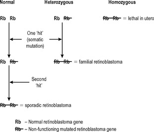
Another characteristic that the retinoblastoma syndrome shows, that is common in familial cancer syndromes, is that it affects more than one tissue. If individuals with the retinoblastoma syndrome survive the development of retinoblastomas (which are usually bilateral) early in childhood, they have a very high incidence of osteosarcoma during adolescence. The retinoblastoma syndrome is used here as an illustration because its genetics are simple and well characterised. However, there are a number of other familial cancer syndromes, many of which, such as familial polyposis coli, are more common. Increasingly, these syndromes are being identified with mutations in genes that are involved in DNA repair such as the BRCA1 gene associated with familial breast cancer. The best known examples are given in Table 5.3.
Table 5.3 Examples of familial cancer syndromes
Syndrome | Gene affected | Resultant neoplasms |
---|---|---|
Li Fraumeni | p53 | Breast, ovarian carcinomas, astrocytomas, sarcomas |
Retinoblastoma | Rb1 | Retinoblastoma, osteosarcoma |
Familial polyposis coli von Hippel-Lindau | APC | GI tract carcinomas, mainly colon |
VHL | Renal carcinoma, phaeochromocytoma, haemangioblastoma | |
Multiple endocrine neoplasia syndromes (I–III) | RET, others | Tumours of pituitary parathyroids, thyroid, pancreas, adrenal (combination depends on which syndrome) |
Familial breast cancer | BRCA 1, BRCA 2 | Breast, ovarian syndrome prostatic carcinomas |
Immune response
Some neoplasms attract large numbers of inflammatory cells, usually lymphocytes, into their substance, and there is evidence that in some tumours this may convey a better prognosis. These observations have led to the development of the major research subspecialty of tumour immunology, but, at present, treating neoplasms by stimulating the host immune response islittle more than a theoretical concept.
However, evidence has been put forward that the immune system can detect and mount a response against neoplasms, principally via NK cells. The activity of these cells can be stimulated by lymphokines such as interleukin 2, and there is some evidence that factors like interleukin 2 could have therapeutic efficacy against some neoplasms. A more convincing example of an immunological treatment that can suppress the development of a neoplasm is the effect of BCG treatment on carcinoma-in-situ of the bladder, although whether this is due to a specific immunological reaction or shedding of unstable transformed urothelium in response to a non-specific inflammatory response is not clear.
The host immune response has another important indirect effect on the development of neoplasms. There is a strong positive link between immunodeficiency and the development of neoplasia. This is illustrated by the frequency of development of lymphomas and Kaposi’s sarcoma in the acquired immune deficiency syndrome (AIDS), and cutaneous and anogenital squamous carcinomas in organ transplant recipients taking immunosuppressive therapy. In these settings the increased risk of neoplasia seems to be due to an inadequate immune response to oncogenic viruses such as HHV-8 in the case of Kaposi’s sarcoma and HPV in the case of transplant-associated squamous carcinomas.
PREMALIGNANT DISEASE
Given that malignant neoplasms usually develop as the result of multiple steps over a period of time, it is perhaps not surprising that many premalignant diseases have been described. Premalignant lesions are discrete identifiable lesions that may progress to become malignant neoplasms. These can be:
Premalignant conditions are non-neoplastic conditions that frequently lead to the development of neoplasms.
(The distinction between benign and malignant neoplasms will be defined below. The term dysplasia was defined in Chapter 4.)
Malignant change in benign neoplasms
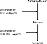
The majority of benign neoplasms do not alter in any way, but some benign neoplasms have the ability to progress to become malignant neoplasms. Probably the best-characterised example of this phenomenon is the adenoma-carcinoma sequence in the colon. Adenomatous polyps of the colon are more numerous than colonic carcinomas, but all adenomatous polyps have the potential to develop into carcinomas, and many (but not all) carcinomas originate from adenomatous polyps. The polyps most likely to undergo malignant change show the greatest degree of histological dysplasia and a sequence of genetic changes that leads to the development of colorectal carcinoma from normal epithelium via adenomatous polyps has now been described (Fig. 5.3).

Fig. 5.3 The sequence of genetic alterations in the colorectal adenoma-carcinoma sequence. APC = adenomatous polyposis coli gene; MCC = mutated in colon cancer gene; DCC = deleted in colon cancer gene; Ras = a cellular oncogene involved in growth factors signal transduction; p53 = a tumour suppressor gene.
Metaplasia-dysplasia sequence
Neoplastic transformation of cells occurs in cells undergoing proliferation, and is particularly likely to occur if the cells are also undergoing metaplasia (defined and described in the previous chapter). Neoplastic transformation of metaplastic epithelium usually follows a predictable and histologically identifiable sequence of low grade dysplasia progressing to high grade dysplasia/in-situ malignancy to invasive malignancy as additional genetic abnormalities are acquired in the neoplastic population. This progression is very well demonstrated in the cervix (Fig. 5.4). Other examples of the metaplasia–dysplasia sequence are shown in Table 5.4.
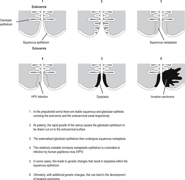
Table 5.4 Examples of the metaplasia-dysplasia sequence
Organ | Form of metaplasia undergoing dysplasia | Resulting malignancy |
---|---|---|
Oesophagus | Barrett’s oesophagus (intestinal metaplasia) | Oesphageal adenocarcinoma |
Stomach | Intestinal metaplasia (associated with achlorhydria) | Gastric adenocarcinoma |
Bronchus | Squamous metaplasia | Brochogenic squamous carcinoma |
Cervix | Squamous metaplasia | Cervical squamous carcinoma |
Premalignant conditions
These are usually conditions characterised by high cell turnover over a sustained period of time, usually resulting from a destructive form of chronic inflammation. Congenital abnormalities can also be premalignant conditions: for example, maldescent of the testis is associated with an increased risk of testicular neoplasia in later life. Some examples of premalignant conditions are given in Table 5.5.
Table 5.5 Examples of premalignant conditions
Premalignant condition | Resulting neoplasm |
---|---|
Ulcerative colitis | Colorectal carcinoma |
Chronic fistulae | Squamous carcinoma |
Epithelial hyperplasia of the breast | Breast carcinoma |
Paget’s disease of bone | Osteosarcoma |
Xeroderma pigmentosum | Skin malignancies |
CARCINOGENIC PROCESS
The carcinogenic process is the chain of events whereby a carcinogenic stimulus leads to theformation of a neoplasm. The principal steps in this process are as follows:
These four steps occur with decreasing frequency: exposure of cells to carcinogens is a very common event, and genetic alterations to growth-controlling genes probably occur quite frequently, but, because of inbuilt defense mechanisms, the latter two steps are relatively uncommon.
The division of the carcinogenic process into the stages of initiation, promotion and persistence is based upon experimental evidence from models of tumour formation in which initiating and promoting stimuli are required. However, our increasing understanding of the molecular genetics of this process indicates that the stages described above simply reflect the requirements for more than one genetic change to occur before neoplasia becomes established.
The precise chains of molecular events in most tumour types have yet to be established.
GENETICS
Chromosomal abnormalities
Very crude DNA abnormalities may manifest themselves as visible changes in chromosomes isolated from neoplastic cells. These abnormalities can occur in a number of forms:
The DNA of many neoplasms, particularly malignant ones, is inherently unstable, and random chromosomal abnormalities are common. However, there are a number of chromosomal abnormalities that are consistently found in certain tumour types, the best known being the ‘Philadelphia chromosome’ (a reciprocal, balanced translocation between chromosomes 9 and 22). At a purely descriptive level, these can be useful for diagnosis, particularly in groups of tumours in which the cells are morphologically similar, such as leukaemias and the ‘small round cell tumours’ of childhood such as neuroblastoma and alveolar rhabdomyosarcoma. Detailed molecular study of these chromosomal abnormalities has yielded some insight into the pathogenesis of some of the neoplasms with specific chromosomal abnormalities. A good example of this is the translocation between chromosomes 14 and 18 that occurs in follicular (low grade) B cell non-Hodgkin’s lymphomas. This translocation results in the bcl-2 gene coming under the control of the immunoglobulin heavy chain gene promoter. As B lymphocytes constitutively express their immunoglobulin genes, this results in inappropriate over-expression of the bcl-2 gene and thus overproduction of the bcl-2 protein. As bcl-2 is an anti-apoptotic protein, this results in the ‘immortalisation’ of the neoplastic B lymphocytes.

Stay updated, free articles. Join our Telegram channel

Full access? Get Clinical Tree

