and John E. Skandalakis1
(1)
Centers for Surgical Anatomy and Technique, Emory University School of Medicine Piedmont Hospital, Atlanta, GA, USA
Abstract
Surgery of the neck includes the thyroid, parathyroid, parotid and submaxillary glands, thoracic ducts, trachea, and branchial remnants. The surgical anatomy and subdivisions of the cervical triangles (anterior and posterior) are presented in detail. The complicated fascial planes and spaces of the neck are described. Knowledge of the overall anatomy and drainage of the lymphatics of the head and neck, crucial for surgery, is presented in detail.
Diagnosis of neck masses follows a well-marked pathway. Step-by-step technique is presented for parotidectomy, thyroidectomy, parathyroidectomy (and their respective reoperation), tracheostomy, and gland resection and cystectomy. Radical neck dissection as a curative procedure follows surgicoanatomical guidelines.
Anatomy
Anterior Cervical Triangle (Fig. 2.1)
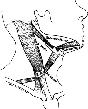
Figure 2.1.
The subdivision of the anterior triangle of the neck (By permission of JE Skandalakis, SW Gray, and JR Rowe. Am Surg 45(9):590–596, 1979).
The boundaries are:
Lateral: sternocleidomastoid muscle
Superior: inferior border of the mandible
Medial: anterior midline of the neck
This large triangle may be subdivided into four more triangles: submandibular, submental, carotid, and muscular.
Submandibular Triangle
The submandibular triangle is demarcated above by the inferior border of the mandible and below by the anterior and posterior bellies of the digastric muscle.
The largest structure in the triangle is the submandibular salivary gland. A number of vessels, nerves, and muscles also are found in the triangle.
For the surgeon, the contents of the triangle are best described in four layers, or surgical planes, starting from the skin. It must be noted that severe inflammation of the submandibular gland can destroy all traces of normal anatomy. When this occurs, identifying the essential nerves becomes a great challenge.
Roof of the Submandibular Triangle
The roof—the first surgical plane—is composed of skin, superficial fascia enclosing platysma muscle and fat, and the mandibular and cervical branches of the facial nerve (VII) (Fig. 2.2).
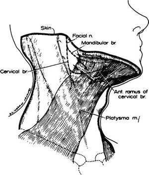

Figure 2.2.
The roof of the submandibular triangle (the first surgical plane). The platysma lies over the mandibular and cervical branches of the facial nerve (By permission of JE Skandalakis, SW Gray, and JR Rowe. Am Surg 45(9):590–596, 1979).
It is important to remember that: (1) the skin should be incised 4–5 cm below the mandibular angle, (2) the platysma and fat compose the superficial fascia, and (3) the cervical branch of the facial nerve (VII) lies just below the angle, superficial to the facial artery (Fig. 2.3).
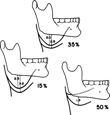

Figure 2.3.
The neural “hammocks” formed by the mandibular branch (upper) and the anterior ramus of the cervical branch (lower) of the facial nerve. The distance below the mandible is given in centimeters, and percentages indicate the frequency found in 80 dissections of these nerves (By permission of JE Skandalakis, SW Gray, and JR Rowe. Am Surg 45(9):590–596, 1979).
The mandibular (or marginal mandibular) nerve passes approximately 3 cm below the angle of the mandible to supply the muscles of the corner of the mouth and lower lip.
The cervical branch of the facial nerve divides to form descending and anterior branches. The descending branch innervates the platysma and communicates with the anterior cutaneous nerve of the neck. The anterior branch—the ramus colli mandibularis—crosses the mandible superficial to the facial artery and vein and joins the mandibular branch to contribute to the innervation of the muscles of the lower lip.
Injury to the mandibular branch results in severe drooling at the corner of the mouth. Injury to the anterior cervical branch produces minimal drooling that will disappear in 4–6 months.
The distance between these two nerves and the lower border of the mandible is shown in Fig. 2.3.
Contents of the Submandibular Triangle
The structures of the second surgical plane, from superficial to deep, are the anterior and posterior facial vein, part of the facial (external maxillary) artery, the submental branch of the facial artery, the superficial layer of the submaxillary fascia (deep cervical fascia), the lymph nodes, the deep layer of the submaxillary fascia (deep cervical fascia), and the hypoglossal nerve (XII) (Fig. 2.4).
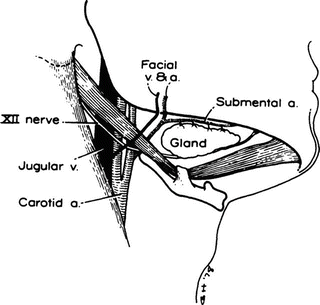

Figure 2.4.
The contents of the submandibular triangle (the second surgical plane). Exposure of the superficial portion of the submandibular gland (By permission of JE Skandalakis, SW Gray, and JR Rowe. Am Surg 45(9): 590–596, 1979).
It is necessary to remember that the facial artery pierces the stylomandibular ligament. Therefore, it must be ligated before it is cut to prevent bleeding after retraction. Also, it is important to remember that the lymph nodes lie within the envelope of the submandibular fascia in close relationship with the gland. Differentiation between gland and lymph node may be difficult.
The anterior and posterior facial veins cross the triangle in front of the submandibular gland and unite close to the angle of the mandible to form the common facial vein, which empties into the internal jugular vein near the greater cornu of the hyoid bone. It is wise to identify, isolate, clamp, and ligate both of these veins.
The facial artery—a branch of the external carotid artery—enters the submandibular triangle under the posterior belly of the digastric muscle and under the stylohyoid muscle. At its entrance into the triangle it is under the submandibular gland. After crossing the gland posteriorly, the artery passes over the mandible, lying always under the platysma. It can be ligated easily.
Floor of the Submandibular Triangle
The structures of the third surgical plane, from superficial to deep, include the mylohyoid muscle with its nerve, the hyoglossus muscle, the middle constrictor muscle covering the lower part of the superior constrictor, and part of the styloglossus muscle (Fig. 2.5).
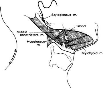

Figure 2.5.
The floor of the submandibular triangle (the third surgical plane). Exposure of mylohyoid and hyoglossus muscles (By permission of JE Skandalakis, SW Gray, and JR Rowe. Am Surg 45(9):590–596, 1979).
The mylohyoid muscles are considered to form a true diaphragm of the floor of the mouth. They arise from the mylohyoid line of the inner surface of the mandible and insert on the body of the hyoid bone into the median raphe. The nerve to the mylohyoid, which arises from the inferior alveolar branch of the mandibular division of the trigeminal nerve (V), lies on the inferior surface of the muscle. The superior surface is in relationship with the lingual and hypoglossal nerves.
Basement of the Submandibular Triangle
The structures of the fourth surgical plane, or basement of the triangle, include the deep portion of the submandibular gland, the submandibular (Wharton’s) duct, lingual nerve, sublingual artery, sublingual vein, sublingual gland, hypoglossal nerve (XII), and the submandibular ganglion (Fig. 2.6).
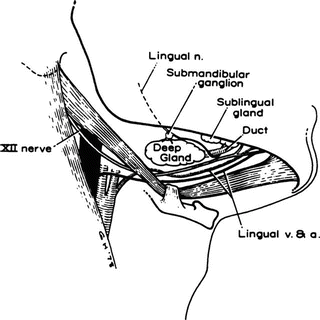

Figure 2.6.
The basement of the submandibular triangle (the fourth surgical plane). Exposure of the deep portion of the submandibular gland, the lingual nerve, and the hypoglossal (XII) nerve (By permission of JE Skandalakis, SW Gray, and JR Rowe. Am Surg 45(9):590–596, 1979).
The submandibular duct lies below the lingual nerve (except where the nerve passes under it) and above the hypoglossal nerve.
Lymphatic Drainage of the Submandibular Triangle
The submandibular lymph nodes receive afferent channels from the submental nodes, oral cavity, and anterior parts of the face. Efferent channels drain primarily into the jugulodigastric, jugulocarotid, and jugulo-omohyoid nodes of the chain accompanying the internal jugular vein (deep cervical chain). A few channels pass by way of the subparotid nodes to the spinal accessory chain.
Submental Triangle (See Fig. 2.1)
The boundaries of this triangle are:
Lateral: anterior belly of digastric muscle
Inferior: hyoid bone
Medial: midline
Floor: mylohyoid muscle
Roof: skin and superficial fascia
The lymph nodes of the submental triangle receive lymph from the skin of the chin, the lower lip, the floor of the mouth, and the tip of the tongue. They send lymph to the submandibular and jugular chains of nodes.
Carotid Triangle (See Fig. 2.1)
The boundaries are:
Posterior: sternocleidomastoid muscle
Anterior: anterior belly of omohyoid muscle
Superior: posterior belly of digastric muscle
Floor: hyoglossus muscle, inferior constrictor of pharynx, thyrohyoid muscle, longus capitis muscle, and middle constrictor of pharynx
Roof: investing layer of deep cervical fascia
Contents of the carotid triangle: bifurcation of carotid artery; internal carotid artery (no branches in the neck); external carotid artery branches, e.g., superficial temporal artery, internal maxillary artery, occipital artery, ascending pharyngeal artery, sternocleidomastoid artery, lingual artery (occasionally), and external maxillary artery (occasionally); jugular vein tributaries, e.g., superior thyroid vein, occipital vein, common facial vein, and pharyngeal vein; and vagus nerve, spinal accessory nerve, hypoglossal nerve, ansa hypoglossi, and sympathetic nerves (partially).
Lymph is received by the jugulodigastric, jugulocarotid, and jugulo-omohyoid nodes and by the nodes along the internal jugular vein from submandibular and submental nodes, deep parotid nodes, and posterior deep cervical nodes. Lymph passes to the supraclavicular nodes.
Muscular Triangle (Fig. 2.1)
The boundaries are:
Superior lateral: anterior belly of omohyoid muscle
Inferior lateral: sternocleidomastoid muscle
Medial: midline of the neck
Floor: prevertebral fascia and prevertebral muscles; sternohyoid and sternothyroid muscles
Roof: investing layer of deep fascia; strap, sternohyoid, and cricothyroid muscles
Contents of the muscular triangle include: thyroid and parathyroid glands, trachea, esophagus, and sympathetic nerve trunk.
Remember that occasionally the strap muscles must be cut to facilitate thyroid surgery. They should be cut across the upper third of their length to avoid sacrificing their nerve supply.
Posterior Cervical Triangle (Fig. 2.7)
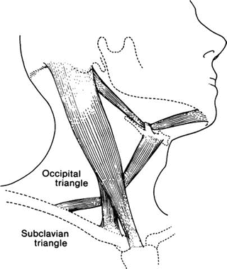
Figure 2.7.
The posterior triangle of the neck. The triangle may be divided into two smaller triangles by the omohyoid muscle (By permission of JE Skandalakis, SW Gray, and JR Rowe. Anatomical Complications in General Surgery. New York: McGraw-Hill, 1983).
The posterior cervical triangle is sometimes considered to be two triangles—occipital and subclavian—divided by the posterior belly of the omohyoid muscle or, perhaps, by the spinal accessory nerve (see Fig. 2.7); we will treat it as one.
The boundaries of the posterior triangle are:
Anterior: sternocleidomastoid muscle
Posterior: anterior border of trapezius muscle
Inferior: clavicle
Floor: prevertebral fascia and muscles, splenius capitis muscle, levator scapulae muscle, and three scalene muscles
Roof: superficial investing layer of the deep cervical fascia
Contents of the posterior cervical triangle include: subclavian artery, subclavian vein, cervical nerves, brachial plexus, phrenic nerve, accessory phrenic nerve, spinal accessory nerve, and lymph nodes.
The superficial occipital lymph nodes receive lymph from the occipital region of the scalp and the back of the neck. The efferent vessels pass to the deep occipital lymph node (usually only one), which drains into the deep cervical nodes along the spinal accessory nerve.
Fasciae of the Neck
Our classification of the rather complicated fascial planes of the neck follows the work of several investigators. It consists of the superficial fascia and three layers that compose the deep fascia.
Superficial Fascia
The superficial fascia lies beneath the skin and is composed of loose connective tissue, fat, the platysma muscle, and small unnamed nerves and blood vessels (Fig. 2.8). The surgeon should remember that the cutaneous nerves of the neck and the anterior and external jugular veins are between the platysma and the deep cervical fascia. If these veins are to be cut, they must first be ligated. Because of their attachment to the platysma above and the fascia below, they do not retract; bleeding from them may be serious. For all practical purposes, there is no space between this layer and the deep fascia.
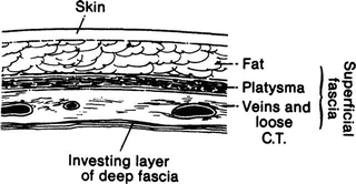

Figure 2.8.
The superficial fascia of the neck lies between the skin and the investing layer of the deep cervical fascia (By permission of JE Skandalakis, SW Gray, and JR Rowe. Anatomical Complications in General Surgery. New York: McGraw-Hill, 1983).
Deep Fascia
Investing, Anterior, or Superficial Layer (Figs. 2.9 and 2.10)
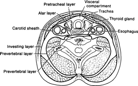
Figure 2.9.
Diagrammatic cross section through the neck below the hyoid bone showing the layers of the deep cervical fascia and the structures that they envelop (By permission of JE Skandalakis, SW Gray, and LJ Skandalakis. In: CG Jamieson (ed), Surgery of the Esophagus, Edinburgh: Churchill Livingstone, 19–35, 1988).
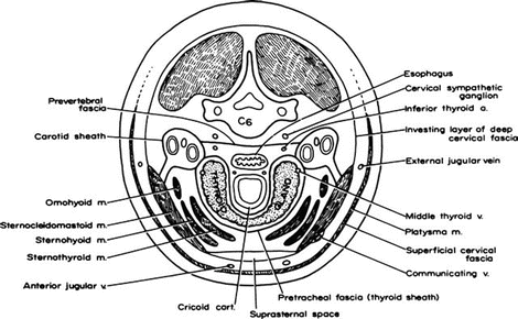
Figure 2.10.
Diagrammatic cross section of the neck through the thyroid gland at the level of the sixth cervical vertebra showing the fascial planes, muscles, and vessels that may be encountered in an incision for thyroidectomy (By permission of JT Akin and JE Skandalakis. Am Surg 42(9):648–652, 1976).
This layer envelops two muscles (the trapezius and the sternocleidomastoid) and two glands (the parotid and the submaxillary) and forms two spaces (the supraclavicular and the suprasternal). It forms the roof of the anterior and posterior cervical triangles and the midline raphe of the strap muscles.
Pretracheal or Middle Layer
The middle layer of the deep fascia splits into an anterior portion that envelops the strap muscles and a posterior layer that envelops the thyroid gland, forming the false capsule of the gland.
Prevertebral, Posterior, or Deep Layer
This layer lies in front of the prevertebral muscles. It covers the cervical spine muscles, including the scalene muscles and vertebral column anteriorly. The fascia divides to form a space in front of the vertebral bodies, the anterior layer being the alar fascia and the posterior layer retaining the designation of prevertebral fascia.
Carotid Sheath
Beneath the sternocleidomastoid muscle, all the layers of the deep fascia contribute to a fascial tube, the carotid sheath. Within this tube lie the common carotid artery, internal jugular vein, vagus nerve, and deep cervical lymph nodes.
Buccopharyngeal Fascia
This layer covers the lateral and posterior surfaces of the pharynx and binds the pharynx to the alar layer of the prevertebral fascia.
Axillary Fascia
This fascia takes its origin from the prevertebral fascia. It is discussed in Chap. 3.
Spaces of the Neck
There are many spaces in the neck defined by the fasciae, but for the general surgeon the visceral compartment is the most important; be very familiar with its boundaries and contents.
The boundaries of the visceral compartment of the neck are:
Anterior: pretracheal fascia
Posterior: prevertebral fascia
Lateral: carotid sheath
Superior: hyoid bone and thyroid cartilage
Posteroinferior: posterior mediastinum
Anteroinferior: bifurcation of the trachea at the level of the fifth thoracic vertebra
Contents of the spaces of the neck include: part of esophagus, larynx, trachea, thyroid gland, and parathyroid glands.
Lymphatics of the Neck/Right and Left Thoracic Ducts
The overall anatomy of the lymphatics of the head and neck may be appreciated from Table 2.1 and Fig. 2.11.
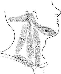
Table 2.1
Lymph nodes and the lymphatic drainage of the head and neck
Lymphatics | |||
|---|---|---|---|
Location | From | To | |
Superior horizontal chain: | |||
Submental nodes | Submental triangle | Skin of chin, lip, floor of mouth, tip of tongue | Submandibular nodes or jugular chain |
Submandibular nodes | Submandibular triangle | Submental nodes, oral cavity, face, except forehead and part of lower lip | Intermediate jugular nodes, deep posterior cervical nodes |
Preauricular (parotid) nodes | In front of tragus | Lateral surface of pinna, side of scalp | Deep cervical nodes |
Postauricular (mastoid) nodes | Mastoid process | Temporal scalp, medial surface of pinna, external auditory meatus | Deep cervical nodes |
Occipital node | Between mastoid process and external occipital protuberance | Back of scalp | Deep cervical nodes |
Vertical chain: | |||
Posterior cervical (posterior triangle) nodes | Subparotid nodes, jugular chain, occipital, and mastoid area | Supraclavicular and deep cervical nodes | |
Superficial | Along external jugular vein | ||
Deep | Along spinal accessory nerve | ||
Intermediate (jugular) nodes | All other nodes of the neck | Lymphatic trunks to left and right thoracic ducts | |
Jugulocarotid (subparotid) nodes | Angle of mandible, near parotid nodes | ||
Jugulodigastric (subdigastric) nodes | Junction of common facial and internal jugular veins | Palatine tonsils | |
Jugulocarotid (bifurcation) nodes | Bifurcation of common carotid artery close to carotid body | Tongue, except tip | |
Jugulo-omohyoid (omohyoid) nodes | Crossing of omohyoid and internal jugular vein | Tip of tongue | |
Anterior (visceral) nodes | |||
Parapharyngeal nodes | Lateral and posterior wall of pharynx | Deep face and esophagus | Intermediate nodes |
Paralaryngeal nodes | Lateral wall of larynx | Larynx and thyroid gland | Deep cervical nodes |
Paratracheal nodes | Lateral wall of trachea | Thyroid gland, trachea, esophagus | Deep cervical and mediastinal nodes |
Prelaryngeal (Delphian) nodes | Cricothyroid ligament | Thyroid gland, pharynx | Deep cervical nodes |
Pretracheal nodes | Anterior wall of trachea below isthmus of thyroid gland | Thyroid gland, trachea, esophagus | Deep cervical and mediastinal nodes |
Inferior horizontal chain: | |||
Supraclavicular and scalene nodes | Subclavian triangle | Axilla, thorax, vertical chain | Jugular or subclavian trunks to right lymphatic duct and thoracic duct |

Figure 2.11.
The lymph nodes of the neck. SH superior horizontal chain, IH inferior horizontal chain, PV posterior vertical chain, IV intermediate vertical chain, AV anterior vertical chain (By permission of JE Skandalakis, SW Gray, and JR Rowe. Anatomical Complications in General Surgery. New York: McGraw-Hill, 1983).
The thoracic duct originates from the cisterna chyli and terminates in the left subclavian vein (Fig. 2.12). It is approximately 38–45 cm long. The duct begins at about the level of the second lumbar vertebra from the cisterna chyli or, if the cisterna is absent (about 50 % of cases), from the junction of the right and left lumbar lymphatic trunks and the intestinal lymph trunk. It ascends to the right of the midline on the anterior surface of the bodies of the thoracic vertebrae. It crosses the midline between the seventh and fifth thoracic vertebrae to lie on the left side, to the left of the esophageal wall. It passes behind the great vessels at the level of the seventh cervical vertebra and descends slightly to enter the left subclavian vein (see Fig. 2.12). The duct may have multiple entrances to the vein, and one or more of the contributing lymphatic trunks may enter the subclavian or the jugular vein independently. It may be ligated with impunity.
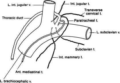

Figure 2.12.
The thoracic duct and main left lymphatic trunks. Trunks are variable and may enter the veins with the thoracic duct or separately (By permission of JE Skandalakis, SW Gray, and JR Rowe. Anatomical Complications in General Surgery. New York: McGraw-Hill, 1983).
The thoracic duct collects lymph from the entire body below the diaphragm, as well as from the left side of the thorax. Lymph nodes may be present at the caudal end, but there are none along its upward course. Injury to the duct in supraclavicular lymph node dissections results in copious lymphorrhea. Ligation is the answer.
The right lymphatic duct is a variable structure about 1 cm long formed by the right jugular, transverse cervical, internal mammary, and mediastinal lymphatic trunks (Fig. 2.13). If these trunks enter the veins separately, there is no right lymphatic duct. When present, the right lymphatic duct enters the superior surface of the right subclavian vein at its junction with the right internal jugular vein and drains most of the right side of the thorax.
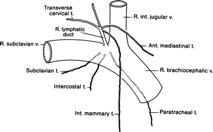

Figure 2.13.
The right lymphatic duct is formed by the junction of several lymphatic trunks. If they enter the veins separately, there may be no right lymphatic duct (By permission of JE Skandalakis, SW Gray, and JR Rowe. Anatomical Complications in General Surgery. New York: McGraw-Hill, 1983).
Anatomy of the Thyroid Gland
The thyroid gland consists typically of two lobes, a connecting isthmus, and an ascending pyramidal lobe. One lobe, usually the right, may be smaller than the other (7 % of cases) or completely absent (1.7 %). The isthmus is absent in about 10 % of thyroid glands, and the pyramidal lobe is absent in about 50 % (Fig. 2.14). A minute epithelial tube or fibrous cord—the thyroglossal duct—almost always extends between the thyroid gland and the foramen cecum of the tongue.
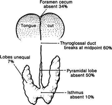

Figure 2.14.
Normal vestiges of thyroid gland development. None are of clinical significance, but their presence may be of concern to the surgeon (By permission of JE Skandalakis, SW Gray, and JR Rowe. Anatomical Complications in General Surgery. New York: McGraw-Hill, 1983).
The thyroid gland normally extends from the level of the fifth cervical vertebra to that of the first thoracic vertebra. It may lie higher (lingual thyroid), but rarely lower.
Capsule of the Thyroid Gland
The thyroid gland has a connective tissue capsule which is continuous with the septa and which makes up the stroma of the organ. This is the true capsule of the thyroid.
External to the true capsule is a well-developed (to a lesser or greater degree) layer of fascia derived from the pretracheal fascia. This is the false capsule, perithyroid sheath, or surgical capsule. The false capsule, or fascia, is not removed with the gland at thyroidectomy.
The superior parathyroid glands normally lie between the true capsule of the thyroid and the fascial false capsule. The inferior parathyroids may be between the true and the false capsules, within the thyroid parenchyma, or lying on the outer surface of the fascia.
Arterial Supply of the Thyroid and Parathyroid Glands
Two paired arteries, the superior and inferior thyroid arteries, and an inconstant midline vessel—the thyroid ima artery—supply the thyroid (Fig. 2.15).
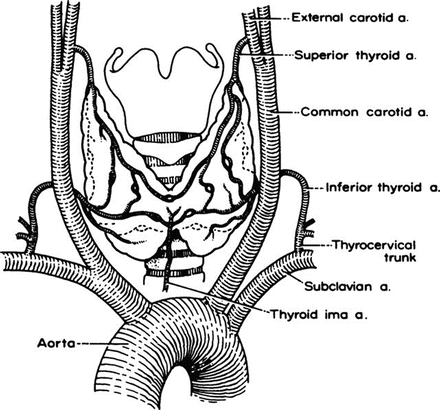

Figure 2.15.
The arterial supply to the thyroid gland. The thyroid ima artery is only occasionally present (By permission of S Tzinas, C Droulias, N Harlaftis, et al. Am Surg 42(9):639–644, 1976).
The superior thyroid artery arises from the external carotid artery just above, at, or just below the bifurcation of the common carotid artery. It passes downward and anteriorly to reach the superior pole of the thyroid gland. Along part of its course, the artery parallels the external branch of the superior laryngeal nerve. At the superior pole the artery divides into anterior and posterior branches. From the posterior branch, a small parathyroid artery passes to the superior parathyroid gland.
The inferior thyroid artery usually arises from the thyrocervical trunk, or from the subclavian artery. It ascends behind the carotid artery and the internal jugular vein, passing medially and posteriorly on the anterior surface of the longus colli muscle. After piercing the prevertebral fascia, the artery divides into two or more branches as it crosses the ascending recurrent laryngeal nerve. The nerve may pass anterior or posterior to the artery, or between its branches (Fig. 2.16). The lowest branch sends a twig to the inferior parathyroid gland. On the right, the inferior thyroid artery is absent in about 2 % of individuals. On the left, it is absent in about 5 %. The artery is occasionally double.
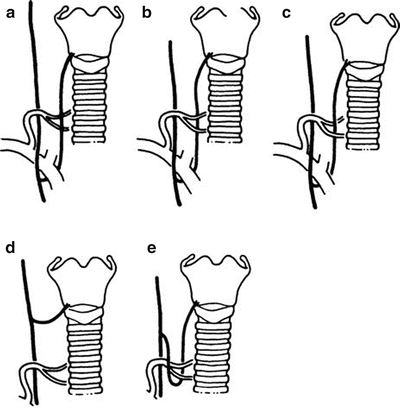

Figure 2.16.
Relations at the crossing of the recurrent laryngeal nerve and the inferior thyroid artery. (a–c) Common variations. Their frequencies are given in Table 2.2. (d) A nonrecurrent nerve is not related to the inferior thyroid artery. (e) The nerve loops beneath the artery (By permission S Tzinas, C Droulias, N Harlaftis, et al. Am Surg 42(9):639–644, 1976).
The arteria thyroidea ima is unpaired and inconstant. It arises from the brachiocephalic artery, the right common carotid artery, or the aortic arch. Its position anterior to the trachea makes it important for tracheostomy.
Venous Drainage
The veins of the thyroid gland form a plexus of vessels lying in the substance and on the surface of the gland. The plexus is drained by three pairs of veins (Fig. 2.17):
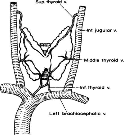

Figure 2.17.
The venous drainage of the thyroid gland. The inferior thyroid veins are quite variable (By permission S Tzinas, C Droulias, N Harlaftis, et al. Am Surg 42(9):639–644, 1976).
The superior thyroid vein accompanies the superior thyroid artery.
The middle thyroid vein arises on the lateral surface of the gland at about two-thirds of its anteroposterior extent. No artery accompanies it. This vein may be absent; occasionally it is double.
The inferior thyroid vein is the largest and most variable of the thyroid veins.
Recurrent Laryngeal Nerves (Figs. 2.16 and 2.18)
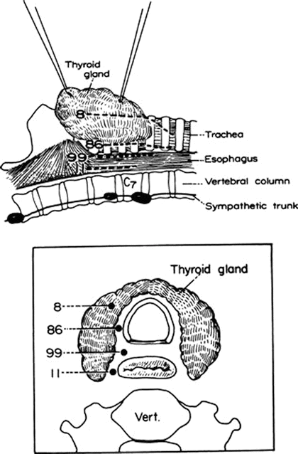
Figure 2.18.
The course of the recurrent laryngeal nerve at the level of the thyroid gland in 102 cadavers. In about one-half of the cases, the nerve lay in the groove between the trachea and the esophagus. (Top) Lateral view. (Bottom) Cross-sectional view (By permission of JE Skandalakis, C Droulias, N Harlaftis, et al. Am Surg 42(9):629–634, 1976).
The right recurrent laryngeal nerve branches from the vagus as it crosses anterior to the right subclavian artery, loops around the artery from posterior to anterior, crosses behind the right common carotid, and ascends in or near the tracheoesophageal groove. It passes posterior to the right lobe of the thyroid gland to enter the larynx behind the cricothyroid articulation and the inferior cornu of the thyroid cartilage.
The left recurrent laryngeal nerve arises where the vagus nerve crosses the aortic arch, just distal to the origin of the left subclavian artery from the aortic arch. It loops under the ligamentum arteriosum and the aorta, and ascends in the same manner as the right nerve. Both nerves cross the inferior thyroid arteries near the lower border of the middle third of the gland.
In about 1 % of patients, the right recurrent nerve arises normally from the vagus, but passes medially almost directly from its origin to the larynx without looping under the subclavian artery. In these cases, the right subclavian artery arises from the descending aorta and passes to the right behind the esophagus. This anomaly is asymptomatic, and the thyroid surgeon will rarely be aware of it prior to operation. Even less common is a nonrecurrent left nerve in the presence of a right aortic arch and a retroesophageal left subclavian artery.
In the lower third of its course, the recurrent laryngeal nerve ascends behind the pretracheal fascia at a slight angle to the tracheoesophageal groove. In the middle third of its course, the nerve may lie in the groove or within the substance of the thyroid gland.
The vulnerability of the recurrent laryngeal nerve may be appreciated from Table 2.2.
Table 2.2
Recurrent laryngeal nerve vulnerability
Cause of vulnerability | Percent encountered |
|---|---|
Lateral and anterior location | 1.5–3.0 |
Tunneling through thyroid tissue | 2.5–15.0 |
Fascial fixation | 2.0–3.0 |
Arterial fixation | 5.0–12.5 |
Close proximity to inferior thyroid vein | 1.5–2.0+ |
Exposure of the Laryngeal Nerves
The recurrent laryngeal nerve forms the medial border of a triangle bounded superiorly by the inferior thyroid artery and laterally by the carotid artery. The nerve can be identified where it enters the larynx just posterior to the inferior cornu of the thyroid cartilage. If the nerve is not found, a nonrecurrent nerve should be suspected, especially on the right.
In the lower portion of its course, the nerve can be palpated as a tight strand over the tracheal surface. There is more connective tissue between the nerve and the trachea on the right than on the left. Visual identification, with avoidance of traction, compression, or stripping the connective tissue, is all that is necessary.
Stay updated, free articles. Join our Telegram channel

Full access? Get Clinical Tree


