8
Nasal Region
The nasal region includes the nose, nasal cavity, and paranasal sinuses. Air is filtered, humidified, and warmed in this region before entering the respiratory tract. The nasal cavity contains the specialized olfactory mucosa responsible for the sense of smell.
External Nose
The superior portion of the external nose is bony and is composed of paired nasal bones and the frontal processes of the maxillae. The inferior expanded portion of the nose is cartilaginous and is formed by the major and minor alar cartilages and a variable number of accessory cartilages (Fig. 8.1).
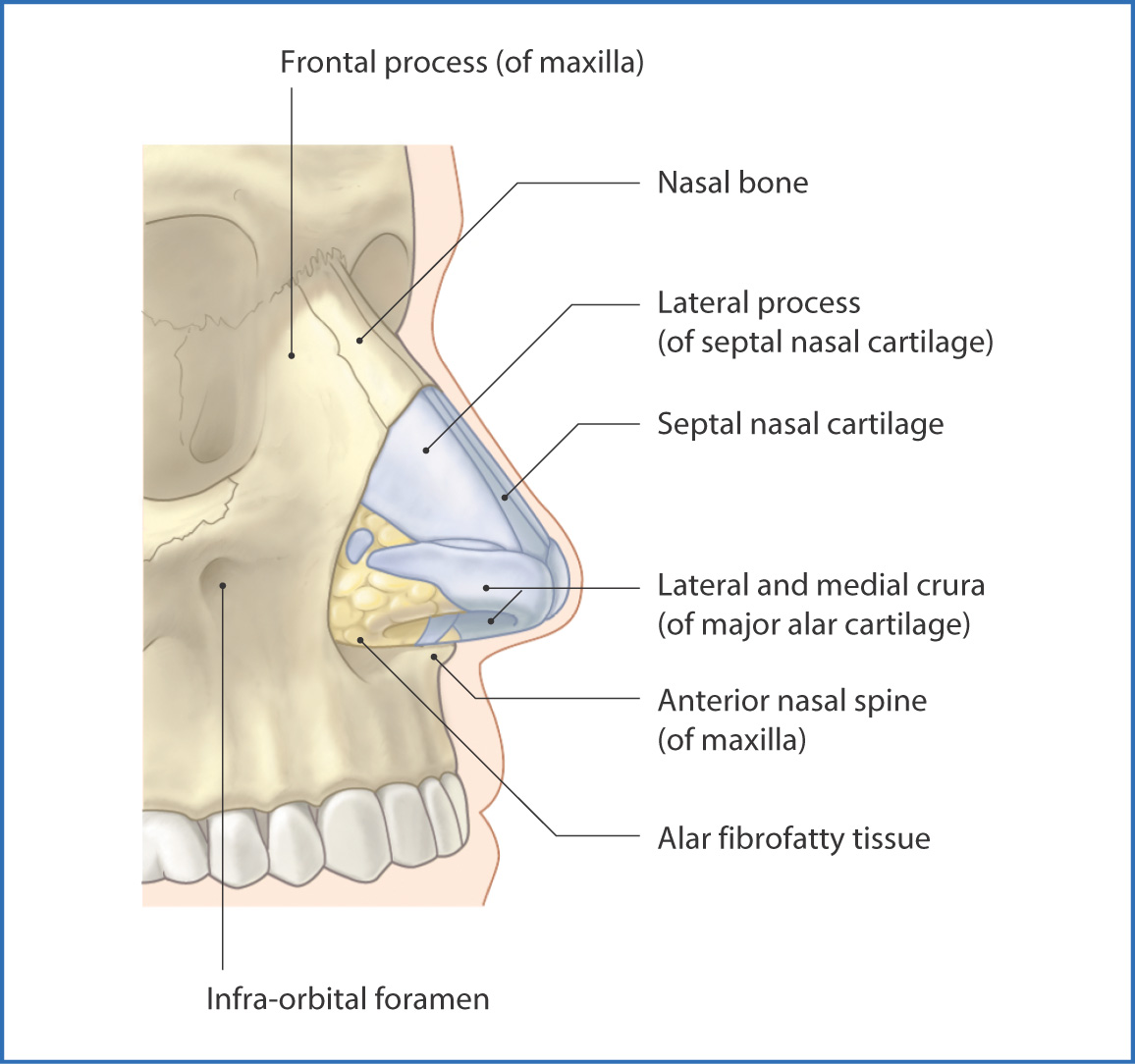
FIGURE 8.1 Nose (anterolateral view).
The triangular shape created by the nasal cartilages is an architecturally stable structure that creates and maintains an open airway. The skin covering the nose is freely movable over the nasal bones but is firmly attached over the cartilages. The external nose has two nares (nostrils) into which air flows during inspiration.
Attached to the external nose are muscles for dilating and flattening the nose—the dilator and compressor muscles, respectively—which are innervated by the facial nerve [VII].
Sensory innervation of the external nose is provided by branches of the ophthalmic [V1] and maxillary [V2] nerve divisions of the trigeminal nerve [V]. Blood is supplied by branches of the ophthalmic artery (a branch of the internal carotid artery) and the facial artery (a branch of the external carotid artery). Venous drainage is important clinically because it is to the facial and ophthalmic veins, which communicate with the cavernous sinus within the cranial cavity; this presents a potential route for infection from the superficial layer of the face to the brain and intracranial structures.
Nasal Cavity
The nasal cavity extends from the vestibule of the nose to the choanae (the posterior nasal apertures) (Table 8.1, p. 88). The nasal cavity has a floor, a roof, two lateral walls, and a midline nasal septum that divides it in half. The walls of the nasal cavity are covered with respiratory epithelium. Olfactory epithelium, involved in the sense of smell, is present on the superior part of the nasal septum and superior conchae.
TABLE 8.1 Walls of the Nasal Cavity
Walls | Components | Relationships |
Roof | Nasal cartilages, nasal and frontal bones, cribriform plate of ethmoid, body of sphenoid, and parts of vomer and palatine bones | Anterior cranial fossa, olfactory nerves [I] and bulb, and sphenoidal sinus |
Floor | Palatine process of maxilla and horizontal plate of palatine bones | Lies between nasal and oral cavities |
Medial wall/septum | Septal cartilage, perpendicular plate of ethmoid and vomer | Separates the two nasal cavities |
Lateral walls | Nasal bone, maxilla, lacrimal, ethmoid (labyrinth and conchae), inferior nasal concha, palatine (perpendicular plate), and sphenoid bones (medial plate of pterygoid process; 3 nasal conchae and their underlying nasal meatuses and spheno-ethmoidal recess) | Medial to orbits, ethmoidal cells, maxillary sinuses, and pterygopalatine fossa |
The roof of the nasal cavity is narrow, arched, and inferior to the anterior cranial fossa. From anterior to posterior it is formed by the nasal cartilages, the nasal and frontal bones, the cribriform plate of the ethmoid, and the body of the sphenoid.
Paired horizontal plates of the palatine bones and the palatine processes of the maxillae form the floor of the nasal cavity and the roof of the oral cavity. Foramina for the greater and lesser palatine nerves and vessels are located posteriorly, with the incisive foramen for the nasopalatine nerve located anteriorly.
The nasal septum is formed anteriorly by the septal cartilage. Posterior to this is the perpendicular plate
Stay updated, free articles. Join our Telegram channel

Full access? Get Clinical Tree


