Fig. 5.1
A typical morbilliform drug eruption, with pink erythematous papules coalescing into larger plaques
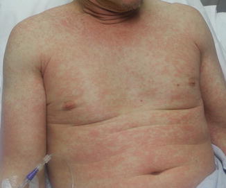
Fig. 5.2
Morbilliform drug eruption in an adult patient with pink erythematous papules coalescing into larger plaques, predominantly affecting the trunk
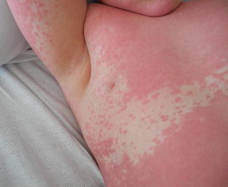
Fig. 5.3
Morbilliform eruption in a male pediatric patient
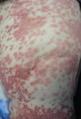
Fig. 5.4
Purpuric appearance of a morbilliform drug eruption in a patient with thrombocytopenia
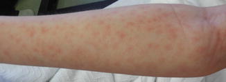
Fig. 5.5
Flat atypical targetoid lesions in a patient with a morbilliform drug eruption
Associated symptoms in patients with morbilliform drug eruptions include pruritus and low-grade fever. Furthermore, mucous membranes are usually spared, which helps to differentiate morbilliform drug eruptions from more severe reactions, such as SJS/TEN or DRESS. Early phases of SJS/TEN, DRESS, and acute graft-versus-host disease (GVHD) may present as a morbilliform eruption. Certain signs or “red flags” suggest a more severe cutaneous hypersensitivity reaction. For example, the timing of the eruption is important, as a later onset of 2–3 weeks is more common in DRESS and SJS/TEN. DRESS is usually caused by allopurinol, sulfa drugs, anti-epileptics, and dapsone. Constitutional symptoms of fever, myalgias and arthralgias, as well as edema of the face, diffuse eruption covering >50 % BSA, infiltrative edematous lesions, purpura, blisters, edema of hands and feet, and lymphadenopathy are all signs of DRESS more often than morbilliform eruptions. Mucosal involvement occurs in 20 % of cases of DRESS and peripheral blood eosinophilia is seen in most patients, unlike morbilliform eruptions. Finally, lesions on the mucous membranes along with painful or dusky skin are seen more commonly with TEN or SJS than with morbilliform eruptions.
Morbilliform eruptions usually disappear spontaneously after 1 or 2 weeks, but if the causative agent is introduced again, the eruption can reappear in a shorter time frame. During resolution of the eruption, desquamation often occurs, and in people with darker skin tones, postinflammatory hyperpigmentation is common.
Rare variants of morbilliform drug eruption include the symmetrical drug-related intertriginous and flexural exanthem (SDRIFE), which has an inverse, or flexural, distribution and is usually induced by aminopenicillins. SDRIFE presents as a maculopapular eruption of the flexural areas (inguinal and perianal skin) with a V-shaped pattern on the medial thighs and diffuse erythema of the buttocks within a few days of exposure to the offending drug (Figs. 5.6 and 5.7). Infrequently, allergic contact dermatitis to nickel or mercury can cause lesions resembling SDRIFE.
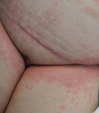
Fig. 5.6
Axillary involvement in a patient with the SDRIFE-variant of a morbilliform drug eruption
Fig. 5.7
Bilateral inguinal involvement in a patient with the SDRIFE-variant of a morbilliform drug eruption
Diagnosis
The differential diagnosis for morbilliform reactions is different for pediatric patients compared to adult patients. Children usually develop a morbilliform rash due to a virus or other infections. Infectious etiologies include viruses (e.g. EBV, enteroviruses, adenovirus, early HIV, cytomegalovirus, human herpes virus type 6, human parvovirus B19, measles); bacteria (scarlet fever, mycoplasma infection); and streptococcal or staphylococcal toxins. The clinical presentations of the viruses are often indistinguishable from a morbilliform drug reaction and although the causative agent is often never found, certain labs including Rapid Strep Test (RST) or rapid antigen detection test (RADT), throat culture, ASO titer, anti-DNase antibody, and heterophile antibody test may be helpful to identify the cause. In the era of declining vaccination rates and increase in measles in the United States, measles IgM and IgG serologies may be useful to obtain in affected patients, particularly those whose vaccination histories cannot be verified. Rare causes of a morbilliform eruption in pediatric patients include Kawasaki Disease, hemophagocytic lymphohistiocytosis (HLH) and Juvenile idiopathic arthritis (Fig. 5.8).
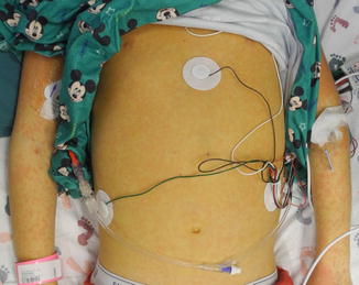
Fig. 5.8
Jaundice and a morbilliform eruption in pediatric patient secondary to primary HLH with liver involvement
In contrast, a drug etiology is more common for adults. The differential diagnosis includes DRESS, TEN, SJS, connective tissue disease (i.e. acute cutaneous lupus erythematous), acute GVHD, pityriasis rosea, allergic contact dermatitis, secondary syphilis, atopic dermatitis, and adult-onset Still disease. Viral and infectious exanthems are less likely for adults.
The diagnosis of a morbilliform drug reaction requires both the cessation of the offending drug and observation of a resolution of the eruption. The Naranjo algorithm or Naranjo Scale is a commonly used questionnaire that allows physicians to assess the likelihood of a drug-induced eruption. The probability of a drug causing the reaction is scored as definite, probable, possible, or doubtful. A lower score corresponds with a lower probability of a drug cause for the eruption.
In complex cases, such as patients with multiple comorbidities taking multiple different drugs, or cases where the culprit drug has not been identified, a detailed patient history should be taken. The patient should be asked about past and current medications, both over-the-counter and prescription, in order to establish a chronologic relationship between drug exposure and onset of the rash. For hospitalized patients, a review of the medical administration record (MAR) is essential. For patients transferred from outside institutions, including nursing homes or outside hospitals, a careful review of outside records should be performed. It is also recommended that records from the patient’s pharmacy be obtained, as often patients may not be familiar with the specific names of their medications. When multiple medications are potential culprits, creating a drug table may be helpful (Table 5.1).
Table 5.1
Drug table
Although laboratory tests are not necessary for morbilliform drug eruptions, they may be useful when the clinical diagnosis is uncertain, in the presence of symptoms suggesting a severe drug reaction or in hospitalized patients. General laboratory tests include a complete blood cell count with differential as well as liver and kidney function tests, including urinalysis. A finding of eosinophilia or atypical lymphocytes is suggestive of DRESS. Similarly, abnormal liver function tests or evidence of renal dysfunction, including proteinuria or hematuria, may be seen in more severe drug reactions. Specific immunologic tests including patch testing, intradermal testing with delayed cutaneous readings, or in vitro tests for delayed reaction (i.e. lymphocyte transformation/activation tests, upregulation of activation markers on T cells, cytokine assays, and drug-induced cytotoxicity assays) can be performed 1–6 months after clinical symptoms have resolved, although this is not usually done in day-to-day practice.
Skin biopsies are often not helpful when evaluating patients with morbilliform eruptions as the histological pattern is often non-specific and does not identify the causative agent or etiology. Nonetheless, biopsy may be indicated when the diagnosis is uncertain. Possible indications for a skin biopsy include, but are not limited to, underlying immunosuppression, severe systemic symptoms, fever >38 °C (100.4 °F), symptoms involving internal organs, presence of erythroderma, blistering, purpura, or pustules, involvement of mucous membranes, and multiple drugs involved without a definite chronological relationship with the morbilliform drug reaction.
Stay updated, free articles. Join our Telegram channel

Full access? Get Clinical Tree


