Microglandular Adenosis
Key Facts
Terminology
Proliferation of small round tubules with a single cell layer (i.e., without both luminal and myoepithelial cells) in an infiltrative pattern
Etiology/Pathogenesis
Associated with invasive carcinoma in about 25% of cases (at presentation or after recurrence)
MGA is molecularly related to invasive carcinoma
MGA in its classic appearance has never been reported to metastasize
Clinical Issues
Treatment: Complete excision with negative margins due to risk of local recurrence
Microscopic Pathology
Negative for estrogen and progesterone receptors
MGA should be considered if a lesion thought to be a well-differentiated carcinoma is estrogen receptor negative
Shows strong cytoplasmic positivity for S100 and is reported to be cathepsin D positive
Tubules are surrounded by basement material as demonstrated by collagen IV and laminin
Top Differential Diagnoses
Invasive tubular carcinoma
Sclerosing adenosis
MGA with atypia or carcinoma arising in MGA
These carcinomas share features with MGA and may be of unusual types (basaloid or metaplastic)
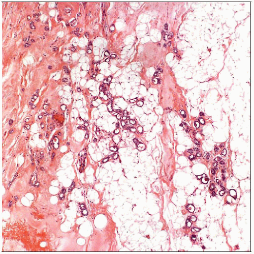
 Microglandular adenosis consists of small round tubules infiltrating through fibrous tissue and adipose tissue and around normal ducts and lobules without an associated stromal response. |
TERMINOLOGY
Abbreviations
Microglandular adenosis (MGA)
Definitions
Proliferation of small round tubules with a single cell layer distributed in an infiltrative pattern
ETIOLOGY/PATHOGENESIS
Pathogenesis
Currently classified as benign lesion
Only benign epithelial lesion that lacks myoepithelial cells
Associated with invasive carcinoma in about 25% of cases (at presentation or after recurrence) and is molecularly related to carcinoma
MGA in its classic appearance has never been reported to metastasize
CLINICAL ISSUES
Presentation
Most commonly presents as palpable mass
Less commonly, MGA forms mammographic density or is incidental finding
Women over wide age range, from 20s to 80s, are affected (mean: Mid 50s)
Treatment
Complete excision with negative margins due to risk of local recurrence
Prognosis
Carcinomas capable of metastasis may arise in association with MGA at presentation or after local recurrence
MACROSCOPIC FEATURES
General Features
Not associated with marked desmoplastic response; lesions may be ill-defined mass or not be apparent
MICROSCOPIC PATHOLOGY
Histologic Features
Consists of haphazard arrangement of small round tubules in and around normal ducts and lobules
Little or no stromal reaction
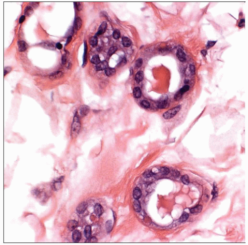
Tubules are round and consist of single cell layer
Tubules contain characteristic eosinophilic (PAS- and mucicarmine-positive) secretory material
Cytologic Features
Cells are cuboidal and have clear &/or foamy cytoplasm
Apocrine snouts are not present
Nuclei are round with small nucleoli and minimal pleomorphism
ANCILLARY TESTS
Immunohistochemistry
Immunoperoxidase studies for myoepithelial markers confirm absence of myoepithelial cells
MGA is negative for estrogen receptor, progesterone receptor, HER2, and epithelial membrane antigen
Should be considered if lesion thought to be well-differentiated carcinoma is negative for hormone receptors
Shows strong cytoplasmic positivity for S100 and is reported to be cathepsin-D positive
Tubules are surrounded by basement material as demonstrated by collagen IV and laminin
Stay updated, free articles. Join our Telegram channel

Full access? Get Clinical Tree


