Melanotic Neuroectodermal Tumor
Steven S. Shen, MD, PhD
Jae Y. Ro, MD, PhD
Key Facts
Terminology
Congenital melanocarcinoma, retinal anlage tumor, melanotic progonoma, melanotic hamartoma
Rare paratesticular (usually epididymis) tumor of neural crest origin in infants and young children
Clinical Issues
Extremely rare; < 1 dozen cases reported involving testis or epididymis
Range: 4 months to 8 years (80% < 1 year old)
Firm mass in epididymis; may be associated with hydrocele
Macroscopic Features
Round to oval homogeneous white-gray to bluish firm nodule; may show areas of dark pigmentation
Microscopic Pathology
Distinct biphasic tumor composed of 2 types of cells
Small neuroblast-like round cells with scant cytoplasm forming sheets or irregularly shaped nests
Large polygonal epithelioid cells with abundant eosinophilic cytoplasm, large vesicular nuclei, small nucleoli, and variable amounts of melanin deposits
Large cells may form nests, cords, and gland-like structures
Typically prominent fibrous and hyalinized stroma
Ancillary Tests
Large cells positive for cytokeratin, vimentin, HMB-45, and S100
Small cells positive for CD56, NSE, and synaptophysin; negative for keratin
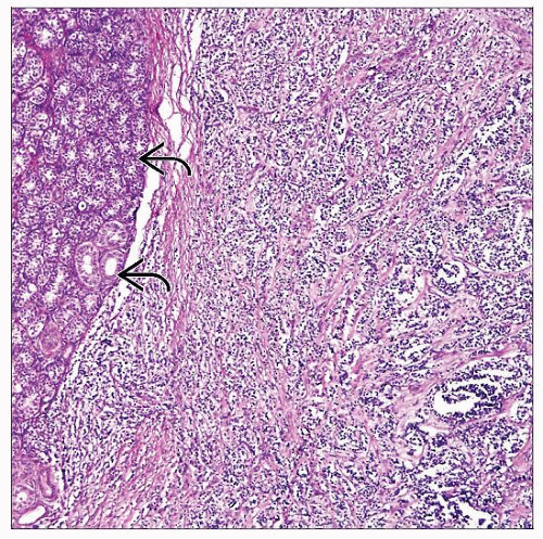 Melanotic neuroectodermal tumor shows a paratesticular cellular tumor, which is well demarcated from the adjacent testis  . It is characterized by sheets of small blue cells in a fibrous background. . It is characterized by sheets of small blue cells in a fibrous background. |
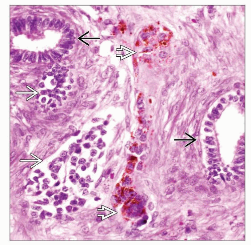 High-power image shows a melanotic neuroectodermal tumor involving epididymis  . There are clusters of small blue cells . There are clusters of small blue cells  and large epithelioid cells with brown pigment and large epithelioid cells with brown pigment  . . |
TERMINOLOGY
Synonyms
Retinal anlage tumor, melanotic progonoma, melanotic hamartoma
Definitions
Rare paratesticular (usually epididymal) tumor of neural crest origin in infants and young children
CLINICAL ISSUES
Epidemiology
Incidence
Extremely rare
< 1 dozen cases reported in testis or epididymis (more common in jaw)
Age
Range: 4 months to 8 years (80% younger than 1 year old)
Presentation
Firm mass in epididymis; may be associated with hydrocele
Laboratory Tests
Mild elevation of serum α-fetoprotein, urine vanillylmandelic acid (VMA), and homovanillic acid in some cases
Treatment
Surgical resection, occasionally with adjuvant therapy (chemotherapy or radiotherapy)
Prognosis
Generally behaves in benign fashion with rare recurrence and metastasis
MACROSCOPIC FEATURES
General Features
Round to oval homogeneous white-gray to bluish firm nodule
May show dark brown or black areas due to pigmentation
Closely apposed to, but usually does not involve, testicular parenchyma
Size
Usually < 4 cm
MICROSCOPIC PATHOLOGY
Histologic Features
Distinct biphasic tumor composed of 2 types of cells
Small neuroblast-like round cells with scant cytoplasm forming sheets or irregularly shaped nests
Large polygonal epithelioid cells with abundant eosinophilic cytoplasm, large vesicular nuclei, small nucleoli, and variable amounts of melanin deposits
Large cells may form nests, cords, and gland-like structures
Typically prominent fibrous and hyalinized stroma
Predominant Pattern/Injury Type
Biphasic with sheets of small round cells and large polygonal cells with melanin pigment
Predominant Cell/Compartment Type
2 cell components: Small cells, and large cells with melanin pigment
ANCILLARY TESTS
Immunohistochemistry
Large cells positive for cytokeratin, vimentin, HMB-45 and S100 (less common), synaptophysin, NSE, GFAP, and desmin; CD99 rarely positive
Stay updated, free articles. Join our Telegram channel

Full access? Get Clinical Tree


