The tumor  is crusted on the scalp of this elderly man. The skin-colored nodule on the left
  is benign, either a neurofibroma or a neurotized intradermal nevus. (Courtesy J. Finch, MD.)
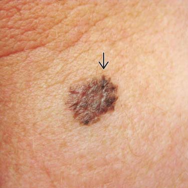
This lesion was located on the posterior neck of a middle-aged man. Although it has a stuck-on appearance, it has uneven, jagged edges
 . The biopsy showed melanoma, nodular type (MM-NT). (Courtesy J. Finch, MD.)
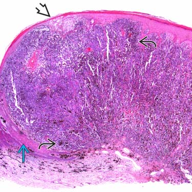
Low-power examination of a melanoma, nodular type shows a large, expansile dermal nodule with irregular pigmentation
 and areas of epidermal thinning and necrosis
  (but no complete ulceration in this section). There is collarette formation
  and no radial growth phase.
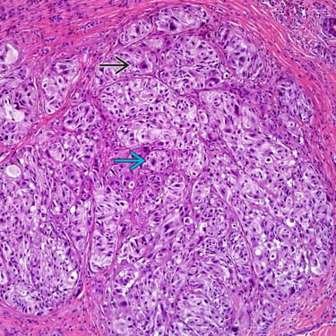
There is proliferation of melanoma cells in nests and nodules. There is significant nuclear pleomorphism and hyperchromasia
 . Mitotic figures
  are easily found.
CLINICAL ISSUES
Epidemiology


Prognosis

• Lead to poor prognosis in most cases


Stay updated, free articles. Join our Telegram channel
Full access? Get Clinical Tree
