Making it better

Chapter Ten Making it better


Ultrasound


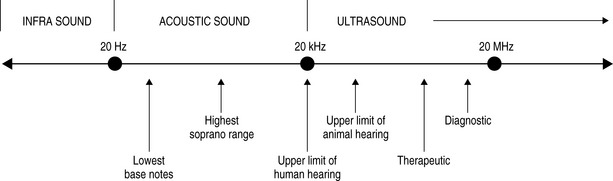
We have already discussed ultrasonography as a tool for diagnostic imaging; however, it is also the oldest and most widely used of the physical modalities. The term ultrasound refers to cyclic sound pressure with any frequency greater than that of the upper limit of human hearing. Although this limit varies between individuals and genders and decreases with age, it is approximately 20 kHz (20 000 hertz) in healthy young adults and this figure serves as a useful lower limit in describing ultrasound (Fig. 10.1).


Although therapeutic ultrasound has been used for over 50 years in physical therapy, its application within the clinical environment has changed significantly over the last 20 years. Whereas, in the past, it was employed primarily for thermal effects, this is no longer the case; now it is much more widely used for its physiological effects, especially in relation to tissue repair and wound healing.


Whether used for diagnostic or therapeutic purposes, ultrasonography begins with the production of the sound waves. This takes place in the transducer, the component of the ultrasound equipment that is placed in direct contact with the patient’s body. It contains piezoelectric material, which converts the electrical impulses received from the pulse generator into sound waves. These can be continuous (therapeutic usage) or pulsed (therapeutic and diagnostic usage). In diagnostic imaging, the transducer also contains a second piezoelectric element that, when echo pulses return to the body surface, are converted back into electrical pulses that are then processed by the system and formed into an image.


As with all waves, the ultrasound wave has a wavelength and reciprocally related frequency. It also has amplitude and a velocity that will vary according to the density of the medium through which it is passing (Table 10.1).


Table 10.1 Velocity of ultrasound waves in selected human tissues


Material Velocity (ms−1)
Fat 1450
Water 1480
Soft tissue 1540
Bone 4100

The power and intensity of the beam are also considerations: you will recall from earlier chapters that power is the rate of energy transfer and is expressed in units of watts. Intensity is the rate at which power passes through a specified area and is expressed in units of watts per square centimetre. Intensity is the rate at which ultrasound energy is applied to a specific tissue location within the patient’s body. It is the quantity that must be considered with respect to producing biological effects and safety. The intensity of most diagnostic ultrasound beams at the transducer surface is on the order of a few milliwatts per square centimetre; this rises to as much as 2 Wcm−2 for therapeutic purposes.


In order to be of medical use, it is necessary to provide a medium through which the ultrasound can freely pass in order to reach the patient’s tissues. Unless this is done, ultrasound will be reflected at the metal/air interface found at the transducer head. This medium is referred to as a coupling medium, and several different types are used in practice including water, various oils, creams and gels. Ideally, the coupling medium should be sufficiently fluid to fill all available spaces, relatively viscous so that it stays in place and should allow the transmission of ultrasound waves with minimal absorption, attenuation or disturbance.


Once the beam enters the body, it can be absorbed, transmitted or reflected by the tissues therein. We have already dealt with this in regard to ultrasonographic imaging in Chapter 9; however, there are additional considerations for therapeutic usage.


In order to have a therapeutic effect, absorption of the applied energy is necessary; therefore the effectiveness of the modality will vary according to a tissue’s capacity to absorb the applied energy. The rate of absorption is related to protein content; tissues with a higher protein content (e.g. ligaments, tendons and scar tissue) will absorb ultrasound to a greater extent whilst tissues with high water and low protein content (e.g. blood and fat) absorb much less energy.


These absorption characteristics have also to be balanced against the wave reflection at the tissue surface. Although cartilage and bone also have a high-protein-low-water profile, a significant proportion of ultrasound energy striking the surface of structures composed of these tissues is likely to be reflected.


Non-thermal effects


A number of trials and meta-analyses produced during the 1980s and 1990s increasingly challenged the routine use of therapeutic ultrasound, showing it to be no better or, in some cases worse, than placebo for the treatment of a number of acute musculoskeletal injuries, as well as more chronic problems such as osteoarthritis, most probably owing to its heating effects, which can increase local inflammation.


At the same time, interest started to become focused on the use of ultrasound as a rehabilitative tool to assist tissues that were healing. The production of scar tissue is a complex series of cascaded, chemically mediated events that results in the deposition of collagen at the site of tissue damage.


The effects of ultrasound during the repair process are thought to vary, according to the primary events that are occurring in the tissues. Once tissue bleeding has ceased (ultrasound can increase blood flow, presumably through heat effects, and thus perpetuate this phase of injury response), it is appropriate to start using ultrasound as soon as is feasible. During the inflammatory phase, ultrasound has a stimulating effect on the mast cells, platelets and white blood cells, which mediate the inflammatory response; however, this intervention is not intended to increase the inflammatory response as such (though if applied with too greater intensity at this stage, this can be a complication) but rather to ‘optimize’ the inflammatory response, which is essential for the effective repair of tissue and inhibition of which can inhibit the repair phases that follow.


Dosage, intensity, pulsation, timing and the preferential absorption of the damaged tissue are all factors that must be considered carefully to make the initial earliest repair phase as efficient as possible, and thus have a promotional effect on the healing process as a whole. Despite the tendency for osseous structures to reflect rather than absorb ultrasound waves, it appears that the modality can also be of use in reducing fracture healing time.


During the proliferative phase, in which scar tissue is produced, ultrasound can also have a stimulative effect; the targets now are the fibroblasts, endothelial cells and myofibroblasts, the cells that activate collagen production in wound healing. Pulsed ultrasound appears to be more effective in encouraging fibroplasia and collagen synthesis than a continuous dose application.


Ultrasound may also have a beneficial role during the remodelling phase of repair, which can last for up to a year after the original injury. The proliferative stage produces a ‘generic’ scar, which is then refined so that it adopts some of the functional characteristics of the tissue that it is repairing. Although a scar in tendon will not become tendon itself, it will behave more like tendinous tissue. This is achieved mainly by the orientation of the collagen fibres in the developing scar and also to the change in collagen type, from predominantly type III to type I collagen, which has greater mobility and higher tensile strength.


It is important to emphasize that therapeutic ultrasound does not appear to alter the body’s normal physiological response, rather it optimizes and enhances it, thus reducing healing time and compensating for inhibition to the normal healing response.


Most recently, it has been discovered that ultrasound has a role in aiding and targeting drug uptake by enhancing membrane transport and increasing vascular and soft tissue transportation.


Cryotherapy


By contrast to ultrasound, the application of cooling agents to an area of injury aims to limit the inflammatory response and its vascular, mechanical and biochemical nociceptive consequences. Cryotherapy is usually applied by means of a gel pack, containing material that remains pliant at the temperatures generated by domestic freezers (−20° to −26°C) or by the application of cooling gel, a substance with a high latent heat of vaporization, which achieves a similar effect by carrying heat away from the injured area as it evaporates.


There is good evidence that this approach, when used in the acute phase of injury, can reduce pain and ischaemia and increase mobility and return to normal activity; this may be at the expense of slightly longer secondary healing phases. Dosage and application need to be carefully monitored, particularly when the procedure is being performed by the patient in an unsupervised manner: 5 minutes for superficial and 15 minutes for deep structures appears to represent the maximum beneficial period for application and a latent period of 30–90 minutes for the local physiology to normalize also appears optimal.


The most common complication of cryotherapy is soft tissue damage from over-cooling. This can produce epidermal damage (‘cold burns’) and local ischaemic effects – it is therefore important for the clinician to monitor the duration and frequency of application as well as ensuring that ice packs are not applied directly to the unprotected skin.


Electromagnetic field therapies


There are a number of techniques in physical therapy that involve the application of an electromagnetic field of varying strengths to a variety of tissues. Generally, these fields are induced by high-frequency alternating currents and the application is either via pads placed on the skin over the area to be treated or, in less portable machines, by a transducer head that can be positioned on or directly above the skin.


Stay updated, free articles. Join our Telegram channel

Apr 4, 2017 | Posted by in GENERAL & FAMILY MEDICINE | Comments Off on Making it better

Full access? Get Clinical Tree

Get Clinical Tree app for offline access