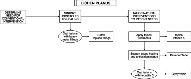
• History: pruritus (can be severe); rash or skin eruption. • Physical examination: skin lesions are flat topped, violaceous to purple, polygonal or oval papules from 1-10 mm wide with edges that are sharply defined and shiny. Lesions may be grouped, linear (Koebner phenomenon), annular, or disseminated when generalized. White lines (Wickham’s striae) are possible. Use handheld lens; apply clear oil over lesion to intensify visibility of Wickham’s striae. In dark-skinned patients appears as postinflammatory hyperpigmentation. Lesion location: flexor surfaces of wrists, lumbar region, eyelids, shin, scalp, glans penis, mouth, but may occur anywhere. An uncommon presentation is plaque-size lesions. Location is on nails and hair follicles, with dystrophic changes and scarring. • Oral lesions: inflammation and leukohyperkeratoses generating reticulated, white puncta or papules and lines in a lacelike pattern. Plaques may occur. Inflammation may cause erosions with blisters. Location is on buccal mucosa; bilateral in nearly all patients on tongue, lips, and gingiva. Skin lesions occur in 10%-60% of oral cases. • Other locations: genitalia; papular, annular, or erosive lesions on penis, scrotum, labia majora, labia minor, and vagina. On the scalp, atrophic skin and scarring alopecia; on the nails, destruction of nail fold and nail bed with longitudinal splintering. • Biopsy: if doubt exists or if blisters, plaques, or erosions occur in the oral form. • Etiology: unknown. Metals (gold, mercury), or infection (hepatitis C virus) altering cell-mediated immunity may play a role. Human leukocyte antigen genetic susceptibility may exist. • Epidemiology: higher prevalence of hepatitis C virus infection in patients with LP, but a pathogenic relation has not been established. LP affects women more than men. Age of onset is 30-60 years. Hypertrophic LP is more common in African Americans. — Hypertrophic: large, thick plaques on foot and shins; more common in African-American men. Hypertrophic lesions may become hyperkeratotic rather than smooth. — Follicular: individual keratotic-follicular papules and plaques leading to cicatricial alopecia. Graham Little syndrome is a complex of spinous follicular lesions, any lichen planus, and cicatricial alopecia. — Vesicular: vesicular or bullous lesions within or independent of lesions. Direct immunofluorescence reveals bullous pemphigoid; patients have bullous pemphigoid immunoglobulin G (IgG) autoantibodies. — Actinicus: papular lesions in sun exposed areas (e.g., dorsum of hands and arms). — Ulcerative: therapy-resistant ulcers (soles) requiring skin grafting. — Oral/mucous membranes: reticular, hyperkeratotic, erosive, plaque type, atrophic, bullous types
Lichen Planus
DIAGNOSTIC SUMMARY
GENERAL CONSIDERATIONS

![]()
Stay updated, free articles. Join our Telegram channel

Full access? Get Clinical Tree


Lichen Planus
