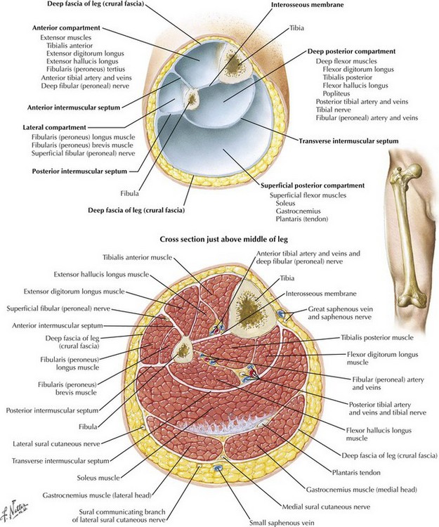
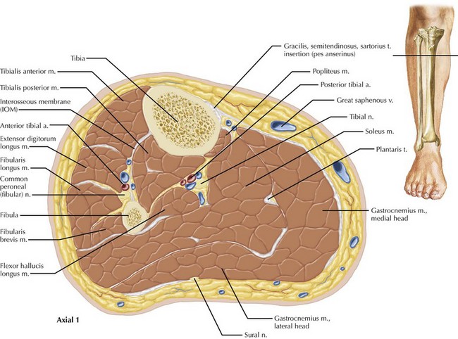
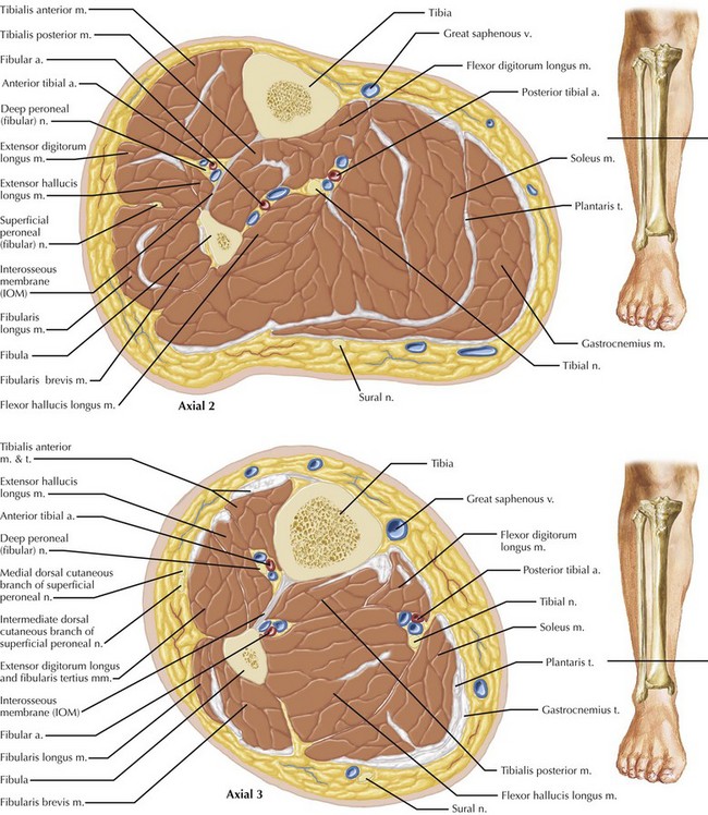
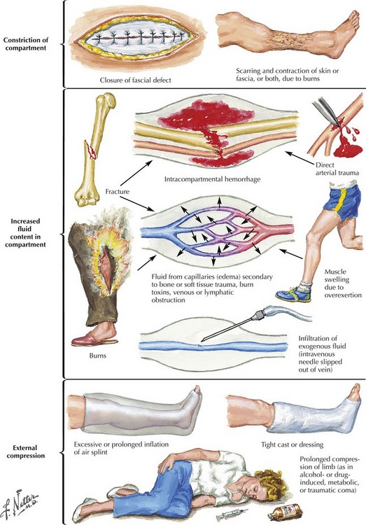
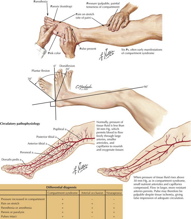
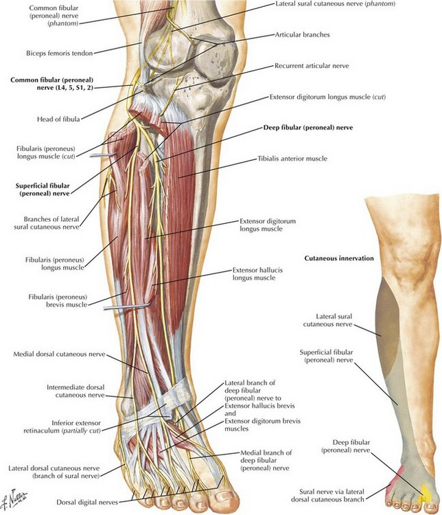
Leg and Forearm Fasciotomy

Chapter 42


Leg and Forearm Fasciotomy


Introduction


Compartment syndrome of the lower extremity is a morbid condition that can lead to limb loss, functional impairment, renal failure, and death. Fasciotomy is required to treat the syndrome and prevent or minimize complications. Compartment syndrome can develop in any anatomic compartment, but the most common site is the lower extremity, in particular the leg. The lower-extremity compartments include those of the buttocks, thigh, leg, and foot.


Compartment syndrome of the forearm and hand is usually seen after blunt trauma. Other conditions that contribute to the development of upper-extremity compartment syndromes include blood dyscrasias, clotting abnormalities, metabolic fluid shifts, longstanding compression, direct fluid injection, and infection. Increased pressure in the forearm compartments results from direct muscle injury, hematoma from soft tissue and osseous injuries, or secondary swelling. The condition can occur in the rapid acute manner or after fluid resuscitation in a seriously injured patient.


Leg Fasciotomy


The lower leg is divided into four compartments: anterior, lateral, superficial posterior, and deep posterior (Fig. 42-1). The anterior compartment is especially prone to ischemic injury because of its relative paucity of collateral arterial supply. Compared with the other compartments, especially the posterior compartments, the anterior compartment is also more firmly constrained. The spatial relationships among the nerve, vascular, muscular, and bony structures change from proximal to distal in the leg, an especially relevant consideration in performing complete lysis of the posterior compartments (Figs. 42-2 and 42-3).


Etiology of Compartment Syndrome


Compartment syndrome occurs when the compartmental pressure rises sufficiently to prevent adequate tissue perfusion. This condition can result from increased volume in the compartment caused by bleeding, infiltration of exogenous fluid, or reperfusion edema (Fig. 42-4). Increased compartment pressures can also result from external constraint on the compartment, such as with casts, braces, or bandages. A compartment pressure greater than 30 mm Hg is accepted as sufficiently elevated to cause compartment syndrome. This pressure is usually sufficient to restrict venous outflow from the compartment, thereby leading to further increases in compartment pressure.


Compartment syndrome can occur at lower compartment pressures, especially in the patient with hypotension. In clinical practice, compartment syndrome more often occurs in the setting of reperfusion after arterial revascularization for acute, limb-threatening ischemia, as well as in the trauma patient. Reperfusion injury after arterial revascularization is more common after a longer period of ischemia, and prophylactic fasciotomy should be considered when acute ischemia lasts longer than 4 to 6 hours. In the injured extremity, contributing factors to development of compartment syndrome include intracompartmental bleeding, crush or blast injury, and arterial insufficiency from direct vascular injury or shock. In the trauma patient, concomitant venous injury may also lead to venous hypertension, further increasing the risk of compartment syndrome.


Clinical Diagnosis and Decision Making


In the appropriate clinical setting, compartment syndrome should always be considered. Pain is the most prevalent symptom, but the patient may report diminished motor strength and altered or reduced sensation (Fig. 42-5). Pain with passive movement and palpation is extremely common, although the absence of pain in the extremity with compromised neurologic function can be misleading.


Findings consistent with arterial insufficiency are not always present, so their absence does not exclude the presence of compartment syndrome. Therefore the presence of palpable pedal pulses (dorsalis pedis artery, posterior tibial artery) does not rule out the diagnosis of compartment syndrome. Because neuronal tissue is sensitive to ischemia, peripheral nerve dysfunction is common in the patient with compartment syndrome and is caused by neuronal ischemia. Diminished motor strength can be tested by assessing dorsiflexion and plantar flexion of the great toe and the ankle, which reflect function of the major muscle groups the leg.


Light touch sensation is often diminished before the development of motor weakness and can best be tested in the web space between the first and second toes. Touch sensation reflects the function of the deep peroneal nerve (Fig. 42-6).


Stay updated, free articles. Join our Telegram channel

Aug 12, 2016 | Posted by in ANATOMY | Comments Off on Leg and Forearm Fasciotomy

Full access? Get Clinical Tree

Get Clinical Tree app for offline access