Reactive epithelial changes in interlobular bile ducts ± neutrophils
Canalicular cholestasis typically earliest change
– May be absent if blockage is partial or intermittent
Copper deposition, fibrosis and even cirrhosis can develop if process is chronic
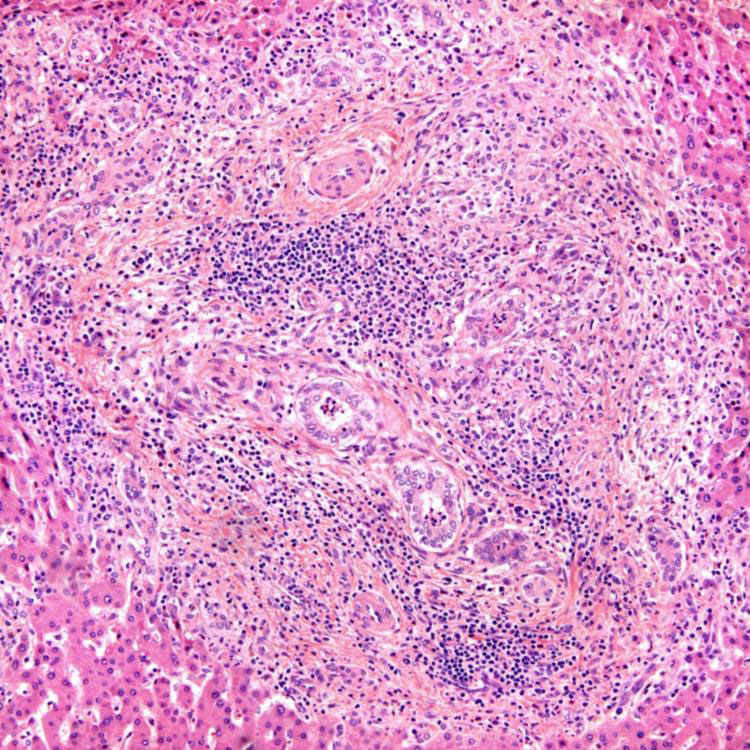
Portal Edema Portal edema is a common feature of large bile duct obstruction. It appears as expansion and pallor of the portal tracts. Note the mixed portal inflammation, ductular reaction, and neutrophils in the lumen of the interlobular bile duct.
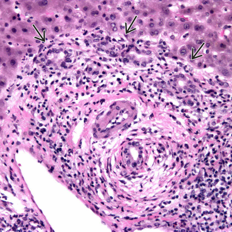
Ductular Reaction This case of large bile duct obstruction shows marked periductal edema, ductular reaction at the periphery of the portal tracts with admixed neutrophils, and a mixed portal inflammatory infiltrate.
Canalicular Cholestasis Canalicular cholestasis, featuring prominent bile plugs throughout the parenchyma , is often the earliest change seen in liver biopsies from patients with large bile duct obstruction.
Mixed Inflammation This portal tract is edematous, and features mixed inflammation with numerous neutrophils and eosinophils, which is typical of large bile duct obstruction. The neutrophils also infiltrate the bile duct , and are present in the lumen.
TERMINOLOGY
Abbreviations
• Large bile duct obstruction (LBDO)
Definitions
• Mechanical blockage of extrahepatic or large intrahepatic bile ducts
Changes on liver biopsy are secondary to obstructive biliary process
ETIOLOGY/PATHOGENESIS
Multifactorial
• Gallstones, neoplasms/masses
Only gold members can continue reading. Log In or Register to continue


 with admixed neutrophils, and a mixed portal inflammatory infiltrate.
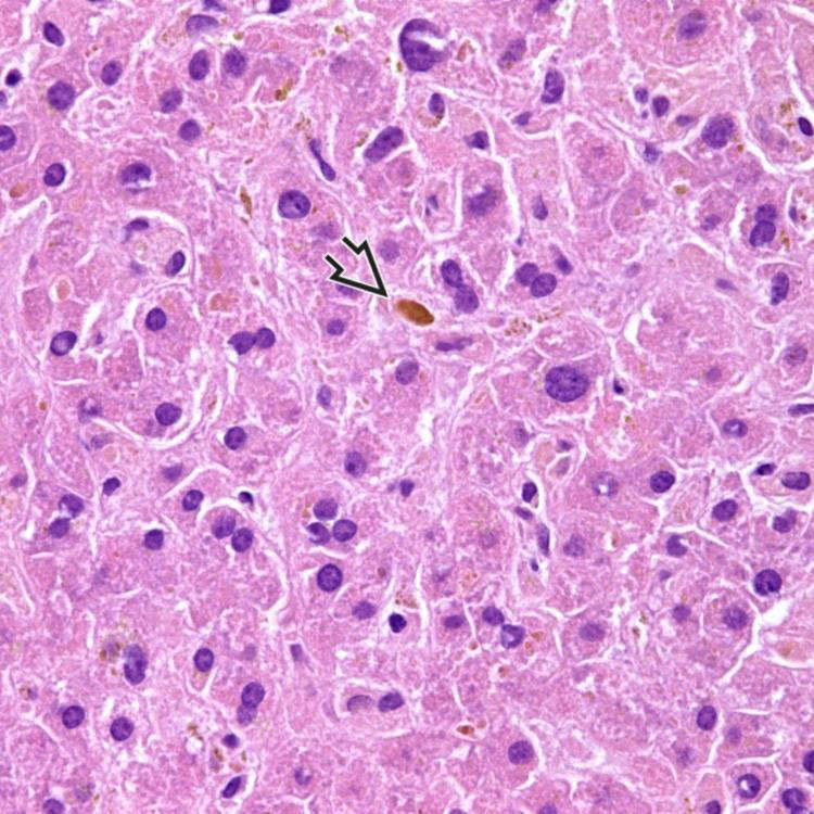
 , is often the earliest change seen in liver biopsies from patients with large bile duct obstruction.
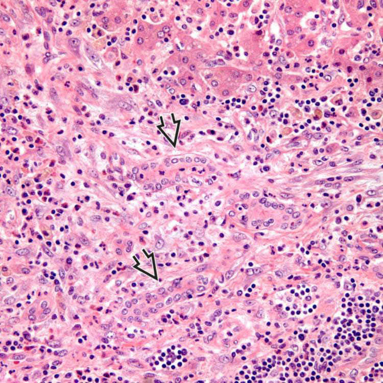
 , and are present in the lumen.


