43
Knee Joint and Popliteal Fossa
The knee joint is a synovial hinge-type joint between the femur and tibia; its range of movement is limited primarily to flexion and extension, but some rotation is possible. The superior part of the knee joint is formed by the condyles of the femur. These enlarged bony prominences transfer weight to the flattened plateaus of the superior surfaces of the tibia and the medial and lateral condyles.
The patella is a large sesamoid bone that supports and protects the anterior knee joint through its attachments to the quadriceps femoris tendon and patellar ligament.
Muscles
The knee joint is supported anteriorly by the aponeurotic expansion of the quadriceps femoris muscle. This large tendon emerges from the inferior quadriceps femoris muscle and attaches to the superior aspect of the patella. At the inferior part of the patella, the patellar ligament (Fig. 43.1) emerges inferiorly to insert onto the tibial tuberosity. With this design, the quadriceps femoris muscle is the primary extensor for the knee joint and supports the anterior portion of the knee via its tendon and the patella. The lateral part of the knee is supported by the iliotibial tract and the tendon of the biceps femoris muscle, whereas the medial side of the knee is supported by the muscle tendons of the pes anserinus.
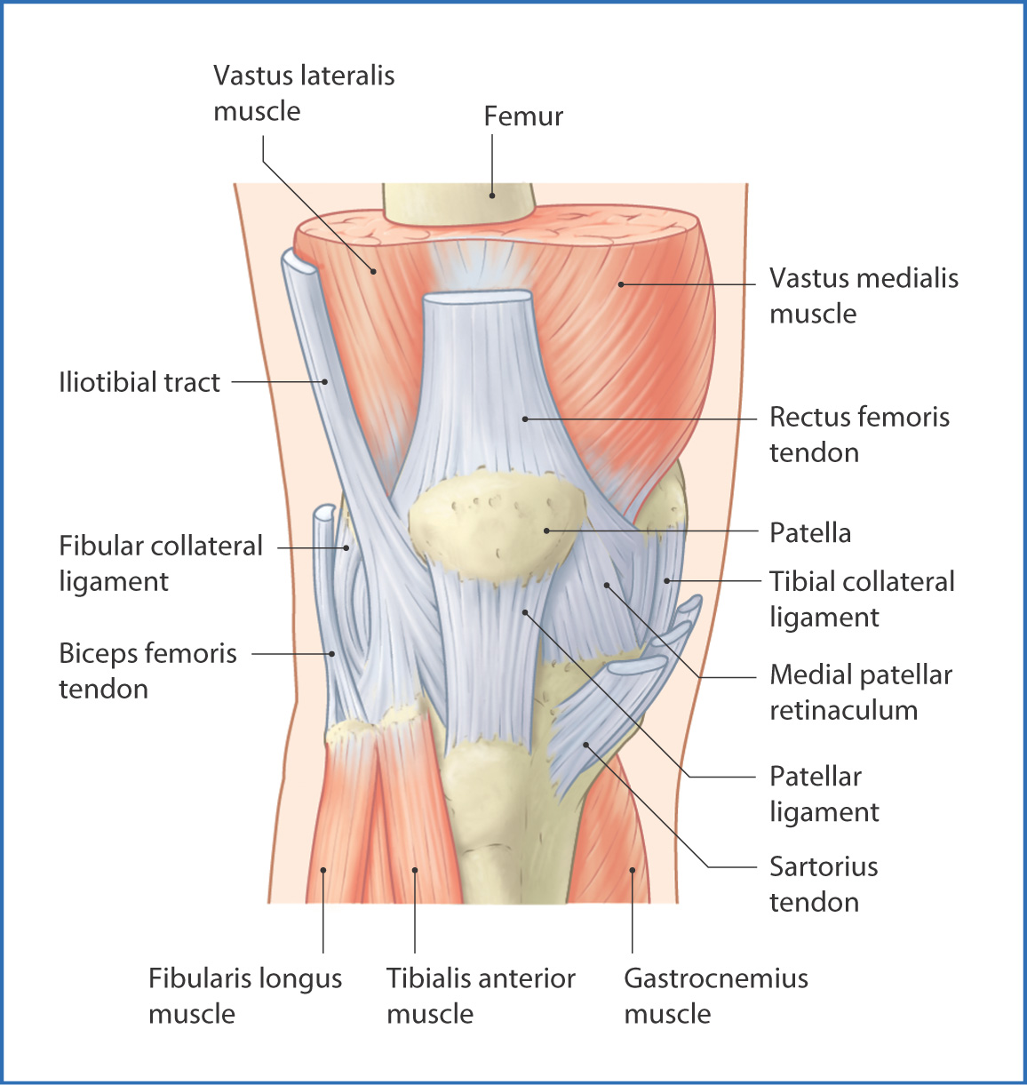
FIGURE 43.1 Knee joint—superficial anterior view.
The hamstring muscles and posterior leg muscles support the posterior knee joint (Fig. 43.2). Medially, the semimembranosus, semitendinosus, gracilis, and sartorius muscles and tendons, which originate in the upper part of the thigh, insert onto points along the proximal medial aspect of the tibia. The lateral part of the posterior knee joint is supported by the biceps femoris, which originates in the thigh and inserts onto the head of the fibula (see Chapter 42).
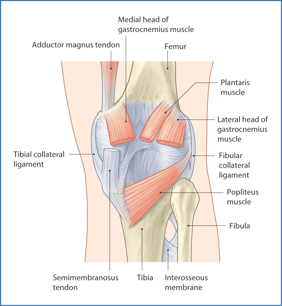
FIGURE 43.2 Knee joint—posterior view.
Nerves
The knee joint is innervated by several nerves that pass the joint, including articular branches from the tibial and common fibular nerves and articular branches from the femoral nerve. The posterior division of the obturator nerve also supplies the knee after supplying the adductor magnus muscle.
Cutaneous innervation of the anterior skin of the knee joint is conveyed by anterior cutaneous branches of the femoral nerve. Posterior cutaneous innervation is conveyed by medial sural cutaneous branches of the tibial nerve (see Chapters 40 and 42).
Arteries
Blood supply to the knee is from genicular anastomoses, which consist of 10 arteries:
- The descending genicular artery and the descending branch of the lateral circumflex femoral artery (both of which are branches of the femoral artery) descend and supply the knee.
- Five branches from the popliteal artery provide blood to the posterior side—the superior medial genicular, superior lateral genicular, middle genicular, inferior medial genicular, and inferior lateral genicular arteries.
- The remaining three vessels arise from the anterior tibial artery of the leg (see Chapter 44)—the posterior tibial recurrent artery, circumflex fibular artery, and anterior tibial recurrent artery.
- Five branches from the popliteal artery provide blood to the posterior side—the superior medial genicular, superior lateral genicular, middle genicular, inferior medial genicular, and inferior lateral genicular arteries.
Veins and Lymphatics
Venous drainage of the superficial structures at the knee joint is to the great saphenous vein and other unnamed superficial veins at the knee. Deep venous drainage of the knee joint follows the arteries that make up the genicular anastomoses named earlier.
Lymphatic drainage of the superficial structures of the knee (skin and soft tissue) follows the great saphenous vein and ends in the inguinal lymph nodes. Deep lymphatic drainage of the knee is to the popliteal nodes, which eventually drain toward the inguinal and gluteal nodes.
Ligaments and Menisci
The knee has a strong fibrous joint capsule that completely surrounds the joint and is strengthened by five intrinsic ligaments (Table 43.1):
- the patellar ligament anteriorly
- the oblique popliteal and arcuate popliteal ligaments posteriorly
- the tibial (medial) and fibular (lateral) collateral ligaments
- the oblique popliteal and arcuate popliteal ligaments posteriorly
TABLE 43.1
Stay updated, free articles. Join our Telegram channel

Full access? Get Clinical Tree


