Causes nonzonal or “geographic” necrosis
Diagnostic Checklist
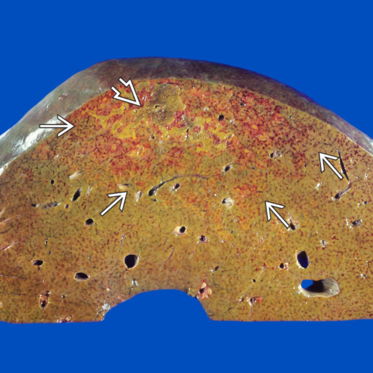
This gross photograph illustrates a subcapsular hepatic infarct. The infarcted area shows variegated red hemorrhagic areas
 admixed with necrosis
  .
.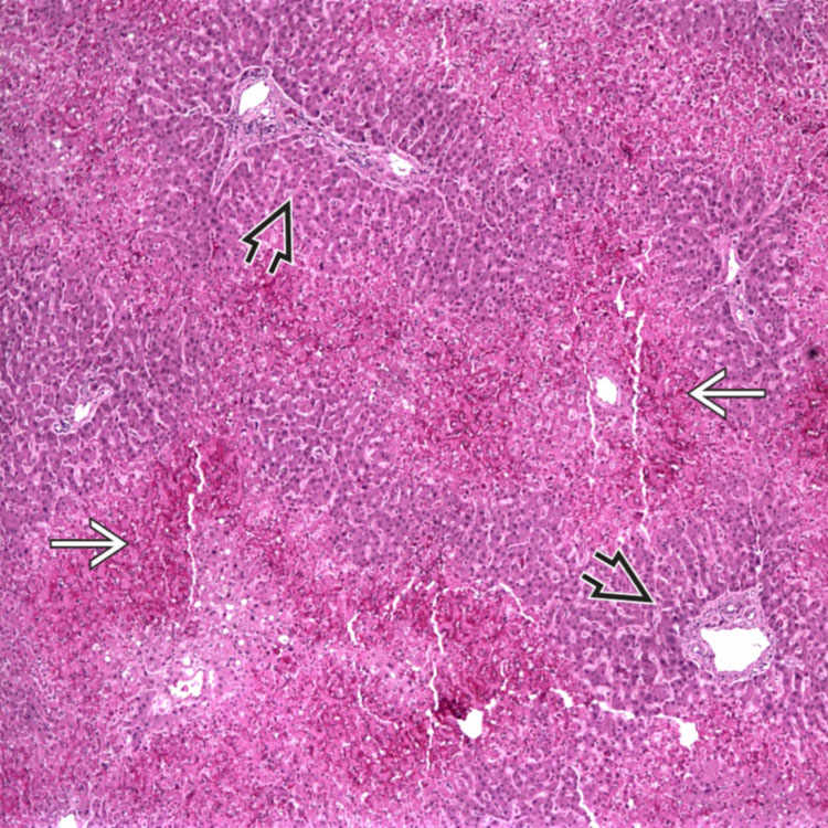
This low-power photomicrograph shows zone 3 perivenular hemorrhage and necrosis
 in a case of hepatic ischemia. The periportal hepatocytes are well preserved
  .
.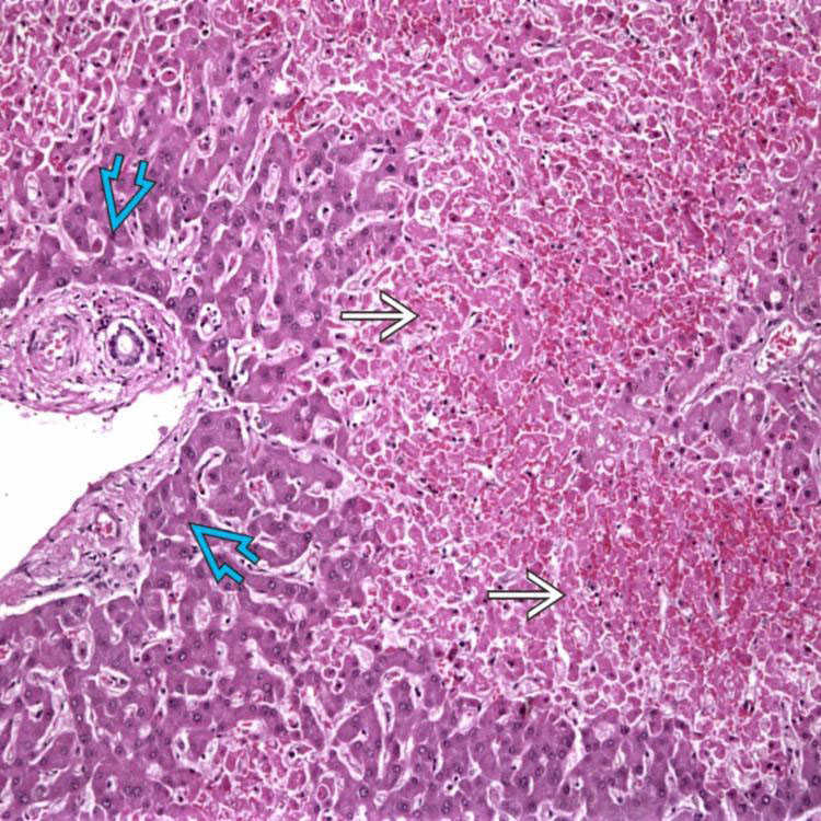
Well-demarcated areas of hepatocyte necrosis and hemorrhage
 are seen in hepatic ischemia. The hemorrhage corresponds to the red areas seen grossly in “nutmeg” liver. Periportal hepatocytes are well preserved
  .
.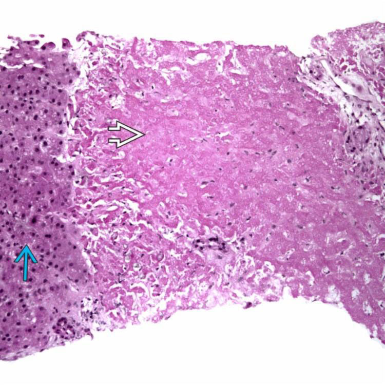
 and preserved adjacent hepatocytes
  .
.ETIOLOGY/PATHOGENESIS
Systemic Hypotension &/or Hypoxemia
Vascular Obstruction

• In native livers, usually require obstruction of 2 vessels to result in clinically significant ischemia
 Greater collateralization present in native livers, but allografts subject to injury with single vessel obstruction


Greater collateralization present in native livers, but allografts subject to injury with single vessel obstructionStay updated, free articles. Join our Telegram channel
Full access? Get Clinical Tree
