Invasive Apocrine Carcinoma
Key Facts
Terminology
Invasive apocrine carcinoma (IAC)
Term is reserved for cases in which nearly all tumor cells show prominent apocrine features
Etiology/Pathogenesis
Molecular studies indicate that IAC may represent a distinct subgroup of breast cancers
Characterized by gene expression pattern largely driven by expression of androgen receptor (AR)
Clinical Issues
Pure IAC incidence varies from < 1% up to 4%
Variability due to lack of well-defined diagnostic criteria
IAC does not have any specific epidemiologic, clinical, or imaging features
Prognosis appears to be similar to carcinomas of no special type
Expression of AR may lead to therapeutic strategies in future
Ancillary Tests
Tumor cells are usually positive for AR, negative for ER and PR, and about 1/2 overexpress HER2
Top Differential Diagnoses
Apocrine metaplasia or DCIS involving adenosis
Granular cell tumor
Cutaneous apocrine carcinoma (sweat gland carcinoma of the skin)
Other special types of invasive carcinoma (oncocytic, lipid-rich, histiocytic, sebaceous)
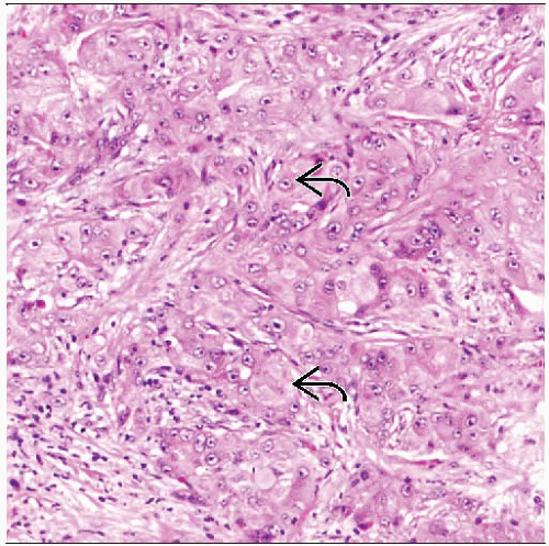 Apocrine carcinoma is a morphologically distinct type of invasive breast cancer characterized by large cells with a low nuclear to cytoplasmic ratio and abundant eosinophilic granular cytoplasm  . . |
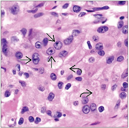 The cytoplasm is abundant with a coarsely granular eosinophilic appearance  . The nuclei are large and pleomorphic with prominent nucleoli . The nuclei are large and pleomorphic with prominent nucleoli  . The growth pattern is typically solid in type. . The growth pattern is typically solid in type. |
TERMINOLOGY
Abbreviations
Invasive apocrine carcinoma (IAC)
Definitions
Breast carcinoma with characteristic morphologic appearance resembling apocrine sweat glands in at least 90% of tumor cells, closely linked with androgen receptor expression
ETIOLOGY/PATHOGENESIS
Androgen Metabolism in IAC
Majority of breast cancers (70-80%) express androgen receptor (AR)
Highest expression is seen in ER/PR/HER2 positive cancers (˜ 80%), followed by ER/PR positive, HER2 negative cancers (˜ 60%) and ER/PR negative, HER2 positive cancers (˜ 50%); lowest expression is in ER/PR/HER2 negative cancers (˜ 35%)
HER2 may induce AR transactivation through MAP kinase pathway
IAC expresses AR in 56-100% of cases
AR expression may help distinguish IAC from basal-like cancers that are also “triple negative”
Expression of AR is also frequent in benign apocrine lesions
Growth of cutaneous apocrine glands is stimulated by androgens
Enhanced metabolism of testosterone precursors has been reported in IAC
Altered androgen metabolism may play a role in pathogenesis and tumor progression
Gene Expression Profiling
Molecular studies indicate that invasive apocrine carcinomas may represent distinct subgroup of breast cancers
IAC is characterized by gene expression pattern largely driven by expression of AR
Majority AR positive; AR may be potential therapeutic target
About 1/2 negative for ER, PR, and HER2; however, IAC does not cluster with basal-like group
About 1/2 negative for ER and PR but HER2 positive
Tumors overlap significantly with HER2 group as defined by intrinsic gene classification
Gene expression data suggests link between HER2 signaling and molecular apocrine phenotype
Gene signature includes increased expression of numerous genes with role in metabolism
Association with Benign Apocrine Lesions
Apocrine glands normally occur in skin
Breast evolved from skin appendages and, thus, has many features in common
Cells with functional apocrine characteristics have been described in fetal breast tissue
Apocrine change is commonly seen in cysts in adult breast tissue and may arise from fetal cells or from metaplasia
Benign apocrine lesions express AR and are negative for ER and PR
Apocrine change without atypia has not been consistently linked to increase in risk of breast cancer
Difficult to define atypia in apocrine lesions
Lesions with high-grade nuclei, cribriform architecture, &/or necrosis would be considered atypical
Additional studies will be necessary to show that these changes increase risk of invasive cancer
Some apocrine lesions show loss of heterozygosity and other genetic changes, supporting that some are clonal lesions and may be nonobligate precursors of apocrine carcinomas
Atypical apocrine proliferations are found more commonly in breasts with apocrine carcinomas
CLINICAL ISSUES


