Introduction to Autonomic and Neuromuscular Pharmacology


Introduction to Autonomic and Neuromuscular Pharmacology


Overview


The nervous system consists of the central and peripheral nervous systems. The central nervous system includes the brain and spinal cord, whereas the peripheral nervous system consists of the autonomic nervous system and the somatic nervous system.


Drugs alter nervous system function primarily by affecting neurotransmitters or their receptors. In some cases drugs alter the synthesis, storage, release, inactivation, or neuronal reuptake of neurotransmitters. In other cases they activate or block neurotransmitter receptors. Most drugs are relatively selective for a particular neurotransmitter or receptor. The effects produced by a drug depend partly on the distribution of the affected neurotransmitters in the central and peripheral nervous systems. The actions of some drugs are localized to either the central or the peripheral nervous system, but other drugs (e.g., cocaine and amphetamine) affect both central and peripheral functions.


This chapter reviews the anatomy and physiology of the peripheral nervous system and introduces the mechanisms by which drugs affect nervous system function. Drugs acting on the central nervous system are discussed in Section IV.


Anatomy and Physiology of the Peripheral Nervous System


The autonomic nervous system involuntarily modifies the activity of smooth muscles, exocrine glands, cardiac tissue, and certain metabolic activities, whereas the somatic nervous system activates skeletal muscle contraction, enabling voluntary body movements. Both the autonomic and the somatic nervous systems are controlled by the central nervous system. The autonomic nervous system is regulated by brain stem centers responsible for cardiovascular, respiratory, and other visceral functions. The somatic nervous system is activated by corticospinal tracts, which originate in the cerebral motor cortex, and by spinal reflexes.


Autonomic Nervous System


The autonomic nervous system consists of sympathetic and parasympathetic divisions. In the sympathetic nervous system, nerves arise from the thoracic and lumbar spinal cord and have a short preganglionic fiber and a long postganglionic fiber. Most of the ganglia are located in the paravertebral chain adjacent to the spinal cord, but a few prevertebral ganglia (the celiac, splanchnic, and mesenteric ganglia) are located more distally to the spinal cord. The parasympathetic nervous system includes portions of cranial nerves III, VII, IX, and X (the oculomotor, facial, glossopharyngeal, and vagus nerves, respectively) and some of the nerves originating from the sacral spinal cord. The parasympathetic nerves have long preganglionic fibers and short postganglionic fibers, with the ganglia often located in the innervated organs.


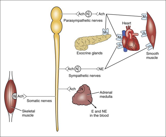
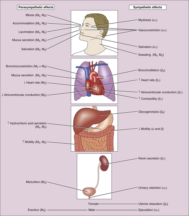
The origins, neurotransmitters, and receptors of the sympathetic and parasympathetic systems are shown in Figure 5-1. The sympathetic nervous system tends to discharge as a unit, producing a diffuse activation of target organs. Preganglionic, sympathetic neurons synapse with a large number of postganglionic neurons, which contributes to widespread activation of the organs during sympathetic stimulation. In addition, the release of epinephrine and norepinephrine from the adrenal medulla into the circulation enables the activation of target tissues throughout the body, including some tissues not directly innervated by sympathetic nerves. In contrast, the parasympathetic system can discretely activate specific target tissues. For example, it is possible for parasympathetic nerves to slow the heart rate without simultaneously stimulating gastrointestinal or bladder function. This is partly because of the low ratio of postganglionic fibers to preganglionic fibers in the parasympathetic system.


As shown in Figure 5-2, the sympathetic and parasympathetic nervous systems often have opposing effects on organ function. Activation of the sympathetic system produces the “fight or flight” reaction in response to threatening situations. In this reaction, cardiovascular stimulation provides muscles with oxygen and fuels required to support vigorous physical activity, while activation of glycogenolysis and lipolysis releases the necessary energy substrates. The parasympathetic system is sometimes called the “rest and digest” system, because it slows the heart rate and promotes more vegetative functions, such as digestion, defecation, and micturition. Many parasympathetic effects (including pupillary constriction, bronchoconstriction, and stimulation of gut and bladder motility) are caused by smooth muscle contraction.


Neurotransmitters and Receptors


Neurotransmitters


The primary neurotransmitters found in the autonomic and somatic nervous systems are acetylcholine and norepinephrine (see Fig. 5-1). The terms adrenergic and cholinergic refer to neurons that release norepinephrine or acetylcholine, respectively.


Acetylcholine is the transmitter at all autonomic ganglia, at parasympathetic neuroeffector junctions, and at somatic neuromuscular junctions. It is also the transmitter at a few sympathetic neuroeffector junctions, including the junctions of nerves in sweat glands and vasodilator fibers in skeletal muscle. The presence of acetylcholine in several types of autonomic and somatic synapses contributes to the lack of specificity of drugs acting on acetylcholine neurotransmission.


Although norepinephrine (noradrenaline) is the primary neurotransmitter at most sympathetic postganglionic neuroeffector junctions, epinephrine (adrenaline) is the principal catecholamine released from the adrenal medulla in response to activation of the sympathetic nervous system.


A number of other neurotransmitters have been identified in autonomic nerves of the ENS of the gastrointestinal tract, as well as in the genitourinary tract and certain blood vessels. The transmitters released by these neurons include neuropeptide Y, vasoactive intestinal polypeptide, enkephalin, substance P, serotonin (5-hydroxytryptamine), adenosine triphosphate, and nitric oxide. In some tissues, adenosine triphosphate released by these neurons is converted to adenosine, which can then activate adenosine receptors in a number of tissues (see Chapter 27). Nitric oxide is an important neurotransmitter that produces vasodilatation in many vascular beds and is also found in the ENS.


Receptors for Acetylcholine, Norepinephrine, and Epinephrine


The acetylcholine receptors have been divided into two types, based on their selective activation by one of two plant alkaloids. Muscarinic (M) receptors, which are acetylcholine receptors activated by muscarine, are primarily located at parasympathetic neuroeffector junctions. Nicotinic receptors are acetylcholine receptors activated by nicotine. They are found in all autonomic ganglia, at somatic neuromuscular junctions, and in the brain. Muscarinic receptors are subdivided based on molecular and pharmacologic criteria. Activation of the M3 receptor produces smooth muscle contraction (except sphincters) and gland secretion. Activation of the M2 receptor mediates cardiac slowing. The M1 receptor is primarily concerned with modulation of neurotransmission at central and peripheral sites. Activation of nicotinic receptors in autonomic ganglia excites neurotransmission, whereas activation of these receptors in skeletal muscle causes muscle contraction.


The receptors for norepinephrine and epinephrine at sympathetic neuroeffector junctions are called adrenoceptors, a term that is derived from adrenaline, another name for epinephrine. The two types of adrenoceptors, called α-adrenoceptors and β-adrenoceptors, can be activated or blocked by drugs known as adrenoceptor agonists and adrenoceptor antagonists, respectively. These receptors have been further divided into several subtypes. The α1-adrenoceptors mediate smooth muscle contraction, whereas β2-adrenoceptors mediate smooth muscle relaxation. Activation of β1-adrenoceptors produces cardiac stimulation.

< div class='tao-gold-member'>

Stay updated, free articles. Join our Telegram channel

Jul 23, 2016 | Posted by in PHARMACY | Comments Off on Introduction to Autonomic and Neuromuscular Pharmacology

Full access? Get Clinical Tree

Get Clinical Tree app for offline access