Chapter 43 Insulin and Drugs Used in the Therapy of Diabetes Mellitus
| Abbreviations | |
|---|---|
| ADP | Adenosine diphosphate |
| ATP | Adenosine triphosphate |
| cAMP | Cyclic adenosine monophosphate |
| DPP-IV | Dipeptidyl peptidase-IV |
| Epi | Epinephrine |
| GI | Gastrointestinal |
| GIP | Glucose-dependent insulinotropic polypeptide |
| GLP-1 | Glucagon-like peptide-1 |
| GLUT | Glucose transporter |
| GS | Glycogen synthase |
| HbA1c | Glycosylated hemoglobin |
| IRS | Insulin receptor substrate |
| IV | Intravenous |
| Kir6.2 | Inward rectifying K+ channel 6.2 subunit of the ATP-sensitive K+ channel |
| PI 3-kinase | Phosphatidylinositol 3-kinase |
| PKA | cAMP-dependent protein kinase A |
| PKC | Protein kinase C |
| PPAR | Peroxisome proliferator-activated receptor |
| SC | Subcutaneous |
| SUR1 | Sulfonylurea subunit of the ATP-sensitive K+ channel |
Therapeutic Overview
To determine how well blood glucose has been managed over a 2- to 3-month period, glycosylated hemoglobin (HbA1c) can be measured. Glycosylated hemoglobin is a molecule in red blood cells that attaches to glucose. HbA1c is normally 5%. An HbA1c of greater than 7% means that blood glucose levels have been poorly controlled, and the individual is at risk for developing problems such as kidney or nerve damage, heart disease, or stroke. The closer that HbA1c is to normal, the less risk for developing complications. It is important to recognize that it is not desirable to reduce glycosylated HbA1c values to the normal range if doing so results in hypoglycemic episodes.
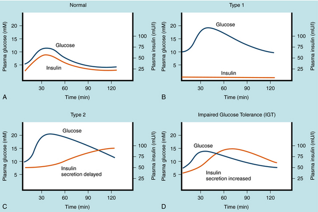
The management of diabetes is highly individualized and must consider the patient’s health, frequency of episodes of diabetic ketoacidosis, hypoglycemia (insulin-induced) and hyperglycemia, need for insulin, and relationship between circulating insulin levels and tissue responsiveness to insulin. There are two primary types of diabetes mellitus, and the pharmacological strategies for treating these differ in several important ways. Plasma glucose and insulin levels after an oral glucose tolerance test in normal individuals, in individuals with both types of diabetes mellitus, and in individuals with impaired glucose tolerance are depicted in Figure 43-1.
Type 1 diabetes mellitus is caused primarily by a T-cell mediated autoimmune response leading to destruction of the pancreatic β cells that produce insulin. Type 1 diabetes has an estimated frequency of 5% to 10% of total cases of diabetes mellitus and occurs predominantly before sexual maturation. Because the incidence of type 1 diabetes in homozygous twins is approximately 50%, factors other than genetic predisposition must be involved. The stimulus that prompts the immune system to attack β cells is still under investigation. The temporal nature of pancreatic β cell destruction can occur over a number of years, but after significant destruction (~90%), the onset of symptoms (polyuria, polydipsia, and polyphagia) can be abrupt. The increased urine volume is caused by osmotic diuresis resulting from increased concentrations of urinary glucose and ultimately ketone bodies. Thirst and hunger are compensatory responses. The development of diabetes mellitus is characterized by weight loss in the untreated disease and premature cessation of growth in children.
At the onset of symptoms, insulin levels are lower than normal and eventually become negligible (Fig. 43-1, B), requiring replacement to prevent metabolic acidosis (ketosis), followed by diabetic coma and premature death. The goal of insulin replacement is the carefully controlled maintenance of blood glucose to prevent or delay the onset of long-term diabetic complications.
Metabolic abnormalities associated with type 2 diabetes mellitus precede the appearance of overt symptoms. The progression of symptoms may evolve from impaired glucose tolerance, to insulin-independent type 2 diabetes mellitus, to insulin-requiring type 2 diabetes mellitus. Detection of impaired glucose tolerance is difficult, because fasting blood glucose and insulin levels can be nearly normal. However, 2 hours after a glucose challenge, above-normal levels of blood sugar and insulin are observed (see Figs. 43-1, C and D). These results are related to insulin resistance at the cellular level leading to diminished glucose transport and metabolism, which promotes hyperglycemia and provokes pancreatic β cell insulin release. Although adequate insulin secretion can occur initially, the amount of insulin release diminishes eventually and is insufficient to reduce hyperglycemia and consequently overcome the effects of insulin resistance. Chronic stimulation of the pancreatic β cell is thought to increase metabolic activity, which can induce cell death and ultimately decreased ability to secrete insulin, leading to the symptoms associated with type 2 diabetes mellitus. The diagnosis of type 2 diabetes mellitus is based on the appearance of hyperglycemia as it meets the criteria discussed. Because the pharmacological agents that are used to manage type 2 diabetes mellitus require insulin, if insulin is lost at this stage, the patient will become a candidate for insulin supplementation.
| Therapeutic Overview |
|---|
| Type 1 Diabetes Mellitus |
| Diabetic diet and exercise |
| Human insulin combination therapy |
| Addition of thiazolidinedione to manage concurrent insulin resistance |
| Management of Diabetic Ketoacidosis |
| Proper replacement of fluid, insulin, Na+, K+ and bicarbonate |
| Type 2 Diabetes Mellitus |
| Obesity management: |
| Diet, increased exercise, and weight reduction |
| Promoters of insulin secretion: |
| Sulfonylureas |
| Meglitinides |
| Incretin analogs |
| Dipeptidyl peptidase-IV inhibitors |
| Insulin supplementation |
| Management of insulin resistance: |
| Metformin |
| Thiazolidinediones |
| Pramlintide |
| α-Glucosidase inhibition to reduce postprandial carbohydrate challenge: |
| Metformin and miglitol |
| Combination therapy with multiple agents |
Mechanisms of Action
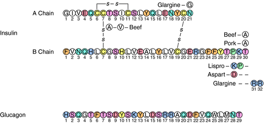
Insulin is a small acidic protein formed from the larger proinsulin precursor. Proinsulin is synthesized and packaged for secretion with trypsin-like proteases in the β cells of the islets of Langerhans. The precursor protein is proteolyzed within the secretory granule to form insulin via cleavage of a sequence of amino acids referred to as the C (connecting) peptide. Insulin is complexed with Zn++ within the granule. The active insulin protein is composed of an A peptide (21 amino acids), which has an intramolecular disulfide bond, and a B peptide (30 amino acids) that are covalently joined by two disulfide bonds (Fig. 43-2). Approximately equimolar amounts of insulin and C peptide are stored in and released from the granule, along with a much smaller amount of proinsulin.
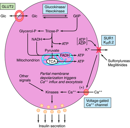
Insulin release is modulated by many factors (Box 43-1) but is controlled primarily by glucose. When blood glucose levels increase, glucose is taken up and metabolized by the pancreatic β cell, generating adenosine triphosphate (ATP) (Fig. 43-3). The increase in the ATP/ADP ratio promotes closure of the inward rectifying potassium channel 6.2 subunit (Kir6.2) of the ATP-sensitive potassium channel (also referred to as the SUR1/Kir6.2 channel). The decreased permeability of potassium ions partially depolarizes the cell membrane, promoting calcium uptake via activation of its voltage-gated channels. The elevated intracellular calcium stimulates exocytosis of the granules, releasing insulin and other components into the circulation.
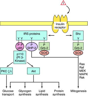
The cellular effects of insulin are initiated after insulin binds to a plasma membrane receptor (Fig. 43-4). The insulin receptor is composed of two α-subunits and two β-subunits. The interaction of insulin with the α-subunit results in changes in the β-subunit configuration, leading to activation of the tyrosine protein kinase that resides in the β-subunit. The phosphorylation of peptide substrates by the insulin receptor tyrosine kinase leads to activation of various anabolic pathways, inhibition of catabolic processes, and subsequently modulation of gene expression. The major peptide substrates for the insulin receptor are insulin receptor substrate (IRS)-1 and IRS-2. Phosphotyrosine residues in the IRS proteins serve as binding sites for intermediaries that trigger signal transduction pathways. The best defined of these pathways involves the small guanosine triphosphate (GTP)-binding protein Ras and leads to cell growth, differentiation, or both. However, the most important acute metabolic effects of insulin are mediated by phosphatidylinositol 3-kinase (PI 3-kinase), which is activated upon binding to phosphorylated IRS proteins. The phospholipid products generated by PI 3-kinase promote activation of several protein kinases including protein kinase B(Akt) and protein kinase C (PKC). These kinases phosphorylate effectors that activate glucose transporters (GLUT) and glycogen synthase (GS) and increase the synthesis of triglyceride and protein. Phosphotyrosine phosphatases are also activated that limit the duration of effects promoted by phosphotyrosines.
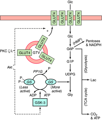
Stimulation of glycogen synthesis is very important in the action of insulin to lower blood glucose concentrations. Glucose taken up in response to insulin is deposited as glycogen in liver and skeletal muscle. Activation of glucose transport and GS contributes to the overall effects of insulin on glycogen synthesis. Increasing glucose transport allows more glucose to enter the muscle fiber, and activation of GS converts the glucose into glycogen. The control of GS activity is very important in this process and involves phosphorylation/dephosphorylation mechanisms (Fig. 43-5). GS is inactivated upon phosphorylation by the kinase GSK-3 and activated upon dephosphorylation by the protein phosphatase PP1G. In addition, phosphorylation of GSK-3 by Akt inactivates the kinase. Thus activation of Akt by insulin (see Fig. 43-4) leads to the inactivation of GSK-3, thereby tipping the balance to favor activation of GS by PP1G. In addition, by stimulating glucose transport via GLUT4, insulin increases the concentration of intracellular glucose 6-phosphate, an allosteric activator of GS. Insulin stimulates glucose transport in skeletal muscle and adipocytes by causing GLUT4 to translocate from intracellular vesicular compartments to the plasma membrane, an action promoted by signals from arising from Akt, PKC, or both (see Fig. 43-5). Increasing the number of glucose transporters at the cell surface increases glucose uptake into the cell. Interruption of any of these processes can be associated with genomic-associated insulin resistance.
Glucagon is a single-chain polypeptide with a molecular weight of approximately 3500 (see Fig. 43-2). It is synthesized in the α cells of the pancreatic islets through processes involving enzymatic cleavage of specific bonds in proglucagon, a large precursor. In the stomach and GI tract, a related molecule, glycentin, is formed from proglucagon. Glucagon is sometimes referred to as a counterregulatory hormone,
Stay updated, free articles. Join our Telegram channel

Full access? Get Clinical Tree


