Histamine and Antihistamines

Chapter 14 Histamine and Antihistamines


Abbreviations
CNS Central nervous system
Epi Epinephrine
IgE Immunoglobulin E


Therapeutic Overview


Histamine is synthesized, stored, and released primarily by mast cells and has profound effects on many organs. It is an important mediator of immediate hypersensitivity reactions and acute inflammatory responses and is a primary stimulator of gastric acid secretion. It is also an important neurotransmitter in the central nervous system (CNS) (see Chapter 27).


The many undesirable effects of histamine preclude its use as a drug. However, drugs that block histamine receptors or prevent its release from mast cells have important clinical uses. The effects of histamine on the heart, vascular, and nonvascular smooth muscle and on the secretion of gastric acid are mediated by at least three distinct receptors: H1, H2, and H3. A fourth histamine receptor (H4) was identified following the sequencing of the human genome, and it is thought to have a role in chemotaxis and mediator release in various types of immune cells. H4 receptor antagonists have antiinflammatory properties and efficacy in models of allergy and in autoimmune disorders.


H1 and H2 receptors have been most widely characterized and mediate well-defined responses in humans, as summarized in Table 14-1. Most responses are mediated by H1 receptors, such as bronchoconstriction, and are selectively antagonized by classical antihistamines, more accurately described as selective H1 receptor blocking drugs such as diphenhydramine. Antihistamines are widely used to treat allergic reactions, motion sickness, and emesis and as over-the-counter sleeping aids. H2-mediated responses such as gastric acid secretion are selectively antagonized by specific H2 receptor blocking drugs such as cimetidine. The H2 receptor antagonists are discussed extensively in Chapter 18; this chapter focuses on histamine and H1 receptor antagonists.


TABLE 14–1 Histamine Receptor Subtypes Mediating Selected Responses in Humans


Subtype Responses
H1 receptor only Basilar, pulmonary, coronary artery constriction; increased permeability of postcapillary venules; contraction of bronchiolar smooth muscle; stimulation of vagal sensory nerve endings promoting bronchospasm and coughing; gastrointestinal smooth muscle relaxation and contraction; Epi release from adrenal medulla
H2 receptor only Acid and pepsin secretion from oxyntic mucosa; facial cutaneous vasodilation; pulmonary and carotid artery relaxation; increased rate and force of cardiac contraction; relaxation of bronchial smooth muscle; inhibition of IgE-dependent degranulation of basophils
H1 and H2 (?) receptors Decreased total peripheral resistance; increased forearm blood flow; increased cardiac atrial and ventricular automaticity; stimulation of cutaneous nerve endings causing pain and itching

H3 receptors have been studied primarily in experimental animals, where they are located on nerve endings and mediate the inhibition of release of histamine and several other transmitters. This receptor remains an attractive target for drug development.


Clinical uses of histamine antagonists are summarized in the Therapeutic Overview Box.


Therapeutic Overview
Histamine and Histamine Receptor Agonists
No significant clinical use
Antihistamines (H1 Receptor Antagonists)
Allergic reactions
Motion sickness
Insomnia
Nausea and vomiting
H2 Receptor Antagonists
Peptic ulcers and gastroesophageal reflux disease (see Chapter 18)


Mechanisms of Action


Synthesis and Metabolism of Histamine


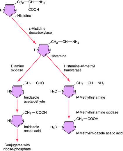
The synthesis and catabolism of histamine are depicted in Figure 14-1. Histamine is synthesized by decarboxylation of the amino acid L-histidine by histidine decarboxylase. Most histamine is stored in an inert form at its site of synthesis, and very little is freely diffusible. After synthesis and release from its storage sites, histamine acts at its targets and is rapidly metabolized through two primary pathways. Oxidative deamination leads to formation of imidazole acetic acid, while methylation, which predominates in the brain, leads to the formation of N-methylimidazole acetic acid. Both metabolites are inactive and subject to further biotransformation.


Storage and Release of Histamine by Mast Cells and Basophils


Histamine is stored and released primarily by mast cells. Although basophils and central neurons also use histamine, their role is not fully understood. Histamine is widely distributed, with the highest concentrations in the skin, lungs, and gastrointestinal tract mucosa, consistent with mast cell densities in these tissues.


Mast cells and basophils have high-affinity immunoglobulin E (IgE) binding sites on their surface membranes and store histamine in secretory granules. Different types of mast cells can be classified by their staining properties, anatomical locations, or susceptibility to degranulation by polyamines. Anatomically, mast cells are classified as being of mucosal or connective tissue origin. However, there are mixed populations in both tissues and additional heterogeneity within these two classes. Human mast cells differ with respect to their proteoglycan structure and content, the types of serine proteases in their storage granules, the eicosanoids synthesized and released on degranulation, and the extent to which degranulation is inhibited by cromolyn Na+.


In mast cell granules, histamine exists as an ionic complex with a proteoglycan, chiefly heparin sulfate, but also chondroitin sulfate E. In basophils, histamine is also stored in granules as an ionic complex, predominantly with proteoglycans. The release of histamine and other mediators from mast cells and basophils is common during allergic reactions but also can be induced by drugs and endogenous compounds to produce pseudoallergic, anaphylactoid reactions as shown in Figure 14-2. The role of mast cells in immediate and delayed hypersensitivity reactions and nonallergic disorders explains the therapeutic utility of antihistamines and degranulation inhibitors.


Histamine is released by noncytolytic or cytolytic degranulation. Cytolytic release occurs when the membrane is damaged, does not require energy or intracellular Ca++, and is accompanied by leakage of cytoplasmic contents. Cytolytic release can be induced by drugs such as phenothiazines, H1 receptor antagonists, and opioids, but the concentrations required are usually greater than therapeutic concentrations.


Noncytolytic release is evoked by binding of a specific ligand to a receptor in the plasma membrane, resulting in exocytosis of secretory granules. Noncytolytic release requires energy, depends on intracellular Ca++, and is not accompanied by leakage of cytoplasmic contents. A classic example is degranulation of sensitized mast cells or basophils induced by cross-bridging of adjacent IgE molecules on the cell surface. This involves activation of various phospholipases, fusion of secretory granules with the plasma membrane, and extrusion of their contents. Such exocytosis results in the release of histamine, heparin, eosinophil, and neutrophil chemotactic factors; neutral proteases; and other enzymes. The release of other mediators, such as eicosanoids and platelet-activating factor, can also occur.


Noncytolytic release can also be produced by other mechanisms, often by highly basic substances. These include the polyamine compound 48/80, polypeptides such as bradykinin, substance P, formylmethionylleucinylphenylalanine, protamine, anaphylatoxins, and a protein present in bee venom. With the exception of protamine, a heparin antagonist, none of these agents has any therapeutic use, but they are likely important in pathological responses.


Noncytolytic degranulation can also be induced by several drugs including d-tubocurarine, succinylcholine, morphine, codeine (in therapeutic doses), doxorubicin, and vancomycin. The mechanism may involve activation of protein kinase A and NF-κB. Histamine release in vivo may also be produced by some plasma expanders, notably those based on cross-linked gelatin, and by radiocontrast media, especially those of high osmotic strength. Intravenous administration of these compounds is most likely to result in histamine release. Life-threatening reactions are rare but occur occasionally.


The most acute and potentially severe allergic reaction is anaphylaxis. In both animals and humans, parenterally administered histamine triggers responses that mimic the early responses of anaphylaxis, including hypotension, vasodilation, myocardial depression, dysrhythmias, urticaria, angioedema, and bronchospasm. These can be partially reversed by H1 and H2 receptor antagonists; however, they are most effective when administered prophylactically rather than after an acute reaction has begun. Histamine is only one of many anaphylactic mediators, but it has important effects on the production and effectiveness of others.


Noncytolytic degranulation of mast cells results in both anaphylactic and anaphylactoid reactions. Many substances can release histamine independently of IgE. However, the clinical signs and symptoms are indistinguishable from those of true anaphylaxis, because the same mediators are involved. The term anaphylactoid is used to refer to a clinical syndrome indistinguishable from anaphylaxis but caused by something other than an immune response.


Many of the solutions and drugs used in general anesthesia can produce mast cell degranulation, especially if administered intravenously. However, skeletal muscle paralysis, the effects of other drugs, and mechanical ventilation can mask signs of histamine release. Cardiac dysrhythmias or hypotension without flush, rashes, or angioedema may be seen. Indeed, most patients undergoing general anesthesia have elevated histamine levels but are relatively asymptomatic. Preoperative prophylaxis with H1 and H2 receptor antagonists remains controversial but is used for certain patients at risk (atopic patients and patients with previous reactions).


A summary of agents that release histamine is presented in Table 14-2. Histamine concentrations of 0.2 to 1.0 ng/mL in humans produce mild signs and symptoms, including metallic taste, headache, and nasal congestion. Concentrations exceeding 1 ng/mL produce moderate effects, including skin reactions, cramping, diarrhea, flushing, tachycardia, cardiac dysrhythmias, and hypotension. Life-threatening hypotension, ventricular fibrillation, and bronchospasm leading to cardiopulmonary arrest can occur when concentrations approach 12 ng/mL.


TABLE 14–2 Common Causal Factors Involved in Anaphylactic and Anaphylactoid Reactions


Only gold members can continue reading. Log In or Register to continue

Stay updated, free articles. Join our Telegram channel

Jun 18, 2016 | Posted by in PHARMACY | Comments Off on Histamine and Antihistamines

Full access? Get Clinical Tree

Get Clinical Tree app for offline access
IgE-Mediated Anaphylaxis Non–IgE-Mediated Anaphylactoid Reactions
Food Drugs
Peanuts, seafood, eggs, milk products, grains Anesthesia related: neuromuscular blocking agents, opioids, plasma expanders
Drugs  
Antibiotics: penicillins, cephalosporins, sulfonamides

  Dyes
Venoms Radiocontrast media, fluorescein
Hymenoptera, fire ants, snakes  
  Other Idiopathic Reactions
 

Foreign Proteins
Nonhuman insulin, corticotropin, serum proteins, seminal proteins, vaccines, antivenoms
Enzymes  
Chymopapain  
Other