Historically, these lesions have poorly defined histologic criteria and have been referred to by variety of names (e.g., fibroinflammatory polyp, fibroepithelial polyp, mucosal hyperplasia)
Microscopic

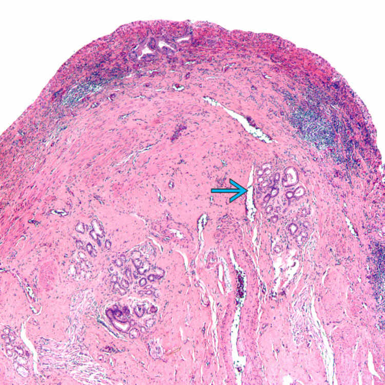
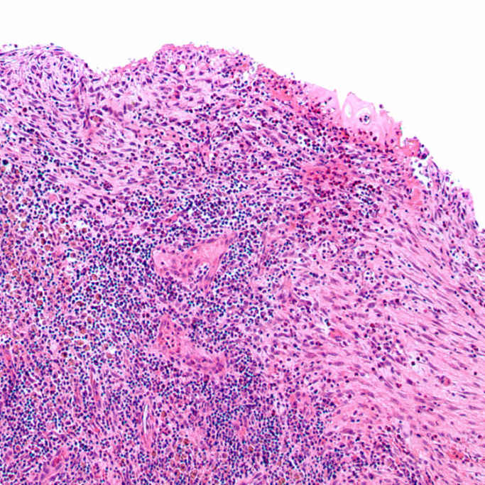
An inflammatory polyp of the gallbladder shows mucosal ulceration and granulation tissue, as well as stromal inflammation and foci of mucosa with pyloric-type metaplasia
 .
.
Inflammatory polyps of the gallbladder are usually secondary to inflammatory processes and thus are composed of inflammation and a proliferation of organizing granulation tissue.

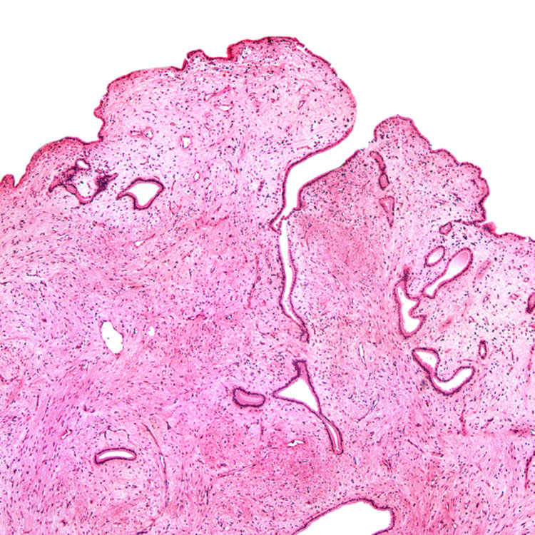
This inflammatory/reactive mucosal polyp shows stromal edema with overlying benign gallbladder epithelium.
TERMINOLOGY
Synonyms
Definitions
• Inflammatory mucosal lesion associated with underlying inflammatory process


Stay updated, free articles. Join our Telegram channel

Full access? Get Clinical Tree


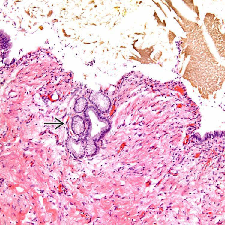
 , which should not be misinterpreted as evidence of a pyloric-type intracholecystic papillary/tubular neoplasm. Note the surrounding inflammation and stromal edema.