Inborn Errors of Metabolism


Inborn Errors of Metabolism


Peter Pytel, M.D.

Selene Koo, M.D., Ph.D.

Darrel Waggoner, M.D.


INTRODUCTION

Over 100 years ago, in 1908, Archibald Garrod delivered his four Croonian Lectures during which he first introduced the expression of “inborn errors of metabolism” (1). He proposed that these result from enzymatic defects in catabolic pathways. Since that time, our understanding of inborn errors of metabolism (IEM) has increased dramatically. Along with improved technology especially in molecular sequencing-based techniques, this has resulted in refinement in diagnosis and classification of IEM. Early diagnosis is increasingly important as treatments, such as dietary management and enzyme replacement, are becoming available for some patients. Although individually rare, the collective incidence of IEM is approximately 1/1500 persons.

Online genetic/metabolic disease databases such as OMIM (Online Mendelian Inheritance in Man, http://www.ncbi.nlm.nih.gov/entrez/query.fcgi?db=OMIM), MetaGene (http://www.metagene.de/index.html), GeneTests (http://www.genetests.org/), GTR (Genetic Testing Registry) (http://www.ncbi.nlm.nih.gov/gtr/) that includes a link to GeneReviews, and NORD (http://www.rarediseases.org/) are ideal sources for current information on the rapidly evolving field of IEM.


CLINICAL PRESENTATION OF INBORN ERRORS OF METABOLISM

Most metabolic diseases are autosomal recessive disorders, some are X linked, and a few are inherited as dominant traits. Carriers of recessive traits are usually asymptomatic, but female carriers of X-linked traits do sometimes show disease manifestations that vary in severity. IEM may present at any age and in a variety of ways even within the same disease entity and within the same family. IEM can lead to organ dysfunction, neurologic and medical complications, skeletal dysplasia, and dysmorphic features (Table 5-1). Symptoms may begin before birth (e.g., with hydrops fetalis or fetal ascites), at birth, with sudden death in infancy, with deterioration after a symptom-free interval, or later in life with milder symptoms and progression. On the one hand, symptoms may sometimes be confused with sepsis, delaying the diagnosis, while on the other hand, infection can lead to decompensation in IEM, becoming the trigger that leads to the first presentation.


DIAGNOSIS OF INBORN ERRORS OF METABOLISM

IEM may be diagnosed by biochemical assays, tissue histology and, more recently, molecular techniques, specifically DNA sequencing. All play an important role depending on the specific disease in question, but molecular testing has grown quickly and may soon be the best, easiest, and fastest method to make a diagnosis. Sometimes, the diagnosis is made based on newborn screening (NBS) as discussed in detail below.

Biochemical studies may allow definitive diagnosis. Routine studies include evaluation of blood glucose, lactate, ammonia, and ketones, urine organic acids, blood and urine amino acids, fatty acids, and carnitine metabolites. Biochemical assays on skin fibroblasts and lymphocytes of specific enzymes can be done for some metabolic pathways.

Morphology of tissues, including liver, muscle, skin, conjunctiva, or placenta (Table 5-2), may show characteristic findings by light microscopy (LM). Brain, lymph nodes, spleen, kidney, and heart often also show findings in IEM, but biopsies of these sites are not performed as commonly. Transmission electron microscopy (EM) of the skin, conjunctiva, mucosal ganglion cells, liver, peripheral nerve, muscle, bone marrow, or peripheral blood leukocytes is important in evaluation of some IEM (Table 5-3). Particularly for lysosomal storage diseases, ultrastructural study of these sites is a sensitive screen that can provide valuable information about stored material.

Many IEM affect the liver (2,3); in some cases (cystinosis, metachromatic leukodystrophy, Fabry disease),
morphologic alterations have no apparent clinical consequence. In other disorders—such as tyrosinemia and glycogen storage disease (GSD) IV—progressive liver disease is common. A variety of histologic alterations—including hepatitis, steatosis, cirrhosis, cholestasis, ductopenia, ductular proliferation, neoplasia, and storage—can be seen in IEM, and many IEM cause similar morphologic alterations (see Chapter 15).


TABLE 5-1 CLINICAL SYMPTOMS IN PATIENTS WITH INBORN ERRORS OF METABOLISM


Nonimmune hydrops fetalis, fetal ascites


Sudden unexpected death in infancy


Acute or episodic presentation of symptoms mimicking sepsis (lethargy, vomiting, tachypnea, shock, coma)


Failure to thrive


Macrocephaly or microcephaly


Medical complications: cardiomyopathy and arrhythmias, acute or chronic liver disease/cirrhosis, organomegaly, deafness


Neurologic complications: hypotonia/hypertonia, seizures, loss of cognitive milestones or motor skills, exercise intolerance, cramps, fatigue, and rhabdomyolysis


Ophthalmologic signs: corneal clouding, macular and retinal changes


Skeletal abnormalities including dysostosis multiplex


Dysmorphic features: coarse facial features, macroglossia


TABLE 5-2 PLACENTAL FINDINGS IN LYSOSOMAL STORAGE DISEASES


Lysosomal Storage Disease


Affected Cells


Hydrops Fetalis


Sialidosis (mucolipidosis type I)


Syncytiotrophoblasts, Hofbauer and stromal cells


Yes


Mucolipidosis II (I-cell disease)


Syncytiotrophoblasts, Hofbauer and stromal, X cells


Yes


Mucolipidosis IV


Hofbauer and stromal cells


Yes


Sialic acid storage disease


Syncytiotrophoblasts, Hofbauer cells, endothelium, amniocytes


Yes


Galactosialidosis


Syncytiotrophoblasts


Yes


MPS I (Hurler)


Villous stromal cells


Yes


MPS III (Sanfilippo)


Syncytiotrophoblasts, Hofbauer cells, and stromal cells


Yes


MPS IVA (Morquio A)


Villous stromal cells


Yes


MPS VII (Sly)


Villous stromal cells


Yes


GM1 gangliosidosis


Syncytiotrophoblasts, Hofbauer cells


Yes


GM2 gangliosidosis (Tay-Sachs)


Syncytiotrophoblasts


Cholesterol ester storage disease


Syncytiotrophoblasts


Wolman disease


Syncytiotrophoblasts


Yes


GSD II


Amniocytes, endothelial cells, stromal cells


GSD IV


Amniocytes


Niemann-Pick A


Syncytiotrophoblasts, Hofbauer cells, fibrocytes in the cord


Yes


Gaucher disease type 2


Vessels


Yes


Fabry disease


Endothelial cells, perithelial cells, vascular smooth muscle cells


Yes


Skeletal muscle biopsy is useful for evaluation of mitochondrial myopathies, glycogen storage diseases, and some lysosomal storage diseases. Increased mitochondria with “ragged red fibers,” subsarcolemmal mitochondria collections, and structurally abnormal mitochondria occur in mitochondrial myopathies. Increased muscle fiber glycogen, sometimes in vacuoles, may be present in the GSDs. Increased lipid occurs with abnormal fatty acid metabolism and with mitochondrial dysfunction (see Chapter 27).


NEWBORN SCREENING

Newborn screening (NBS) began in the 1960s with testing for phenylketonuria, and additional tests have been added since then. With the development of new technology, specifically tandem mass spectrometry (MS/MS) for NBS, it has become possible to detect many more metabolic disorders via a rapid-throughput methodology. As of January 2013, 47 states in the United States have added expanded NBS using MS/MS and screen for the majority of the recommended core conditions. Information on each state’s NBS program is available through Genes-R-Us (http://genes-r-us.uthscsa.edu). The Discretionary Advisory Committee on Heritable Disorders in Newborns and Children (DACHDNC) was established under the Public Health Service Act and fulfills the functions previously undertaken by the Secretary’s Advisory Committee on Heritable Disorders in Newborns and Children (SACHDNC). The committee recommends
that every NBS program includes a uniform screening panel (Table 5-4) and advises on the addition of new tests to the screening panel (http://www.hrsa.gov/advisorycommittees/mchbadvisory/heritabledisorders/). The Web site has a list of conditions that have been nominated and are under current review.


TABLE 5-3 ULTRASTRUCTURAL APPEARANCE OF LYSOSOMAL STORAGE DISEASES IN SKIN AND CONJUNCTIVAL BIOPSIES


Disorder


Ultrastructural Appearance of Predominant Storage


Stored Material


Cells Affected


Disorders with ultrastructurally characteristic storage


Pompe disease


Electron-dense glycogen granules


Glycogen


Lymphocytes, endothelium, fibroblasts, epithelium, nerves


Cholesterol ester storage disease, Wolman disease


Cholesterol, lipids


Cholesterol ester


Fibroblasts


Infantile ceroid lipofuscinosis


Granular osmiophilic material


Saposin A, D


Perithelial and endothelial cells, smooth muscle, sweat gland epithelial cells, lymphocytes


Late infantile ceroid lipofuscinosis


Curvilinear profiles


Mitochondrial subunit c of ATPase synthase


Perithelial and endothelial cells, smooth muscle, sweat gland epithelial cells, lymphocytes


Juvenile ceroid lipofuscinosis


Curvilinear and finger-print profiles


Mitochondrial subunit c of ATPase synthase


Perithelial and endothelial cells, smooth muscle, sweat gland epithelial cells, lymphocytes


Cystinosis


Rectangular, rhomboid, polymorphic crystals


Cystine


Fibroblasts


Metachromatic leukodystrophy


Tuffstone or herringbone bodies


Sulfatide, cholesterol, phosphatides


Peripheral nerve Schwann cells, histiocytes


Krabbe disease (globoid leukodystrophy)


Crystals with sharp corners


Galactosylceramide


Peripheral nerve Schwann cells


Fabry disease


Pleomorphic leaflets, lamellae, tubular structures


Globotriaosylceramide-containing substrates


Epithelium, fibroblasts, endothelium, lymphocytes


Disorders with ultrastructurally nonspecific storage


Mucopolysaccharidoses (MPS)


Mucolipidoses


Aspartylglycosaminuria


Alpha-mannosidosis


Beta-mannosidosis, GM1


Gangliosidosis, I-cell disease


Lucent or fine fibrillogranular material


Oligosaccharides, glycosaminoglycans


Eccrine gland epithelium, endothelial cells (may be spared in MPS), fibroblasts, lymphocytes, macrophages, pericytes, Schwann cells, smooth muscle cells. Mucolipidosis III affects primarily fibroblasts with normal lymphocytes.


Fucosidosis, GM1 and GM2 gangliosidosis, galactosialidosis, MPS, mucolipidosis IV, and sialidosis


Fine lamellated membranous cytoplasmic bodies, zebra bodies


Gangliosides and glycolipids


Peripheral nerve Schwann cells, endothelial cells, pericytes, smooth muscle cells in Fabry disease. In Niemann-Pick and ML IV lymphocytes have storage


Molecular testing for DNA mutations has been incorporated into NBS for several disorders, and this will likely increase over time. There has been discussion about the use of whole genome sequencing in NBS, which is controversial, but there is little doubt that molecular techniques will play a larger role in NBS in the future (4). Cystic fibrosis screening
includes the use of molecular techniques to detect a panel of common mutations. The initial screen is done by measurement of immunoreactive trypsinogen and then followed by molecular genetic testing of the CFTR gene and sweat testing. The genetic testing involves determination of a panel of common mutations, and a diagnosis is made based on a combination of sweat test results and genetic testing (5).


TABLE 5-4 RECOMMENDED UNIFORM SCREENING PANEL FOR ALL STATES


Metabolic Disorders


Organic acid disorders


  • Core Conditions: Propionic acidemia; methylmalonic acidemia; isovaleric acidemia; 3-methylcrotonyl-CoA carboxylase deficiency; 3-hydroxy-3-methylglutaric aciduria; holocarboxylase synthase deficiency; β-ketothiolase deficiency; glutaric aciduria


  • Secondary Conditions: Methylmalonic acidemia with homocystinuria; malonic acidemia; isobutyrylglycinuria; 2-methylbutyrylglycinuria; 2-methylglutaconic aciduria; 2-methyl-3-hydroxybutyric aciduria


Fatty acid oxidation disorders


  • Core Conditions: Carnitine uptake defect/carnitine transport defect; medium-chain acyl-CoA dehydrogenase deficiency; very long-chain acyl-CoA dehydrogenase deficiency; long-chain L-3 hydroxyacyl-CoA dehydrogenase deficiency; trifunctional protein deficiency


  • Secondary Conditions: Short-chain acyl-CoA dehydrogenase deficiency; medium-/short-chain L-3 hydroxyacyl-CoA dehydrogenase deficiency; glutaric acidemia type II; medium-chain ketoacyl-CoA thiolase deficiency; carnitine palmitoyltransferase type I and II deficiencies; carnitine-acylcarnitine translocase deficiency


Amino acid disorders


  • Core Conditions: Argininosuccinic aciduria; citrullinemia; maple syrup urine disease; homocystinuria; phenylketonuria; tyrosinemia, type I


  • Secondary Conditions: Argininemia; hypermethioninemia; tyrosinemia types II and III


Endocrine Disorder


Congenital adrenal hyperplasia


Primary congenital hypothyroidism


Hemoglobin Disorder


S, S disease (sickle cell anemia)


S, beta-thalassemia


S, C disease


Other Disorders


Biotinidase deficiency


Critical congenital heart disease


Cystic fibrosis


Classic galactosemia


Hearing loss


Severe combined immunodeficiencies


Several states have recently implemented screening for lysosomal storage disorders including Krabbe, Gaucher, Pompe, Fabry, and Niemann-Pick diseases and mucopolysaccharidoses I and II. Molecular testing is an important component for the follow-up of screen-positive individuals and can be used in predicting problems such as severity of disease (Gaucher) (6) and pseudodeficiency (Hurler) (7). Krabbe testing utilizes full gene sequencing in addition to enzyme determination as part of the NBS process. Detection of specific mutations associated with the severe infantile presentation of Krabbe disease is used when recommending treatment and follow-up protocols (8).

The genetic basis for most IEM is known, and Sanger sequencing has been utilized to define the mutations in affected individuals and has typically been done after biochemical or enzyme testing established the diagnosis. Although this has been a powerful tool for diagnosis, molecular techniques have been sparingly used for primary diagnosis as the technology limits how many genes can reasonably be sequenced in a single individual, and hence the need to know which gene to sequence. Next-generation sequencing (NGS) is the newest technology that utilizes massive parallel sequencing to generate large-scale sequence data, which can be used to sequence the entire exome or genome. NGS is also used to offer panel testing where many genes related to a common phenotype can be sequenced at one time. This is useful in the diagnosis of IEM and has resulted in NGS panels becoming a more common primary diagnostic technique. Three examples are reviewed below, but many diseases could illustrate this approach.


Glycogen storage disorders (GSDs) are a group of disorders of glycogen metabolism that primarily affect the liver and muscle. GSDs are classified on the basis of specific enzyme defects in glycogen production or breakdown and the various disorders lead to overlapping features of hypoglycemia, hepatomegaly, and muscle symptoms. The diagnosis of GSD once relied on invasive liver or muscle biopsies and biochemical assays to determine the specific subtype important for counseling and treatment (8). Congenital disorders of glycosylation (CDG) are a group of over 60 different disorders in glycosylation of proteins. CDG is characterized by multiorgan dysfunction with significant morbidity and mortality. Biochemical testing of serum transferrin is a useful screening tool for CDG but cannot diagnose the specific form of the disease (9). Disorders of mitochondrial dysfunction show clinical and genetic heterogeneity and are difficult to diagnose due to extreme locus and allelic heterogeneity. These diseases develop as a result of dysfunction of more than 900 genes in the nuclear and mitochondrial genome (10). NGS of a panel of genes implicated in GSDs, CDG, and mitochondrial dysfunction is now available and can be used successfully to diagnose these disorders (8,9,10).


LYSOSOMAL STORAGE DISEASES

The first lysosomal storage disease described was Pompe disease (11). Historically, lysosomal storage diseases were defined by the storage product, clinical features, histomorphology, and biochemical abnormalities. Nowadays, there is a shift to a molecular classification of these diseases. In this chapter, lysosomal storage diseases are defined broadly to also include diseases like neuronal ceroid lipofuscinoses (12). As such, lysosomal storage diseases are currently a group of some 50 to 70 genetic disorders with a combined incidence of approximately 1/5000 (13,14). There is an overrepresentation of some populations including Ashkenazi Jews and those of Finnish ancestry (14,15). As a group, lysosomal storage diseases are among the most commonly diagnosed metabolic disorders. Most are monogenic diseases that are inherited as autosomal recessive disorders except for three X-linked disorders (Fabry, Danon, and Hunter diseases). Many of the mutations affect lysosomal enzymes, but others disrupt lysosomal function in different ways as discussed in detail below. Table 5-5 summarizes important lysosomal storage diseases and groups them based on the function of the mutated gene. The next passages describe common themes in disease phenotype, pathophysiology, diagnosis, and treatment in this large group of diseases. Individual entities will subsequently be described following the overall order of Table 5-5.


Disease Phenotype

Lysosomal storage diseases can present at virtually any age but are most commonly encountered as pediatric diseases. Many affected patients present in early childhood. At one end of the spectrum, these diseases are a cause of nonimmune hydrops fetalis accounting overall for some 6% to 15% of such cases (16,17). On the other end of the spectrum are adult-onset presentations that may mimic other myopathies or psychiatric diseases and are therefore easily misdiagnosed.


TABLE 5-5 MASON CATEGORIES OF LYSOSOMAL STORAGE DISORDERS


Diseases caused by mutations disrupting enzyme function


(1) Sphingolipidoses


Gaucher (types I, II, III), GBA gene encoding acid beta-glycosidase


Fabry, GLA gene encoding alpha-galactosidase A


Gangliosidosis GM1, GLB1 gene encoding beta-galactosidase (allelic with MPS IV; see above)


Gangliosidosis GM2, Tay-Sachs variant, HEXA gene encoding beta-hexosaminidase A


Gangliosidosis GM2, Sandhoff variant, HEXB gene encoding beta-hexosaminidase B


Niemann-Pick (types A and B), SMDP1 gene encoding acid sphingomyelinase


Metachromatic leukodystrophy, ARSA gene encoding arylsulfatase A


Krabbe, GALC gene encoding galactosylceramidase


Farber, ASAH1 gene encoding acid ceramidase


(2) Disorders of glycoprotein degradation (oligosaccharidoses/glycoproteinoses)


Alpha-mannosidosis, MAN2B1 gene encoding alpha-mannosidase


Beta-mannosidosis, MANBA gene encoding beta-mannosidase


Fucosidosis, FUCA1 gene encoding alpha-l-fucosidase


Sialidosis, NEU1 gene encoding neuraminidase 1


Aspartylglucosaminuria, AGA gene encoding aspartylglucosaminidase


(3) Glycogen storage diseases—Pompe disease/adult-onset acid maltase disease


(4) Mucopolysaccharidoses


MPS I—Hurler, Scheie, Hurler-Scheie, IDUA gene encoding alpha-l-iduronidase


MPS II—Hunter, IDS gene encoding iduronate-2-sulfatase


MPS IIIA—Sanfilippo A, SGSH gene encoding sulfamidase


MPS IIIB—Sanfilippo B, NAGLU gene encoding alpha-N-acetylglucosaminidase


MPS IIIC—Sanfilippo C, HGSNAT gene encoding heparan-alpha-glucosaminide N-acetyltransferase


MPS IIID—Sanfilippo D, GNS gene encoding N-acetylglucosamine-6-sulfatase


MPS IV—Morquio A, GALNS gene encoding N-acetylgalacosamine-6-sulfatase


MPSIV—Morquio B, GLB1 gene encoding beta-galactosidase (allelic with GM1 gangliosidosis; causative genes disrupt breakdown of keratin sulfate without affecting degradation of GM1 gangliosides)


MPS VI—Maroteaux-Lamy, ARSB gene encoding arylsulfatase B


MPS VII—Sly, GUSB gene encoding beta-glucuronidase


MPS IX—Natowicz, HYAL2 gene encoding hyaluronoglucosaminidase 1


(5) Lipidosis-Wolman disease and cholesterol ester storage disease (CESD)


Nonenzymatic protein deficiencies—defects affecting, for example, trafficking, membrane proteins, lysosomal enzyme protection, or structural proteins


Mucolipidosis II alpha/beta, III alpha/beta; GNPTAB gene encoding the alpha- and beta-subunits for GlcNAc-1-phosphotransferase


Mucolipidosis III gamma; GNPTG, the gamma subunit for GlcNAc-1-phosphotransferase


Mucolipidosis IV, MCOLN1 encoding mucolipin-1


Cystinosis, CTNS gene encoding cystinosin


Danon, LAMP2 encoding lysosomal-associated membrane protein-2


Gaucher, atypical; PSAP gene encoding saposin C


Gangliosidosis GM2, AB variant; GM2A gene encoding GM2 ganglioside activator protein


Niemann-Pick type C1, NPC2 gene encoding NPC1 protein


Niemann-Pick type C2, NPC2 gene encoding NPC2 protein


Krabbe, atypical; PSAP gene encoding saposin A


Metachromatic leukodystrophy, atypical; PSAP gene encoding saposin B


Ceroid lipofuscinoses


CLN1—Haltia-Santavouri; CLN1 gene encoding PPT-1


CLN2—Jansky-Bielschowsky; CLN2 gene encoding TPP-1


CLN3—Spielmeyer-Sjöegren; CLN3 gene encoding CLN3 protein (battenin)


CLN4—Parry; CLN4 (DNAJC5) gene encoding DnaJ homologue subfamily C member 5


CLN5; CLN5 gene encoding CLN5


CLN6; CLN6 gene encoding CLN6


CLN7; CLN7 (MFSD8) gene encoding motility factor superfamily domain containing protein 8


CLN8; CLN8 gene encoding CLN8


CLN10; CLN10 (CTSD) gene encoding cathepsin D


CLN11; CLN11 (PRN) gene encoding progranulin


CLN12; CLN12 gene encoding CLN12/ATPase


CLN13; CLN13 (CTSF) gene encoding cathepsin F


CLN14; CLN14 (KCTD7) gene encoding potassium channel tetramerization domain containing protein 7


The pattern of organ involvement in a specific lysosomal storage disease depends on the nature of the accumulating substrate, its anatomic distribution, and the cell turnover rate in individual organs. Three recurring themes among these patterns of organ involvement are nervous system, liver, and skeletal involvement.


  • The central and peripheral nervous systems are particularly vulnerable to lysosomal storage diseases and therefore often affected. Neurologic manifestations include developmental delay, dementia, speech impairment, deafness, visual loss, ophthalmoplegia, leukodystrophy, seizures, ataxia, peripheral neuropathy, and macrocephaly (15).


  • Hepatosplenomegaly is a feature of many of these diseases including Gaucher disease, Niemann-Pick disease, and mucopolysaccharidosis (MPS). To what extent Kupffer cells or hepatocytes are affected varies depending on the disease process.


  • Deposition of storage material like glycosaminoglycans in growth plates and cartilage disrupts normal development and maturation of the skeleton. As a result, these patients often have coarse, dysmorphic facial features. “Dysostosis multiplex” is a term that is often used for the skeletal manifestations that include coarse, dysmorphic facial features, short stature, as well as bone and joint abnormalities.

Beyond these three organ systems, there is a plethora of other organ manifestations as illustrated by the following examples: In Gaucher disease, involvement of the reticuloendothelial system may lead to hypersplenism causing anemia. Cardiac involvement with deposition of glycosaminoglycans in the heart valves and connective tissue is a feature of mucopolysaccharidoses (esp. MPS VI) and may be associated with valvular disease, conduction defects, and cardiomyopathy. Deposition of glycosaminoglycans in the dura and ligaments can cause spinal cord compression symptoms or carpal tunnel syndrome. Involvement of adenoids, tonsils, and epiglottis can contribute to upper airway obstruction. Deposition in the cornea can cause cataracts.


Diagnosis of Lysosomal Storage Diseases

The diagnosis and classification of lysosomal storage diseases have evolved and changed. Historically, morphologic studies and biochemical documentation of enzyme deficiency in leukocytes, fibroblasts, or amniocytes were key components of the diagnostic workup. In that setting, the ultrastructural evaluation of storage material often could add valuable insight into the disease process. Histomorphologic and ultrastructural confirmation of storage material may be performed on the skin, conjunctiva, peripheral blood lymphocytes, liver, bone marrow, skeletal muscle, rectal suction biopsies sampling mucosal ganglion cells, or placenta, depending on the suspected clinical diagnosis (17,19,20,21,22,23,24). Some of the storage material may require special handling of the tissue since it may be soluble in solutions used for routine processing. The appropriate triage of a specimen may therefore include setting aside tissue for alcohol fixation (e.g., for cysteine crystals or glycogen), formalin fixation, glutaraldehyde fixation for electron microscopy, and freezing. Stored lipids can be visualized by oil red O or Sudan black-stained sections on frozen tissue or on Epon-embedded sections. The Schultz adaptation of the Liebermann-Burchard reaction has been used to stain cholesterol (25). The sugar moieties of glycosylated storage material or accumulated carbohydrates can be highlighted by PAS staining with and without diastase. Colloidal iron or Alcian blue can be utilized to stain glycosaminogly-can accumulation as seen with mucopolysaccharidoses.

Nowadays, however, our understanding of and our approach toward lysosomal storage diseases have changed dramatically with the advent of new molecular techniques (13). Genetic testing has rapidly become the main diagnostic tool. Targeted treatment options have changed the role and importance of an accurate classification from a purely prognostic aspect to a crucial step in patient management. An understanding of the morphologic manifestations of these diseases is therefore less critical for daily diagnostic practice than in the past. But studying the morphologic changes of these diseases still conveys important insights into the underlying pathophysiologic concepts. And in some unexpected cases, the diagnostic workup may start with the specimen that ends up on the workbench or under the microscope of a pathologist who will then still have to recognize the key morphologic features.


Lysosomal Storage Diseases Linked to Defects in Specific Enzymes


Sphingolipidoses


Gaucher Disease

Gaucher disease is the most common lysosomal storage disease (31). There are three allelic types that represent mutations in the beta-glucocerebrosidase gene (32). Type 1 Gaucher disease is the most common form and is particularly frequent in individuals of Ashkenazi Jewish heritage (33). Type 1 is nonneuronopathic and causes hepatosplenomegaly, hypersplenism, lung disease, and bony involvement. This is in contrast to types 2 and 3 that are known as neuronopathic forms because they additionally result in seizures and brain injury. Type 2 typically presents as life-threatening disease in infancy while type 3 is milder.

The enzyme defect in beta-glucocerebrosidase leads to failure of cleavage of glucose from ceramide. Glucocerebrosides are derived from glycolipids in white and red cell membranes and accumulate mainly in lysosomes of cells of the reticuloendothelial system. Characteristic Gaucher cells are large eosinophilic phagocytes with wrinkled or striated cytoplasm (Figure 5-1A-C) that are found in the liver, bone marrow, spleen, lymph nodes, tonsils, thymus, Peyer patches, alveolar septae, and Virchow-Robin space (28,31,34). Gaucher cells are capable of erythrophagocytosis and are acid phosphatase positive and label with antibody to CD68. The striations can be highlighted with Masson trichrome, aldehyde fuchsin, and PAS after diastase. Electron microscopy shows lysosomal rod-shaped or tubular lipid bilayer stacks (Figure 5-1D, E).

The liver is enlarged in all three types with accumulation of storage material in the Kupffer cells, especially in zone 3, but not the hepatocytes (34). Fibrosis may progress to cirrhosis. The spleen is enlarged, weighing as much as 10 kg, and may be uniformly pale or mottled due to storage accumulation. In the bone marrow, infiltrating Gaucher cells lead to osteopenia, sclerosis, necrosis, and pathologic fractures (35). Erlenmeyer flask deformity of the distal femur is considered diagnostic of Gaucher disease (35). No neuronal storage material is seen in the brain, but there are phagocytic cells in the Virchow-Robin spaces (28). The neuronal loss observed in patients with type 2 and 3 diseases is thought to result from lipids that are toxic to neurons. The placenta may be involved with Gaucher cells in villous vessels (17).

Diagnosis is based on quantitating beta-glucocerebrosidase activity in leukocytes or fibroblasts or by DNA analysis. There is some genotype-phenotype correlation with specific mutations associated with certain patterns of presentation. Splenectomy is performed to reduce thrombocytopenia and anemia (26). Enzyme replacement therapy effectively treats the pancytopenia and hepatosplenomegaly in type 1 patients, but the bone disease responds slowly, if at all (13,26,28).


Fabry Disease (Angiokeratoma Corporis Diffusum Universale)

Fabry disease is the result of alpha-galactosidase A (ceramide trihexosidase) deficiency leading to disordered glycosphingolipid metabolism with accumulation of globotriaosylceramide (ceramide trihexoside, ceramide digalactoside, blood group B glycolipid) containing substrates (36). Fabry disease is X linked, and 1:40,000 to 1:60,000 males are affected (37). Over 150 mutations have been identified. Clinical manifestations include extremity pain and paresthesias (acroparesthesias), hearing loss, skin and mucous membrane angiokeratomas, and corneal opacities (36). Renal impairment leads to end-stage renal disease by 20 to 40 years of age. Early death may result from renal failure,
cardiac disease, or cerebrovascular disease (38). Late-onset manifestations with cardiac disease with or without renal involvement can be seen even into the 6th decade, and the above cited incidence may underestimate the overall disease burden (37,39). In contrast to other X-linked diseases, the phenotype in female heterozygotes is variable, but can be associated with significant medical problems mimicking those of the classic disease seen in males (40). Hemizygotes and heterozygotes with B or AB blood type are more severely affected due to the additional accumulation of B-specific glycolipid.


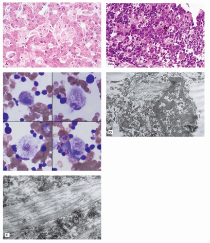
FIGURE 5-1 • Gaucher disease. A: The liver of a patient with Gaucher disease has prominent Kupffer cells due to pale eosinophilic expansion of the cytoplasm by lysosomal glucocerebroside storage material (H&E). B: Enlarged phagocytes in the spleen have a “wrinkled tissue paper” appearance of their cytoplasm because of the glucocerebroside storage (H&E). C: Wright-stained bone marrow aspirate from a patient with Gaucher disease showing “Gaucher cells” with a “wrinkled tissue paper” appearance of cytoplasm due to glucocerebroside storage (Wright). D: Ultrastructural appearance of Gaucher cell from the spleen, obtained at autopsy, showing cytoplasmic storage in upper middle and lower middle of the image, to the left of the nucleus (Uranyl acetate, lead citrate). E: Ultrastructurally, glucocerebroside storage material in Gaucher disease comprises rod-shaped or tubular lipid bilayer stacks with a diameter of up to 4 µm (Uranyl acetate, lead citrate).

PAS-positive glycolipid and cholesterol accumulate in lysosomes of endothelial cells, reticuloendothelial cells, and macrophages. By EM, osmiophilic lamellated leaflets
and tubules are seen in endothelial, perithelial, and smooth muscle cells (41,42). Glomerular podocytes, endothelial, mesangial, interstitial, and tubular epithelial cells all contain storage material. Podocyte storage causes cellular injury, followed by glomerular capillary wall thickening, progressive mesangial matrix expansion, glomerulosclerosis, and, eventually, end-stage renal disease (Figure 5-2A to C). Some patients develop hypertrophic obstructive cardiomyopathy. Storage material in arteries can lead to luminal obstruction and ischemic lesions that may, for example, be found in the brain. There is typically no clinically significant liver involvement, but morphologic studies may show Kupffer cells that have a tan appearance in H&E sections and storage that is birefringent and crystalline in frozen sections stained with the Schultz method.

Diagnosis is based on identifying decreased alpha-galactosidase A in leukocytes or fibroblasts or by DNA analysis for the mutation. Enzyme activity is not a reliable diagnostic tool for female carriers. Their diagnosis requires mutational analysis. Enzyme replacement therapy may have benefit in Fabry patients (13,38).


Gangliosidoses

These autosomal recessive disorders all show lysosomal accumulation of glycosphingolipids (gangliosides).


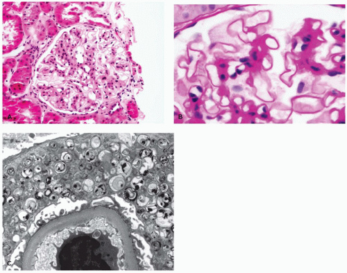
FIGURE 5-2 • Fabry disease. A: By LM, glomeruli in Fabry disease show mesangial expansion with prominence of pale-staining visceral epithelial cells (podocytes) (H&E). B: At higher magnification, podocyte cytoplasm is markedly expanded by PAS-positive storage material (PAS). C: Ultrastructurally, visceral epithelial cell cytoplasm is expanded by osmiophilic, lamellated leaflets and tubules, representing glycolipid and cholesterol storage (Uranyl acetate, lead citrate). (Images courtesy of Helen Liapis, M.D., Nephropath, Little Rock, AR.)


GM1 Gangliosidosis

GM1 gangliosidosis and mucopolysaccharidosis IVB both are the result of mutation in the GLB1 gene encoding beta-galactosidase (43,44). The former is associated with CNS involvement, while the latter is characterized by skeletal involvement, cardiac valvular disease, and corneal clouding but normal intellect. CNS disease is the manifestation of ganglioside accumulation, while systemic manifestations result from galactosyl oligosaccharides and keratan sulfate storage. Differences in the mutation may spare the ability of the enzyme to break down gangliosides in MPS IVB. The phenotype of patients with GM1 gangliosidosis resembles that of MPS by showing coarse facies, dysostosis multiplex, and hepatosplenomegaly, but these patients also develop rapid neurologic deterioration and seizures. Infants may have hydrops fetalis, and most patients die by the age of 2 years. A late-onset form of infantile/juvenile GM1 gangliosidosis that presents at 1 year of age is clinically similar to the early-onset form but with milder dysostosis and death by 5 years of age.

In the nervous system, sudanophilic gangliosides accumulate in the neurons causing cellular ballooning and resulting in neuronal loss, gliosis, and atrophy. By EM, the storage material includes membranous cytoplasmic bodies. Peripheral nerves are also affected. In systemic organs, PAS-positive GAG accumulation causes vacuolization of Kupffer cells, hepatocytes, glomerular visceral epithelial cells and
endothelial cells, placental syncytiotrophoblasts, marrow histiocytes, lymphocytes, lymph nodes, thymus, lung, intestine, pancreas, pituitary, thyroid, salivary gland, skin (including sweat glands), and conjunctiva (45,46). Visceral storage material is fibrillogranular (45,46). Definitive diagnosis rests on demonstrating beta-galactosidase deficiency in leukocytes, fibroblasts, or amniocytes or on DNA analysis.


GM2 Gangliosidosis

These gangliosidoses are due to autosomal recessive defects in lysosomal hexosaminidase with resultant accumulation of GM2 gangliosides mainly in neurons. Hexosaminidase A is a heterodimer composed of an alpha and a beta subunit. Hexosaminidase B is a homodimer composed of two beta subunits. Only hexosaminidase A can hydrolyze GM2 gangliosides.

GM2 gangliosidosis type 1 (Tay-Sachs disease, B variant): This form of GM2 gangliosidosis is due to hexosaminidase A deficiency caused by mutations in the gene encoding the alpha subunit (43,47). Hexosaminidase B is expressed. GM2-containing gangliosides accumulate particularly in the CNS. The incidence is increased in Ashkenazi Jewish populations. Psychomotor deterioration, seizures, blindness, and death by 3 to 5 years of age characterize most patients, although milder juvenile and adult forms are recognized (48). The brain is atrophic with neuronal loss and secondary gliosis; cholesterol, phospholipid, and GM2 ganglioside accumulate as sudanophilic storage in essentially all neurons. The stored material is PAS positive in frozen but not in paraffin sections. By EM, stored material in the CNS is concentrically lamellated, membranous, and granular (45,49). More pleomorphic inclusions are present in the glia. The liver appears normal by LM, but, by EM, there is granular and zebra body storage. Diagnosis is based on hexosaminidase A (decreased) and B (normal) assay in serum, leukocytes, or fibroblasts. Alternatively, DNA analysis by whole genome sequencing or targeted testing for common mutations can confirm the diagnosis.


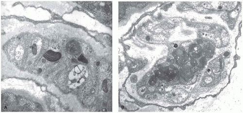
FIGURE 5-3 • Gangliosidosis. A, B: Membranous cytoplasmic bodies in GM2 (AB variant) gangliosidosis are heterogeneous and can show concentric or parallel structure, here in peripheral nerve axons. Though the morphology of the stored gangliosides is often not helpful in distinguishing the gangliosidoses, location of the storage material can be helpful (Uranyl acetate, lead citrate). (Image B from Vogler C, Rosenberg HS, Williams JC, et al. Electron microscopy in the diagnosis of lysosomal storage diseases. Am J Med Genet Suppl 1987;3:243-255, with permission.)

GM2 gangliosidosis type 2 (Sandhoff disease, O variant): Patients have a mutation in the beta subunit of hexosaminidase shared by A and B, resulting in deficiency of both enzymes (hence “O” variant) (43). Clinically affected patients are indistinguishable from Tay-Sachs disease patients. The cerebral cortex is atrophic and yellowed by accumulated asialoganglioside. PAS-positive sphingolipids and glycoprotein accumulate in hepatocytes, Kupffer cells, lymphocytes, pancreatic acinar cells and histiocytes of the spleen, lymph nodes, as well as bone marrow. By EM, storage material similar to that of Tay-Sachs disease is found with prominent membranous cytoplasmic bodies in brain and heterogeneous material in viscera (45). Diagnosis can be determined by enzyme assay or DNA analysis.

GM2 activator protein deficiency (AB variant) results from mutations in the GM2A gene that encodes the GM2 activator protein, which acts as a substrate-specific cofactor for hexosaminidase A (43,50). The designation as “AB variant” stems from the normal expression levels of hexosaminidase A and B, which are not functional because of the defective activator protein. Clinically and pathologically, the AB variant resembles infantile Tay-Sachs and Sandhoff diseases (50,51). Visceral organs are not involved. Zebra and membranous cytoplasmic bodies accumulate (Figure 5-3A,B), and heterogeneous storage affects glial cells. Diagnosis is based on reduced activator protein level in fibroblasts and DNA analysis.


Niemann-Pick Disease (Sphingolipidoses, Sphingomyelin Lipidosis, Sphingomyelin-Cholesterol Lipidosis, NPD)

Two basic variants of Niemann-Pick disease that are discussed further below are those with acid sphingomyelinase deficiency and those with mutations in NPC1 or NPC2 (52). All of these are autosomal recessive. NPD A and B are the result of sphingomyelinase deficiency and are distinguished by phenotype severity and CNS involvement. Niemann-Pick disease type C with NPC1 or NPC2 mutation is not the result of a specific enzyme deficiency but is caused by more general impairment of intracellular lipid trafficking and membrane lipid composition (53).

Niemann-Pick disease (NPD) type A and B: Both of these variants are caused by mutations in the SMPD1 gene that encodes sphingomyelin phosphodiesterase 1. Type A is the more common (85% of cases) and most severe, infantile, neuronopathic form of NPD (54,55). Hydrops fetalis, failure to thrive, hepatosplenomegaly, hypotonia, and progressive neurologic deterioration end with death by 3 to 4 years of age. The disease incidence is higher in the Ashkenazi Jewish populations, among which the carrier frequency is 1:80. Type B is phenotypically variable, more chronic, and non-neuronopathic. This disease variant presents in older infants or later with hepatosplenomegaly. Progressive pulmonary disease may become a major complication.

The pathologic hallmark of NPD is the Niemann-Pick (NP) cell (Figure 5-4A), though NP cells may be infrequent in very young children. These foamy lipid-laden histiocytes have pale yellow or tan cytoplasmic pigmentation on the H&E stain, the result of lipofuscin, sphingomyelin, ganglioside, and cholesterol storage. The vacuoles are birefringent with polarized light and stain with Sudan black B, oil red O (ORO), and Schultz reaction but stain poorly with PAS and for acid phosphatase (56). The blue green cytoplasm of histiocytes with storage stained with Wright-Giemsa stain led to the term sea-blue histiocytes (57).

Kupffer cells (Figure 5-4B) (and, in some cases, hepatocytes) have progressive increase in foamy cytoplasm. Portal fibrosis and cholestasis are observed, but cirrhosis is rare. Infants with NPD A may have cholestasis, bile duct paucity, pseudoglandular formation, and giant cell transformation with a neonatal hepatitis pattern. The spleen may be as much as ten times normal size with extensive infiltrate and replacement of red pulp by NP cells, some of which show erythrophagocytosis. The brain is atrophic with neuron loss, gliosis, and demyelination. Vacuolated neurons have sudanophilic, ORO-positive, and Luxol fast blue-positive storage, and foam cells and lipid-laden glia are in brain parenchyma and Virchow-Robin space (58). Ultrastructural studies can demonstrate storage of lipid with membranous lamellar or concentrically laminated myelin-like features and lipofuscin storage in many tissues including the liver, spleen, lung, bone marrow, kidney, and lymph node (Figure 5-4C to F) (56).

Diagnosis rests with identifying sphingomyelinase deficiency in leukocytes or fibroblasts. In families with a known molecular lesion, heterozygote status can be determined by DNA analysis.

Niemann-Pick disease type C: NPD C is the result of a defect of cholesterol esterification and intracellular trafficking that leads to lysosomal accumulation of sphingomyelin and unesterified cholesterol as well as secondary reduction in sphingomyelinase activity. NPD C is due to mutations in the NPC-1 gene or less commonly the NPC-2 gene. A type D form has been described (“NPD D”) in cases from Nova Scotia with neurologic disease developing in childhood (59,60). On a genetic basis, NPD D is a variant of NPD C occurring in people of that particular heritage. The protein product of NPC-1 is thought to facilitate the egress of cholesterol and other lipids from the late endosomes and lysosomes to other cellular compartments (53). Protean manifestations can begin any time from intrauterine life to adulthood (61,62). Patients may present with fetal ascites or with transient neonatal jaundice and hepatitis (63). Hepatosplenomegaly can occur in some patients but usually regresses over time and, in general, is less severe than that seen with NPD A or B. Neurologic disease is progressive with spasticity and seizures.

Neurovisceral storage is prominent with vacuolated cells in viscera and storage in neurons and glia. Vacuolated cells stain with Luxol fast blue, PAS, and Sudan black B and are positive for cholesterol with the Schultz reaction and for acid phosphatase. EM identifies membrane-bound whorled and dense osmiophilic lysosomal storage in skin and conjunctival cells, endothelial and perithelial cells, keratinocytes, retinal ganglion cells, retinal pigment epithelium, Schwann cells, smooth muscle cells, and fibroblasts (64). In the central nervous system, accumulation of storage material can be associated with the development of Tau-positive neurofibrillary tangles, the formation of meganeurites, and the presence of axonal spheroids.

NPD C may cause a neonatal hepatitis-like histology with giant cell transformation, fibrosis, or cirrhosis (63,65). The pathogenesis of this injury is unknown. Storage in liver is inconspicuous and easily overlooked, particularly in the setting of hepatitis. With time, whorled and irregular lamellar inclusions, clefts, and lipid storage accumulate in macrophages and Kupffer cells and to a lesser extent in hepatocytes (66). A screening test involves staining cultivated cells with filipin to detect free cholesterol. Diagnosis is based on measurement of cholesterol esterification in fibroblasts during LDL uptake and molecular analysis of the NPC-1 or NPC-2 genes.


Metachromatic Leukodystrophy (Sulfatide Lipidosis, MLD)

This is an autosomal recessive disease caused by mutations in the ARSA gene resulting in deficiency of arylsulfatase A, an enzyme that hydrolyzes galactocerebroside sulfate to galactocerebroside (67,68). The enzyme deficiency leads to accumulation of sulfated glycolipids primarily in the CNS but also in extraneural sites. The sulfated glycolipids are responsible for the phenomenon of metachromasia observed with certain

aniline dyes that gave rise to the historical naming of this entity. An atypical form is caused by mutations in the cofactor saposin B (69). Different mutations have been described, and patients have a variable course with progressive neurologic disease resulting in several clinical forms with infantile, juvenile, and adult types. The central and peripheral nervous systems show demyelination, and the cerebellum is atrophic with Purkinje and granular cell loss. Spherical aggregates of metachromatic material that are 15 to 20 µm in diameter occur in oligodendrocytes, in macrophages within Virchow-Robin spaces, and in Schwann cells. This material comprises sulfatide, cholesterol, and phosphatides. In frozen sections, it stains positive with PAS, Alcian blue, and colloidal iron, and it is brown metachromatic (with 1% cresyl violet at low pH) and stains purple with toluidine blue. On electron microscopic studies, the storage material in oligodendrocytes, astrocytes, and Schwann cells is composed of closely packed, lamellar, amorphous, or prismatic material with alternating leaflets and tubules giving it a “herringbone” or “tuffstone” pattern (Figure 5-5A,B).


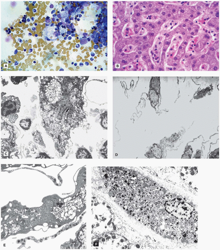
FIGURE 5-4 • Niemann-Pick disease. A: Several vacuolated “Niemann-Pick” cells, the pathologic hallmark of types A and B Niemann-Pick disease, are present and show “sea blue” coloration by Wright-Giemsa stain in this smear of a bone marrow aspirate. Niemann-Pick cells are capable of erythrophagocytosis and emperipolesis. B: Enlarged, foamy Kupffer cells in Niemann-Pick disease, as shown here, may be absent in very young children but become more prominent with time (H&E). C-F: Ultrastructurally, storage material in Niemann-Pick disease is a heterogeneous mix of membranous lamellar material, concentrically lamellated myelin-like material, and lipofuscin (C-F: Uranyl acetate, lead citrate).

The gallbladder may be small and fibrotic with multiple mucosal papillomas and radiolucent choleliths; lamina propria macrophages, gallbladder epithelial cells, and intrahepatic bile ducts have storage. However, patients only rarely present with cholecystitis or pancreatitis. Liver macrophages, Kupffer cells, hepatocytes, and renal tubular epithelial cells also contain metachromatic storage.

Diagnosis is based on measuring arylsulfatase A activity. However, a low level does not prove MLD nor does a normal level exclude the diagnosis. A deficiency of the sphingolipid activator protein saposin B can result in a normal or heterozygous range arylsulfatase A level in an affected patient. Pseudoarylsulfatase A deficiency occurs when an abnormal allele that encodes only 5% to 15% of residual activity leads to low arylsulfatase A activity in a person who does not have MLD. Excessive urine sulfatides can confirm the diagnosis of MLD; a sulfatide-loading test allows distinction between patients homozygous for the pseudodeficiency allele and MLD patients.


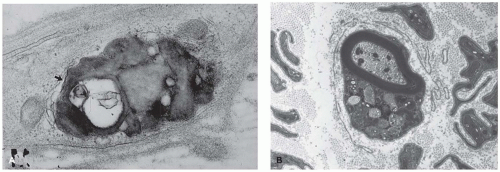
FIGURE 5-5 • Metachromatic leukodystrophy. A: This unmyelinated nerve from a conjunctival biopsy contains an inclusion of variable electron density. In some foci (arrow), closely approximated osmiophilic lamellae contribute to a subtle herringbone pattern. B: Myelinated nerve with pleomorphic lysosomes of variable density, “tuffstone” inclusions from a sural nerve of a patient with metachromatic leukodystrophy. (A, B: Uranyl acetate, lead citrate, A: Used from Vogler C, Rosenberg HS, Williams JC, et al. Electron microscopy in the diagnosis of lysosomal storage diseases. Am J Med Genet Suppl 1987;3:243-255, with permission.)


Krabbe Disease (Galactosylceramide Lipidosis, Globoid Cell Leukodystrophy)

Autosomal recessive deficiency of galactocerebroside beta-galactosidase activity results in rapidly progressive neurologic deterioration in affected infants (67,70). An atypical variant is the result of a mutation in the cofactor saposin A. The pathology is limited largely to the nervous system. Grossly, the brain shows atrophy and discoloration of the white matter. Histologically, there is myelin loss, neuronal degeneration, and gliosis. The distinctive globoid cells are derived from monocyte-macrophage bone marrow stem cells and represent cells distended by PAS-positive and acid phosphatase-positive undigested psychosine (galactosylsphingosine) and galactosylceramide. The globoid cells accumulate in white matter and perivascular spaces. Psychosine accumulation causes oligodendroglia destruction and is an example of a lysosomal storage disease in which accumulated metabolites have distinct toxic effects (12). On electron microscopic studies, the storage material is composed of electron-dense, straight or curved, hollow tubular profiles in longitudinal section with crystalloid profiles in cross section (Figure 5-6A,B). Peripheral nerves have endoneural fibrosis, demyelination, and infiltration of PAS-positive macrophages, similar to the globoid cells seen in the CNS. Storage material also occurs in sweat gland epithelium. Diagnosis is based on DNA analysis or on identifying galactocerebroside beta-galactosidase deficiency in leukocytes, fibroblasts, amniotic, or chorionic cells. Specific mutations are associated with the infantile-onset form and are used in predicting outcome and therapeutic decisions about stem cell transplantation.


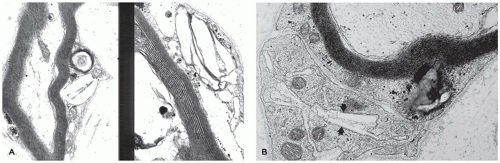
FIGURE 5-6 • Krabbe disease. A, B: Electron-lucent, angulated, and needle-shaped inclusions in conjunctival myelinated nerve Schwann cells, characteristic of Krabbe disease. (A, B: Uranyl acetate, lead citrate; B: Used from Vogler C, Rosenberg HS, Williams JC, et al. Electron microscopy in the diagnosis of lysosomal storage diseases. Am J Med Genet Suppl 1987;3:243-255, with permission.)


Farber Disease (Disseminated or Farber Lipogranulomatosis)

This rare autosomal recessive deficiency of acid ceramidase leads to accumulation of ceramide, which is formed from turnover of sphingolipids in lymph nodes, liver, kidney, and lung. Mucopolysaccharides and gangliosides also accumulate (71). Symptoms begin in infancy and include failure to thrive, vomiting, painful and progressively deformed joints, subcutaneous nodules that are especially prominent near joints, and laryngeal involvement with hoarseness and respiratory insufficiency. Clinically, histiocytosis is often in the differential diagnosis. Farber disease may present in utero with hydrops fetalis.

Lymph nodes, lung, larynx, spleen, liver, heart, subcutaneous, and periarticular tissues show nodular lipogranulomas containing PAS-positive storage in foam cells and multinucleated giant cells. Storage is also present in endothelial cells, pericytes, Schwann cells, hepatocytes, renal tubular epithelium, and glomerular visceral epithelial cells. Neurons are distended with PAS-positive ceramides and gangliosides. By electron microscopy, storage material consists of membrane-bound, comma-shaped curvilinear tubular profiles, termed banana bodies or Farber bodies, along with concentric lamellar, zebra body, and fibrillogranular material (72). Diagnosis is confirmed by demonstration of decreased acid ceramidase activity in leukocytes, fibroblasts, or amniocytes.


Disorders of Glycoprotein Degradation (Oligosaccharidoses/Glycoproteinoses)

These autosomal recessive disorders are due to deficiency of a lysosomal enzyme that degrades glycoprotein oligosaccharide side chains of glycoproteins (73). Defective function in lysosomal exopeptidases involved in the stepwise breakdown of glycoproteins can result in blockage of the entire pathway. Tissues accumulate glycoproteins and oligosaccharides, resulting in a phenotype that resembles that of mucopolysaccharidoses.


Alpha-Mannosidosis

Affected patients have deficient alpha-mannosidase and increased plasma levels as well as excretion of small mannose-rich oligosaccharides (73). The phenotype is variable, but in principle, this disease presents with immune deficiency manifested by recurrent infections, skeletal abnormalities, hearing impairment, speech problems, gradual impairment of cognitive function, and neuromuscular abnormalities with weakness and psychosis (74). Hepatocytes have granular or foamy cytoplasm, and Kupffer cells and hepatocytes contain reticulogranular, amorphous, or membranous storage by EM. In the brain, neurons have marked and widespread ballooning with membrane-bound vacuoles containing reticulogranular material. Diagnosis can be based on ultra-structural morphology of the skin, conjunctiva, or peripheral blood lymphocytes; demonstration of oligosacchariuria; and measurement of tissue alpha-mannosidase activity and gene testing (74).


Beta-Mannosidosis

The clinical phenotype associated with deficiency of beta-mannosidase is variable (73). Cognitive impairment is the most common presentation, but patients may also have deafness, speech problems, susceptibility to infections, hypotonia, epilepsy, peripheral neuropathy, facial dysmorphism, skeletal abnormalities, and angiokeratoma corporis diffusum (75,76). Cytoplasmic vacuoles are described in skin and bone marrow in isolated patients. The diagnosis rests on measurement of beta-mannosidase in leukocytes or fibroblasts and genetic testing.


Fucosidosis

This lysosomal storage disease, due to deficient alpha-l-fucosidase, causes accumulation of fucoside moiety-containing glycolipids, glycoproteins, and oligosaccharides (73,77). Most patients are of Italian or Spanish descent or from the southwestern United States. The presentation includes progressive
mental retardation, seizures, recurrent infections, coarse facies, dysostosis multiplex and growth retardation, angiokeratoma corporis diffusum, and visceromegaly (73,77). The clinical course is variable. Most patients reach the second decade of life. The CNS, conjunctiva, muscle, skin and sweat gland epithelium, and peripheral blood lymphocytes all show granular lysosomal storage by EM (Figure 5-7). Diagnosis is based on demonstrating deficient alpha-l-fucosidase in leukocytes and fibroblasts. Some clinically normal individuals have low alpha-l-fucosidase levels in plasma.


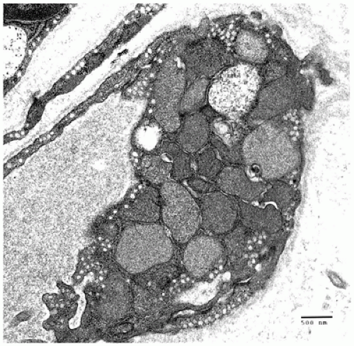
FIGURE 5-7 • Fucosidosis. Granular storage material distends the cytoplasm of this endomysial endothelial cell from a muscle biopsy of a 2-year-old girl with fucosidosis (Uranyl acetate, lead citrate).


Sialidosis

Sialidosis Types I and II are distinguished based on phenotype severity, but both result from mutations in the NEU1 gene encoding lysosomal sialidase (78,79). Milder forms are associated with visual defects, cherry-red macular spots, ataxia, hyperreflexia, and seizures. More severe cases have dysostosis multiplex, cognitive impairment, hepatosplenomegaly, and Hurler-like phenotype. Definitive diagnosis is based on measurement of neuraminidase activity in cultivated fibroblasts or leukocytes.


Aspartylglycosaminuria

This autosomal recessive glycoprotein degradation defect occurs predominantly in Finland and is due to lack of aspartylglucosaminidase (80,81). Decreased cognitive function and facial and skeletal manifestations are most notable. The progression of disease is often slow with many patients living into their 30s, 40s, and 50s. Enlarged lysosomes contain aspartylglucosamine that appears as fibrillogranular storage in skin, conjunctiva, rectal mucosa, peripheral blood lymphocytes, neurons, hepatocytes, and Kupffer cells. In the CNS, delayed myelination, white matter gliosis, and gray matter atrophy are seen. Storage material may be evident in the liver, kidney, skin, and placenta as early as by the 20th week of gestation. Diagnosis is based on enzyme assay or DNA molecular analysis.


Glycogen Storage Disease: Pompe Disease (Glycogen Storage Disease Type II, GSD-II) and Adult Acid Maltase Deficiency

This glycogen storage disease is the result of mutations in the GAA gene encoding the lysosomal enzyme alpha-1,4-glucosidase (acid maltase) (82,83,84,85). A range of phenotypes occurs in GSD II patients, reflecting the variety of residual enzyme activity and tissue-specific isoenzymes. All are autosomal recessive. Patients with severe disease have hypotonia and cardiomegaly. These infants with classical Pompe disease die of cardiac or respiratory failure in the first few years of life. Adult-onset skeletal muscle disease is seen in late-onset cases without solid organ involvement and often results in muscle weakness in a distinct truncal distribution pattern that includes respiratory muscle weakness (86).

PAS-positive, diastase-digestible glycogen lysosomal storage is generalized but most severe in the skeletal muscle, heart, liver, and brain. Increased acid phosphatase activity indicates lysosomal distention and secondary elevation of other lysosomal enzymes (Figure 5-8A). A vacuolar myopathy with disruption of cytoplasmic structure affects skeletal, cardiac, and smooth muscle (Figure 5-8B,C). Cardiac involvement includes cardiac hypertrophy (Figure 5-8D), endocardial fibroelastosis, and arrhythmia from changes in the conduction system.

Glycogen is increased in Schwann cells, anterior horn cells, brainstem motor nuclei and spinal ganglia, myenteric plexus, astrocytes, oligodendroglia, endothelial cells, and pericytes with relative sparing of cortical neurons. Hepatocytes are only slightly enlarged with delicate glycogen-containing vacuoles. Liver lacks the mosaic pattern and nuclear glycogenation seen in other GSD (Figure 5-8E). In the kidney, glycogen accumulates in the epithelium of loops of Henle and collecting tubules, and the adrenal zona fasciculata has prominent storage.

Skin, conjunctiva, liver, muscle, lymphocytes, and placenta can show diagnostic lysosomal glycogen accumulation by EM (Figure 5-8F) (83). Glycogen in muscle is both lysosomal and cytoplasmic. Diagnosis is confirmed by demonstrating absent enzyme in dried blood spots, leukocytes, muscle, liver, or fibroblasts. In addition, DNA analysis can be utilized, which is also useful in defining patients with pseudodeficiency. Enzyme replacement therapy is available (84).


Mucopolysaccharidoses

The mucopolysaccharidoses (MPSs) are systemic diseases due to deficiency of one of the enzymes required for catabolism of glycosaminoglycans (GAG) including dermatan,
heparan, chondroitin, and keratan sulfate, with resultant storage of undegraded GAG in lysosomes in a variety of cell types (12). The clinical presentation is variable but may include facial dysmorphism (Figure 5-9A,B), bone/joint dysplasia (Figure 5-9C), hepatosplenomegaly, neurologic impairment, developmental regression, and short life span to mild clinical phenotype with normal life expectancy (Table 5-6) (87,88). All MPS are autosomal recessive except X-linked Hunter syndrome. Hurler (type I) and Hunter (type II) syndromes are the most common types. Scheie and Hurler-Scheie syndromes are subtypes of MPS I with a milder disease. Some infants, particularly with MPS VII, present with hydrops (Table 5-2).


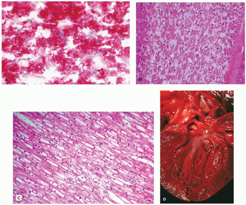
FIGURE 5-8 • Pompe disease (type II glycogen storage disease). A: Glycogen storage in Pompe disease is lysosomal (in contrast to glycogen storage in the other types of glycogen storage disease). The distended lysosomes also contain abundant acid phosphatase activity, which can be demonstrated histochemically, here in skeletal muscle by acid phosphatase staining (acid phosphatase stain). B: Vacuolar myopathy, though not specific for Pompe disease, is nonetheless characteristic and often striking in this LSD. The pale vacuoles in skeletal muscle fibers (representing glycogen storage) seen with H&E stain can be highlighted by PAS stain (not shown) (H&E). C: Histologically, cardiac myocytes are enlarged due to sarcoplasmic expansion by pale, often vacuolar material (H&E). D: Cardiac myocyte enlargement can lead to a hypertrophic gross appearance of myocardium, as seen in this image of the left ventricle from an infant who died with Pompe disease.

The stored GAG can be highlighted with colloidal iron and Alcian blue stains and is digested by hyaluronidase. Adding 10% cetyltrimethylammonium bromide to formalin may help preserve tissue GAG. By EM, visceral lysosomal storage is fine fibrillogranular material (Figure 5-9D).

In MPS, many organs and tissue types have lysosomal storage. The liver is typically enlarged and firm. Vacuolization is more prominent in Kupffer cells than hepatocytes. Fibrosis of the space of Disse occurs late in the disease; rarely, more severe fibrosis can develop in older patients. Coarse facial features, short stature, and bone and joint abnormalities (dysostosis multiplex) are the result of skeletal involvement. The corneas may show cloudiness. Airway obstruction and

nerve or cord compression can be manifestations of soft tissue involvement (Figure 5-9C, G, and H) (Table 5-6) (89). Vessel walls and heart valves are often affected with storage with resultant sclerosis (Figure 5-9E,F), and endocardial fibroelastosis may occur. Neurons store both GAG and gangliosides leading to formation of membranous cytoplasmic bodies, zebra bodies, and fibrillogranular storage material (Figure 5-9I). Neuronal loss and gliosis are seen in some patients, and meningeal storage may contribute to hydrocephalus.
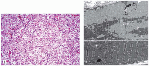


FIGURE 5-8(Continued) E: The histologic appearance of hepatocytes in Pompe disease is usually less striking than that of skeletal muscle. Hepatocytes are slightly enlarged with somewhat rarefied, vacuolar cytoplasm. Note the absence of glycogenated nuclei, which are typically not seen in the liver in Pompe disease but are observed in several other types of glycogen storage disease (H&E). F: Even though the primary defect in Pompe disease is lysosomal (in contrast to other glycogen storage diseases), glycogen accumulation is seen by ultrastructural analysis of skeletal muscle in the cytoplasm as well as within membrane-bound structures representing lysosomes.


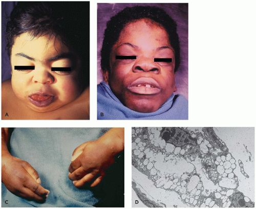
FIGURE 5-9 • Mucopolysaccharidosis. A, B: MPS patients have a characteristic facial appearance with coarse facial appearance, thick doughy skin, coarse hair, flattened midface, wide nasal bridge, and macroglossia, here seen in two children who died with MPS. C: The hands in MPS patients have joint stiffness and are held in a flexed position, a function of periarticular altered connective tissue and altered bone formation. D: In mucopolysaccharidosis, stored GAGs (previously called mucopolysaccharides) have the ultrastructural appearance of fine fibrillogranular material and clear membrane-bound vacuoles. Distinguishing different types of mucopolysaccharidosis based on ultrastructural morphologic characteristics is not possible.
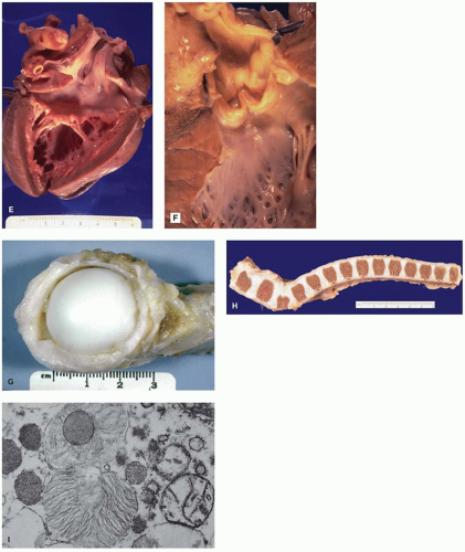


FIGURE 5-9(Continued) E, F: The heart in patients with MPS typically has thickened sclerotic valves, due to GAG storage in heart valve stromal cells and altered extracellular connective tissue in the valve. Endocardial thickening is also frequent. G, H: The femoral head of a patient from MPS shows articular synechiae and thick, poorly pliable periarticular connective tissue. These joint changes cause marked joint stiffness and make normal movement impossible. The vertebral column from an MPS patient shows characteristic anterior inferior breaking of the lower thoracic and upper lumbar areas caused by hypoplasia of the anterior superior aspect. This change results in the dorsal kyphosis or gibbus deformation often seen in MPS patients, and it is part of the widespread dysostosis multiplex. I: Though storage material in neurons can resemble that seen in other organs, it can also take the form of “zebra bodies,” as shown in this case of Hurler syndrome (D, I: Uranyl acetate, lead citrate).

Diagnosis is suggested by increased urine GAG, the presence of vacuoles, and metachromatic Alder-Reilly granules in peripheral blood leukocytes. EM of the skin, conjunctiva, buffy coat, or liver can show characteristic fibrillogranular
lysosomal storage. LM of thick sections of tissue prepared for EM is useful for identifying the multiple clear cytoplasmic vacuoles indicative of lysosomal storage. Enzyme assay of serum, leukocytes, or fibroblast culture provides definitive diagnosis, and carrier testing using DNA analysis is practical. Enzyme replacement therapy is now available for some forms (87,88).


TABLE 5-6 MUCOPOLYSACCHARIDOSES


MPS


Eponym


Enzyme Deficiency (Chromosome locus)


Stored Material


Clinical and Pathologic Findings


I


Hurler, Scheie, Hurler-Scheie


Alpha-l-iduronidase (4p16.3)


Dermatan sulfate


Heparan sulfate


Corneal clouding, dysostosis multiplex, hepatosplenomegaly, cardiac valve sclerosis, cognitive impairment, premature death (Scheie has milder phenotype without cognitive impairment), rarely hydrops fetalis


II


Hunter


Iduronate sulfatase (Xq28)


Dermatan sulfate


Heparan sulfate


Dysostosis multiplex, hepatosplenomegaly, cardiac valve sclerosis, cognitive impairment, X linked


IIIA


Sanfilippo A


Heparan N-sulfatase (sulfamidase) (17q25.3)


Heparan sulfate


cognitive impairment, mild somatic disease


IIIB


Sanfilippo B N-acetyl-alpha-d-glucosaminidase (17q21.2)


Heparan sulfate


Similar to IIIA


IIIC


Sanfilippo C


Acetyl-CoA-alpha-glucosaminide N-acetyltransferase (8p11.21)


Heparan sulfate


Similar to IIIA


IIID


Sanfilippo D


N-acetylglucosamine-6-sulfatase (12q14.3)


Heparan sulfate


Similar to IIIA


IVA


Morquio A


Galactosamine-6-sulfatase (16q24.3)


Keratan sulfate


Chondroitin-6-sulfate


Skeletal abnormalities, corneal clouding, odontoid hypoplasia, hydrops fetalis


IVB


Morquio B


Beta galactosidase (3p21.33)


Keratan sulfate


Similar to IVA


VI


Maroteaux-Lamy


N-acetylgalactosamine-4-sulfatase (arylsulfatase B) (5q14.1)


Dermatan sulfate


Dysostosis multiplex, corneal clouding, normal intelligence


VII


Sly


Beta glucuronidase (7q11.21)


Dermatan sulfate


Heparan sulfate


Chondroitin-4,6-sulfates


Dysostosis multiplex, hepatosplenomegaly, cognitive impairment, hydrops fetalis


IX


Hyaluronidase (3p21.2-21.3)


Hyaluronan


Periarticular soft tissue masses (nodular histiocyte aggregates), short stature


Lipidoses: Wolman Disease and Cholesterol Ester Storage Disease (CESD)

These are phenotypical variants of autosomal recessive mutations in the LIPA gene encoding lysosomal acid lipase and resulting in the accumulation of cholesterol esters and triglyceride (90,91,92). Severe deficiency leads to the phenotype described as Wolman disease that is characterized by failure to thrive, hepatosplenomegaly, and early death. Milder deficiencies are associated with dyslipidemia, hepatic fibrosis, and early atherosclerosis.

The enlarged liver is a distinctive bright orange-yellow with a greasy consistency. Bile duct proliferation and cholestasis are described, and periportal fibrosis with portal bridging may progress to cirrhosis. In viscera—including liver, spleen, adrenal, lymph nodes, lymphocytes, bone marrow, and intestine—cholesterol esters and triglycerides accumulate as cholesterol crystals in foamy histiocytes (Figure 5-10A,B). This storage can be identified by viewing sections of unfixed frozen tissue with polarized light (Figure 5-10C).
Cholesterol and triglycerides in these cells can also be highlighted histochemically with the Schultz modification of the Lieberman-Burchard reaction. EM shows lipid droplets and membrane-bound angular cholesterol clefts in hepatocytes, Kupffer cells, fibroblasts, and macrophages (Figure 5-10D). The mucosa of the small intestine, particularly duodenum and ileum, is velvety yellow due to lamina propria storage. Adrenal glands are large, hard, and bright yellow, with dystrophic calcification and necrosis of the inner fasciculata and residual fetal cortex. Oligodendroglia, some neurons of the CNS, and Schwann cells of the peripheral nervous system contain lipid. Placental syncytiotrophoblasts may be affected. Demonstration of acid lipase deficiency in tissue, cultivated fibroblasts, or leukocytes confirms the diagnosis.


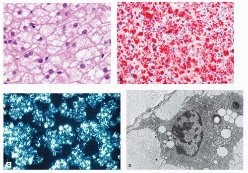
FIGURE 5-10 • Cholesterol ester storage disease. A: In cholesterol ester storage disease, there is widespread vacuolization of hepatocytes (H&E). B: Widespread cytoplasmic lipid can be demonstrated in frozen section analysis of liver tissue in cholesterol storage disease, here stained with oil red O. C: Hepatocellular cholesterol ester crystals are birefringent in frozen sections when viewed with polarized light. D: This conjunctival macrophage does not show needle-shaped clefts but does show many sharply demarcated electron-lucent vacuoles, some of which have peripheral osmiophilia, characteristic of lipid following fixation. (D: Uranyl acetate, lead citrate, Used from Vogler C, Rosenberg HS, Williams JC, et al. Electron microscopy in the diagnosis of lysosomal storage diseases. Am J Med Genet Suppl 1987;3:243-255, with permission.)


Lysosomal Storage Diseases Linked to Nonenzymatic Protein Deficiencies

Some of these diseases are variants of historically and clinically discussed entities mentioned in previous sections and are therefore omitted here (AB variant of GM2 gangliosidosis, or the Niemann-Pick type C variants caused by NPC1 or NPC2 mutations).


Mucolipidoses

I-cell disease (ML II) and pseudo-Hurler polydystrophy (ML III) are autosomal recessive diseases that result from mutations that disrupt the hexameric complex of GlcNAc-1-phosphotransferase, which is composed of 2 alpha, 2 beta, and 2 gamma units (93). This enzyme complex tags newly
synthesized lysosomal hydrolases for their appropriate targeting toward lysosomes by adding mannose-6-phosphate as a signal in the Golgi network (93). Phosphotransferase deficiency results in abnormal lysosomal enzyme transport. As a result, newly synthesized enzymes are secreted out of the cell instead of being transferred to lysosomes. This results in elevated plasma levels of lysosomal enzymes. The GNPTAB gene affected in ML II encodes the alpha- and beta-subunits of the hexameric enzyme complex, and the GNPTG gene affected in ML III encodes the gamma subunit.

Affected patients have features of both MPS and sphingolipidoses, hence the designation mucolipidoses. Clinical and radiologic findings (coarse facial features, psychomotor retardation, failure to thrive, hepatomegaly, dysostosis multiplex) are similar to those seen in MPS I, but earlier onset, a more rapid course, marked gingival hyperplasia, and absence of mucopolysacchariduria help distinguish ML II and III clinically from MPS I (94,95).

The term I-cell disease was coined because cultured fibroblasts from affected patients contain dense inclusions. PAS-positive and Hale colloidal iron-positive vacuoles are prominent in endothelial cells and fibroblasts and occur in lymphocytes, Kupffer cells, glomerular visceral epithelial cells, satellite cells in the muscle, myocardium, and pancreatic acinar cells (96). Storage in stromal fibroblasts of the heart is associated with valve thickening. Granulomas with finely vacuolated histiocytes may occur in lung and portal areas. Hepatocytes are normal or only mildly altered and contain triglyceride droplets.

The CNS may be normal morphologically, except for lamellar bodies in spinal ganglia neurons and anterior horn cells, or may have cerebral cortical atrophy with neuronal loss. Storage may be apparent in affected fetuses and their placentas. I-cell disease can present as nonimmune hydrops (see Table 5-1). By EM, the storage material is electron lucent or fibrillogranular and includes oligosaccharides, mucopolysaccharides, and lipids (96). EM of skin or conjunctiva can be used for diagnostic evaluation (Figure 5-11). Increased serum levels of lysosomal enzymes and decreased levels of N-acetylglucosamine-1-phosphotransferase provide biochemical confirmation.

ML III (pseudo-Hurler polydystrophy) symptoms are similar to ML II but milder with growth restriction, coarse facial features, cardiac valve disease, dysostosis multiplex, and corneal clouding (94,95). The pathology of ML III is not as well documented as that of ML II. Storage is identified in skin fibroblasts, but lymphocytes are normal.

Mucolipidosis type IV (ML IV, sialolipidosis, ganglioside-sialidase deficiency) results from mutations in the gene MCOLN1, which codes for the TRP family ion membrane channel, mucolipin 1. This putative lysosomal ion channel is thought to be involved in Fe2+, Ca2+, and Zn2+ transport and with that critical for still poorly understood aspects of lysosomal function (97). As a result, there is abnormal intracellular membrane trafficking. This disorder is classified as a mucolipidosis because of the storage of both lipids and mucopolysaccharides. Although panethnic, ML IV is more common among Ashkenazi Jews (98). Patients have severe psychomotor retardation, ophthalmologic abnormalities with corneal clouding, retinal degeneration, and optic nerve atrophy, but they do not have dysostosis multiplex seen in MPS. The overall incidence may be underestimated because of milder cases that can have a presentation mimicking cerebral palsy (98).


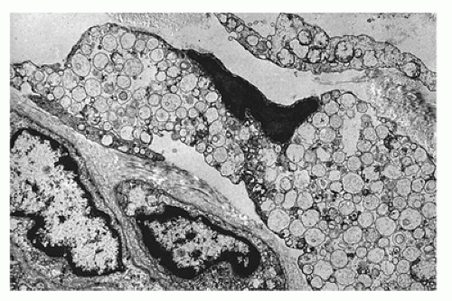
FIGURE 5-11 • I-cell disease. In I-cell disease, fibroblast cytoplasm is expanded by numerous membrane-bound vacuoles containing electron lucent to fibrillogranular material (Uranyl acetate, lead citrate).

Widespread storage affects the brain and viscera including the liver, pancreas, kidney, marrow, conjunctiva, cornea, skin, muscle, peripheral nerve, rectum, and placenta (Figure 5-12A,B). In neurons and glia, ganglioside, phospholipid, and GAG accumulation is variably PAS positive and sudanophilic and is associated with neuronal loss and astrocytosis. By EM, lysosomes contain heterogeneous material with fibrillogranular and concentric membranous bodies (Figure 5-12C).

Hypergastrinemia and achlorhydria are described (99). Chronic atrophic gastritis and enterochromaffin-like cell hyperplasia are seen along with cytoplasmic vacuolization of parietal cells due to lysosomal storage. Confirmatory diagnosis of ML IV should include screening for mutations in MCOLN1.

Only gold members can continue reading. Log In or Register to continue

Stay updated, free articles. Join our Telegram channel

Sep 23, 2016 | Posted by in PATHOLOGY & LABORATORY MEDICINE | Comments Off on Inborn Errors of Metabolism

Full access? Get Clinical Tree

Get Clinical Tree app for offline access