Immunity
Learning Objectives
After studying this chapter, the student is expected to:
1. Describe the normal immune response.
2. List the components of the immune system and the purpose of each.
3. Explain the four methods of acquiring immunity.
4. Discuss tissue transplant rejection and how it is treated.
5. Describe the mechanism and clinical effects of each of the four types of hypersensitivity reactions.
6. Explain the effects of anaphylaxis.
7. Discuss the mechanism of autoimmune disorders.
9. Explain the causes and effects of immunodeficiency.
11. Describe the course, effects, and complications of HIV-AIDS.
Key Terms
antibiotic
antimicrobial
antiviral
autoantibodies
bronchoconstriction
colostrum
complement
cytotoxic
encephalopathy
erythema
fetus
glycoprotein
hypogammaglobulinemia
hypoproteinemia
mast cells
monocytes
mononuclear phagocytic system
mutate
opportunistic
placenta
polymerase chain reaction
prophylactic
pruritic
replication
retrovirus
splenectomy
stem cells
thymus
titer
vesicles
Review of the Immune System
The immune system is responsible for the body’s defenses. The system has a nonspecific response as shown in Chapter 5 and a specific response mechanism discussed in this chapter. In specific defense the immune system is responding to particular substances, cells, toxins, or proteins, which are perceived as foreign to the body and therefore unwanted or potentially dangerous. The specific immune response is intended to recognize and remove undesirable material from cells, tissues, and organs.
Components of the Immune System
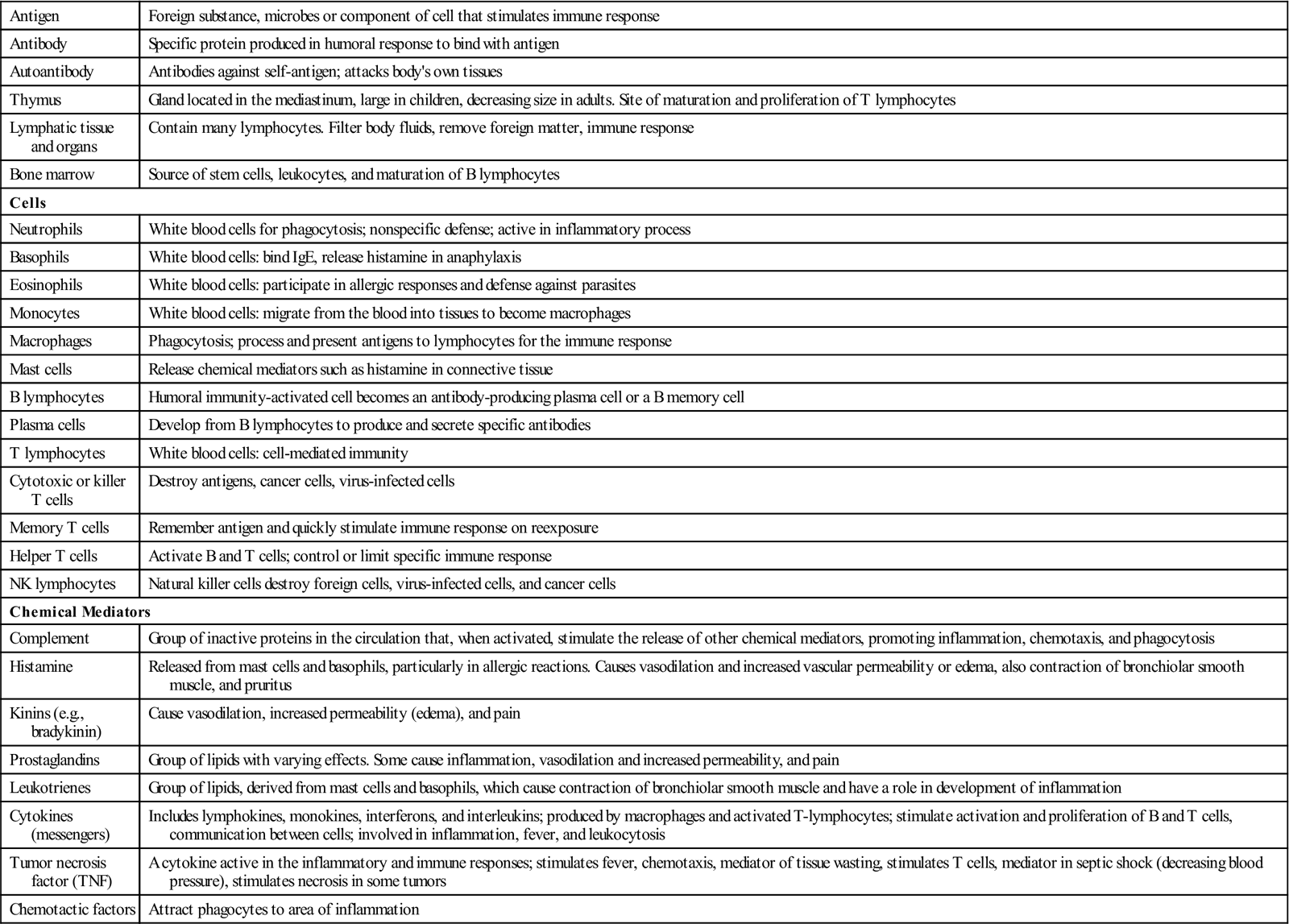
The immune system consists of lymphoid structures, immune cells, tissues concerned with immune cell development, and chemical mediators. The lymphoid structures, including the lymph nodes, the spleen and tonsils, the intestinal lymphoid tissue, and the lymphatic circulation, form the basic structure within which the immune response can function (Fig. 7-1). The immune cells, or lymphocytes, as well as macrophages provide the specific mechanism for the identification and removal of foreign material. All immune cells originate in the bone marrow, and the bone marrow and thymus have roles in the maturation of the cells. The thymus is significant during fetal development in that it programs the immune system to ignore self-antigens. When this process is faulty, the body may attack its own tissues as non–self. The blood and circulatory system provide a major transportation and communication network for the immune system. The major components of the immune system and their functions are summarized in Table 7-1.
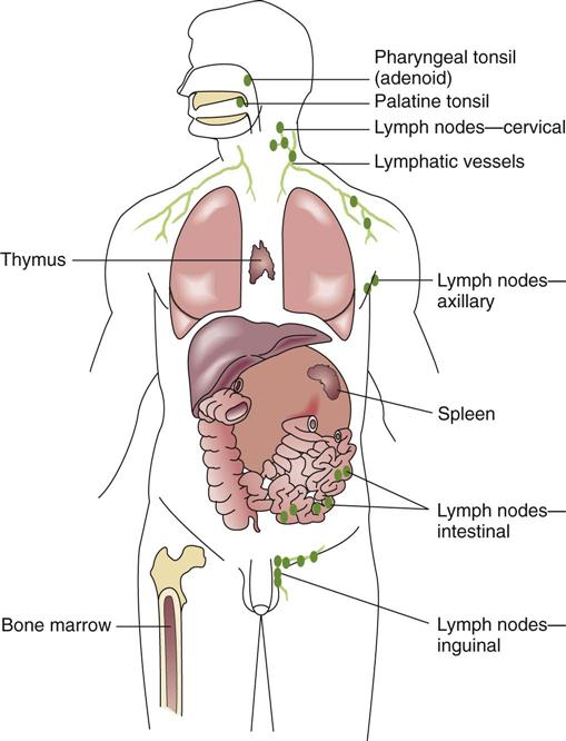
TABLE 7-1
Major Components of the Immune System and Their Functions
| Antigen | Foreign substance, microbes or component of cell that stimulates immune response |
| Antibody | Specific protein produced in humoral response to bind with antigen |
| Autoantibody | Antibodies against self-antigen; attacks body’s own tissues |
| Thymus | Gland located in the mediastinum, large in children, decreasing size in adults. Site of maturation and proliferation of T lymphocytes |
| Lymphatic tissue and organs | Contain many lymphocytes. Filter body fluids, remove foreign matter, immune response |
| Bone marrow | Source of stem cells, leukocytes, and maturation of B lymphocytes |
| Cells | |
| Neutrophils | White blood cells for phagocytosis; nonspecific defense; active in inflammatory process |
| Basophils | White blood cells: bind IgE, release histamine in anaphylaxis |
| Eosinophils | White blood cells: participate in allergic responses and defense against parasites |
| Monocytes | White blood cells: migrate from the blood into tissues to become macrophages |
| Macrophages | Phagocytosis; process and present antigens to lymphocytes for the immune response |
| Mast cells | Release chemical mediators such as histamine in connective tissue |
| B lymphocytes | Humoral immunity-activated cell becomes an antibody-producing plasma cell or a B memory cell |
| Plasma cells | Develop from B lymphocytes to produce and secrete specific antibodies |
| T lymphocytes | White blood cells: cell-mediated immunity |
| Cytotoxic or killer T cells | Destroy antigens, cancer cells, virus-infected cells |
| Memory T cells | Remember antigen and quickly stimulate immune response on reexposure |
| Helper T cells | Activate B and T cells; control or limit specific immune response |
| NK lymphocytes | Natural killer cells destroy foreign cells, virus-infected cells, and cancer cells |
| Chemical Mediators | |
| Complement | Group of inactive proteins in the circulation that, when activated, stimulate the release of other chemical mediators, promoting inflammation, chemotaxis, and phagocytosis |
| Histamine | Released from mast cells and basophils, particularly in allergic reactions. Causes vasodilation and increased vascular permeability or edema, also contraction of bronchiolar smooth muscle, and pruritus |
| Kinins (e.g., bradykinin) | Cause vasodilation, increased permeability (edema), and pain |
| Prostaglandins | Group of lipids with varying effects. Some cause inflammation, vasodilation and increased permeability, and pain |
| Leukotrienes | Group of lipids, derived from mast cells and basophils, which cause contraction of bronchiolar smooth muscle and have a role in development of inflammation |
| Cytokines (messengers) | Includes lymphokines, monokines, interferons, and interleukins; produced by macrophages and activated T-lymphocytes; stimulate activation and proliferation of B and T cells, communication between cells; involved in inflammation, fever, and leukocytosis |
| Tumor necrosis factor (TNF) | A cytokine active in the inflammatory and immune responses; stimulates fever, chemotaxis, mediator of tissue wasting, stimulates T cells, mediator in septic shock (decreasing blood pressure), stimulates necrosis in some tumors |
| Chemotactic factors | Attract phagocytes to area of inflammation |
Elements of the Immune System
Antigens
Antigens (or immunogens) are either foreign substances or human cell surface antigens that are unique (except in identical twins) in each individual. They are usually composed of complex proteins or polysaccharides, or a combination of molecules such as glycoproteins. Antigens activate the immune system to produce specific produce specific antibodies.
The antigens representing self are present on an individual’s plasma membranes. These antigen molecules are coded by a group of genes inherited from the parents, called major histocompatibility complex (MHC) located on chromosome 6. Owing to the large number of possible combinations of genes that may be inherited from the parents, it is unlikely that two individuals would ever have the same antigens, with the exception of identical twins. Major histocompatibility complex has an essential role in the activation and regulation of the immune response as well as intercellular communications. Major histocompatibility complex molecules are useful in detecting changes in cell membranes altered by viruses or cancerous changes and alerting the immune system to their presence. Human MHC is also known as human leukocyte antigen (HLA), because it was first detected on the cell membranes of leukocytes. These antigens are also used to provide the close match for a tissue transplant; the immune system will be activated by the presence of cells with different MHC molecules. The immune system generally tolerates self-antigens on its cells, thus no immune response is initiated against the own cells. Autoimmune diseases are an exception in which the immune system no longer recognizes self from non–self and begins to attack its own cells/structures or organs.
Cells
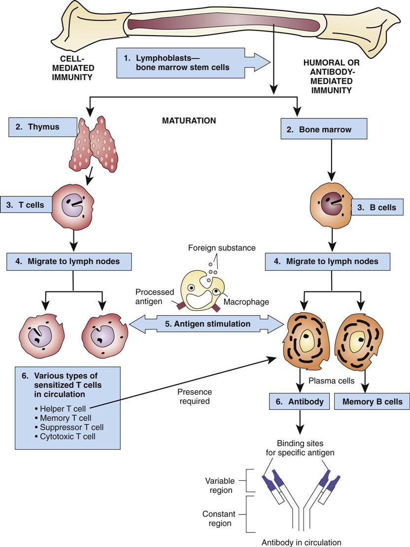
The macrophage is critical in the initiation of the immune response. Macrophages develop from monocytes (see Chapter 10), part of the mononuclear phagocytic system that was formerly known as the reticuloendothelial system. Macrophages occur throughout the body in such tissues as the liver, lungs, and lymph nodes. They are large phagocytic cells that intercept and engulf foreign material and then process and display the antigens from the foreign material on their cell membranes; the lymphocytes respond to this display, thus initiating the immune response (Fig. 7-2). Macrophages also secrete chemicals such as monokines and interleukins (see Table 7-1) that play a role in the activation of additional lymphocytes and in the inflammatory response, which accompanies a secondary immune response.
The primary cell in the immune response is the lymphocyte, one of the leukocytes or white blood cells produced by the bone marrow (see Chapter 11). Mature lymphocytes are termed immunocompetent cells—cells that have the special function of recognizing and reacting with antigens in the body. The two groups of lymphocytes, B lymphocytes and T lymphocytes, determine which type of immunity will be initiated, either cell-mediated immunity or humoral immunity, respectively.
T lymphocytes (T cells) arise from stem cells, which are incompletely differentiated cells held in reserve in the bone marrow and then travel to the thymus for further differentiation and development of cell membrane receptors. Cell-mediated immunity develops when T lymphocytes with protein receptors on the cell surface recognize antigens on the surface of target cells and directly destroy the invading antigens (Fig. 7-2). These specially programmed T cells then reproduce, creating an “army” to battle the invader, and they also activate other T and B lymphocytes. T cells are primarily effective against virus-infected cells, fungal and protozoal infections, cancer cells, and foreign cells such as transplants. There are a number of subgroups of T cells, marked by different surface receptor molecules, each of which has a specialized function in the immune response (see Table 7-1). The cytotoxic CD8 positive T-killer cells destroy the target cell by binding to the antigen and releasing damaging enzymes or chemicals, such as monokines and lymphokines, which may destroy foreign cell membranes or cause an inflammatory response, attract macrophages to the site, stimulate the proliferation of more lymphocytes, and stimulate hematopoiesis. Phagocytic cells then clean up the debris. The helper CD4 positive T cell facilitates the immune response. A subgroup, the memory T cells, remains in the lymph nodes for years, ready to activate the response again if the same invader returns.
Two subgroups of T cells have gained prominence as markers in patients with acquired immunodeficiency syndrome (AIDS). T-helper cells have “CD4” molecules as receptors on the cell membrane, and the killer T cells have “CD8” molecules. These receptors are important in T-cell activation. Although the CD8+ killer cells are primarily cytotoxic, CD4+ T cells regulate all the cells in the immune system, the B and T lymphocytes, macrophages, and natural killer (NK) cells, by secreting the “messenger” cytokines. The human immunodeficiency virus (HIV) destroys the CD4 cells, thus crippling the entire immune system. The ratio of CD4 to CD8 T cells (normal ratio, 2 : 1) is closely monitored in people infected with HIV as a reflection of the progress of the infection.
The B lymphocytes or B cells are responsible for humoral immunity through the production of antibodies or immunoglobulins. B cells are thought to mature in the bone marrow and then proceed to the spleen and lymphoid tissue. After exposure to antigens, and with the assistance of T lymphocytes, they become antibody-producing plasma cells (see Fig. 7-2). B lymphocytes act primarily against bacteria and viruses that are outside body cells. B-memory cells that provide for repeated production of antibodies also form in humoral immune responses.
Natural killer cells are lymphocytes distinct from the T and B lymphocytes. They destroy, without any prior exposure and sensitization, tumor cells and cells infected with viruses.
Antibodies or Immunoglobulins
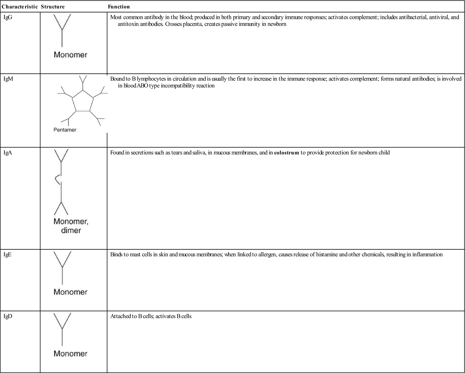
Antibodies are a specific class of proteins termed immunoglobulins. Each has a unique sequence of amino acids (variable portion, which binds to antigen) attached to a common base (constant region that attaches to macrophages). Antibodies bind to the specific matching antigen, destroying it. This specificity of antigen for antibody, similar to a key opening a lock, is a significant factor in the development of immunity to various diseases. Antibodies are found in the general circulation, forming the globulin portion of the plasma proteins, as well as in lymphoid structures. Immunoglobulins are divided into five classes, each of which has a special structure and function (Table 7-2). Specific immunoglobulins may be administered to treat diseases such as Guillain-Barré syndrome an autoimmune disease that attacks the peripheral nervous system that can cause progressive paralysis.
TABLE 7-2
Immunoglobulins and Their Functions
| Characteristic | Structure | Function |
| IgG |  | Most common antibody in the blood; produced in both primary and secondary immune responses; activates complement; includes antibacterial, antiviral, and antitoxin antibodies. Crosses placenta, creates passive immunity in newborn |
| IgM | 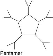 | Bound to B lymphocytes in circulation and is usually the first to increase in the immune response; activates complement; forms natural antibodies; is involved in blood ABO type incompatibility reaction |
| IgA |  | Found in secretions such as tears and saliva, in mucous membranes, and in colostrum to provide protection for newborn child |
| IgE |  | Binds to mast cells in skin and mucous membranes; when linked to allergen, causes release of histamine and other chemicals, resulting in inflammation |
| IgD |  | Attached to B cells; activates B cells |
Complement System
The complement system is frequently activated during an immune reaction with IgG or IgM class immunoglobulins. Complement involves a group of inactive proteins, numbered C1 to C9, circulating in the blood. When an antigen–antibody complex binds to the first complement component, C1, a sequence of activating steps occurs (similar to a blood clotting cascade). Eventually this activation of the complement system results in the destruction of the antigen by lysis when the cell membrane is damaged, or some complement fragment may attach to a microorganism, marking it for phagocytosis. Complement activation also initiates an inflammatory response.
Chemical Mediators
A number of chemical mediators such as histamine or interleukins may be involved in an immune reaction, depending on the particular circumstances. These chemicals have a variety of functions, such as signaling a cellular response or causing cellular damage (see Chapter 5). A brief summary is provided in Table 7-1.
The Immune Response
Because of unique antigens, often a protein, on the surface of an individual’s cells, that person’s immune system can distinguish self from non–self (foreign) and can thus detect and destroy unknown material. Normally the immune system ignores “self” cells, demonstrating tolerance. When the immune system recognizes a specific non–self-antigen as foreign, it develops a specific response to that particular antigen and stores that particular response in its memory cells for future reference if the antigen reappears in the body. It is similar to a surveillance system warning of attack and the subsequent mobilization of an army for defense. For example, lymphoid tissue in the pharynx, such as tonsils and adenoids, can capture antigens in material that is inhaled or ingested and process the immune response. Note that a person must have been exposed to the specific foreign antigen and must have developed specific immunity to it (such as antibodies) before this defense is effective. This response is usually repeated each time the person is exposed to a particular substance (antigen) because the immune system has memory cells. Immune responses that occur after the first introduction of the antigen are usually more rapid and intense than the initial response. In destroying foreign material, the immune system is assisted by nonspecific defense mechanisms such as phagocytosis and the inflammatory response. By removing the foreign material, the immune system also plays a role in preparing injured tissue for healing.
When the plasma membrane of malignant neoplastic cells is abnormal, the immune system may be able to identify these cells as “foreign” and remove them, thus playing an important role in the prevention of cancer (see Chapter 20). It has been noted that individuals frequently develop cancer when the immune system is depressed as with an infection or increased stress. However, not all cancer cells are identifiable as foreign; therefore the immune system may not remove them. The immune system directs the response to an antigen, but also has built-in controls to prevent excessive response. Two limiting factors are the removal of the causative antigen during the response, and the short lifespan of the chemical messengers. Tolerance to self-antigens prevents improper responses.
Diagnostic Tests
Many new and improved techniques are emerging, and more details on these techniques may be found in reference works on serology or diagnostic methods.
Tests may assess the levels and functional quality (qualitative and quantitative) of serum immunoglobulins or the titer (measure) of specific antibodies. Identification of antibodies may be required for such purposes as detecting Rh blood incompatibility (indirect Coombs’ test) or screening for HIV infection (by enzyme-linked immunosorbent assay [ELISA]). Pregnant women are checked for levels of antibodies, particularly for German measles to establish their potential for complications if they are exposed to this disease while pregnancy which could result in fetal death. During hepatitis B infection, changes in the levels of antigens and antibodies take place, and these changes can be used to monitor the course of the infection and level of immunity (see Chapter 17). The number and characteristics of the lymphocytes in the circulation can be examined as well. Extensive HLA (MHC) typing is required to complete tissue matching before transplant procedures.
The Process of Acquiring Immunity
Natural immunity is species specific. For example, humans are not usually susceptible to infections common to many other animals. Innate immunity is gene specific and is related to ethnicity, as evident from the increased susceptibility of North American aboriginal people to tuberculosis.
The immune response consists of two steps:
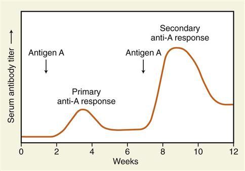
• A primary response occurs when a person is first exposed to an antigen. During exposure the antigen is recognized and processed, and subsequent development of antibodies or sensitized T lymphocytes is initiated (Fig. 7-3). This process usually takes 1 to 2 weeks and can be monitored by testing serum antibody titer. Following the initial rise in seroconversion the level of antibody falls.
When a single strain of bacteria or virus causes a disease, the affected person usually has only one episode of the disease because the specific antibody is retained in the memory. Young children are subject to many infections until they establish a pool of antibodies. As one ages, the number of infections declines. However, when there are many strains of bacteria or viruses causing a disease, for example, the common cold (which has more than 200 causative organisms, each with slightly different antigens), an individual never develops antibodies to all the organisms, and therefore he or she has recurrent colds. The influenza virus, which affects the respiratory tract, has several antigenic forms, for example, type A and type B. These viruses have various strains that mutate or change slightly over time. For this reason, a new influenza vaccine is manufactured each year, its composition based on the current antigenic forms of the virus most likely to cause an epidemic of the infection.
Immunity is acquired four ways (Table 7-3). Active immunity develops when the person’s own body develops antibodies or T cells in response to a specific antigen introduced into the body. This process takes a few weeks, but the result usually lasts for years because memory B and T cells are retained in the body.
TABLE 7-3
| Type | Mechanism | Memory | Example |
| Natural active | Pathogens enter body and cause illness; antibodies form in host | Yes | Person has chickenpox once |
| Artificial active | Vaccine (live or attenuated organisms) is injected into person. No illness results, but antibodies form | Yes | Person has measles vaccine and gains immunity |
| Natural passive | Antibodies passed directly from mother to child to provide temporary protection | No | Placental passage during pregnancy or ingestion of breast milk |
| Artificial passive | Antibodies injected into person (antiserum) to provide temporary protection or minimize severity of infection | No | Gammaglobulin if recent exposure to microbe |

Outcome of Infectious Disease
The occurrence of many infectious diseases, such as polio and measles, has declined where vaccination rates have been high, creating “herd immunity”—a phenomenon in which a high percentage of a population is vaccinated, thus decreasing chances of acquiring and spreading an infectious disease. Believing smallpox (variola) had been eradicated in many countries by the mid-1950s, the United States discontinued the smallpox vaccine in 1972. The WHO worked toward worldwide eradication and the last case of naturally occurring smallpox was recorded in 1977.
Polio vaccination was implemented in 1954, and cases are a rare occurrence today in developed areas of the world. Recent outbreaks of measles and mumps in North America are the result of inadequate revaccination of teens.
The search continues for additional vaccines against AIDS and malaria, tuberculosis, and other widespread infections. Research is also continuing on genetic vaccines, in which only a strand of bacterial DNA forms the vaccine, thus reducing any risks from injection of the microorganism itself. Immunotherapy is an expanding area of cancer research in the search for new and more specific therapies.
Emerging and Re-emerging Infectious Diseases and Immunity
Emerging infectious diseases are those newly identified in a population. Re-emerging infectious diseases were previously under control but unfortunately not all individuals participate in immunization programs; therefore infectious disease outbreaks persist. These infectious diseases are on the rise due to globalization, drug resistance, and many other factors.
Bioterrorism
Biological warfare and bioterrorism use biological agents to attack civilians or military personnel. Concern continues about the possibility of bioterrorism using altered antigenic forms of common viruses or bacteria. Such bioweapons would have widespread impact on populations because current immunizations do not protect against such agents. It is important to recognize that large outbreaks of diseases formerly controlled by vaccines may represent acts of bioterrorism. Such outbreaks should be reported to the local authorities as soon as they are recognized.
Passive immunity occurs when antibodies are transferred from one person to another. These are effective immediately, but offer only temporary protection because memory has not been established in the recipient, and the antibodies are gradually removed from the circulation. There are also two forms of passive immunity:
Tissue and Organ Transplant Rejection
Replacement of damaged organs or tissues by healthy tissues from donors is occurring more frequently as the success of such transplants improves. Skin, corneas, bone, kidneys, lungs, hearts, and bone marrow are among the more common transplants. Transplants differ according to donor characteristics, as indicated in Table 7-4. In most cases transplants, or grafts, involve the introduction of foreign tissue from one human, the donor, into the body of the human recipient (allograft). Because the genetic makeup of cells is the same only in identical twins, the obstacle to complete success of transplantation has been that the immune system of the recipient responds to the HLAs (MHCs) in foreign tissue, rejecting and destroying the graft tissue.
TABLE 7-4
Types of Tissue or Organ Transplants
| Allograft (homograft) | Tissue transferred between members of the same species but may differ genetically; e.g., one human to another human |
| Isograft | Tissue transferred between two genetically identical bodies; e.g., identical twins |
| Autograft | Tissue transferred from one part of the body to another part on the same individual; e.g., skin or bone |
| Xenograft (heterograft) | Tissue transferred from a member of one species to a different species; e.g., pig to human |
Stay updated, free articles. Join our Telegram channel

Full access? Get Clinical Tree


