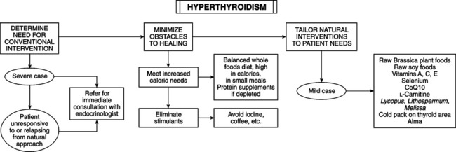
• Weakness, sweating, weight loss, nervousness, loose stools, heat intolerance, irritability, fatigue • Tachycardia; warm, thin, moist skin; stare; tremor • Increased thyroxine (T4), free T4, and free T4 index • Failure of thyroid suppression with triiodothyronine (T3) admin-istration • In Graves’ disease: goiter (often with bruit), ophthalmopathy Following are patterns of susceptibility in autoimmune disease (especially Graves’ disease): • Gender: female/male ratio is 7:1 to 10:1; the ratio in those with ophthalmic complications is 1:1. • Stress: recent stress is a precipitating factor. The most common precipitating event is “actual or threatened separation from person on whom patient is emotionally dependent.” The onset of Graves’ disease often follows emotional shock—divorce, death, or difficult separation. • Genetics: Graves’ disease is more prevalent in some human leukocyte antigen (HLA) haplotypes (HLA-B8 and HLA-DR3 in whites, HLA-Bw35 in Japanese, HLA-Bw46 in Chinese). HLA identical twins have a 50% chance of manifesting Graves’ disease if one twin is affected; fraternal twins have a 9% chance. Some haplotypes are protective against Graves’ disease. Genetic haplotype does not affect clinical course of disease or response to treatment. • Left-handedness: A statistical trend exists for left-handed people to manifest Graves’ disease and other autoimmune diseases. Seventy percent of male Graves’ patients had some degree of left-handedness compared with 24% of control subjects. Some evidence exists for a higher rate of dyslexia in Graves’ patients. • Smoking: a statistical correlation exists between smoking and Graves’ disease, especially with ophthalmic complications. The smoking risk estimate (1.5) is less than heredity (3.6) and negative life events (6.3). The group at highest risk of increase for severe endocrine ophthalmopathy involves those with eye manifestation who continue to smoke. • Iodine supplementation: dietary iodine supplementation in iodine-sufficient areas can increase incidence of thyrotoxicosis (elevated T4 and suppressed TSH with both nodular and diffuse goiters) in susceptible individuals. The rate of increase for women (8.03) was much greater than for men (1.34). • Mercury and cadmium exposure: exposing animals to toxic levels of cadmium (Cd) or mercury (Hg) induces immediate hyperthyroidism. Confirmatory anecdotal human clinical cases have been reported. • Drugs: in older patients with hyperthyroidism, consider toxic reaction to prescription drugs. The most common causes of hyperthyroidism in the elderly are low iodine intake and higher use of amiodarone (an antihypertensive drug). Symptoms of hyperthyroidism vary in the elderly; apathy, tachycardia, and weight loss are more common. • Graves’ disease in young adult women: nervousness, irritability, sweating, palpitations, insomnia, tremor, frequent stools, weight loss despite good appetite. • Physical examination: smooth, diffuse, nontender goiter; tachycardia, especially after exercise; loud heart sounds (often systolic murmur); mild proptosis, lid retraction, lid lag, and tremor. • Other signs and symptoms: muscle weakness and fatigue, anxiety, heat intolerance, pretibial myxedema. • Atypical cases (elderly): in apathetic thyrotoxicosis, the above symptoms are absent. In the elderly, only symptom may be depression. • Screening lab test for suppressed TSH. • Increased thyroid hormone can present as worsening of already present cardiac symptoms (angina pectoris, congestive heart failure, and atrial fibrillation). • Characteristic skin changes: moist, warm, and finely textured; perspiration increases in response to increased body temperature; pigment changes (vitiligo) and increased pigmentation of areas (skin creases, knuckles). • Hair may thin or fall out in patches or altogether (alopecia). • Nails may separate prematurely from nail bed (onycholysis). • Localized, nonpitting edema along shins (myxedema) or elsewhere, such as extensor surfaces. It is often pruritic and red. • Other symptoms of Graves’ disease: glucose intolerance, osteoporosis, dyspnea, polyurea and polydipsia, myopathy, paralysis, Parkinsonian or choreoathetoid affect. • Graves’ disease is the most common diagnosis of hyperthy-roidism. • Several types of thyroiditis cause hyperthyroidism: Hashimoto’s thyroiditis (early stage), subacute thyroiditis, painless thyroiditis, and radiation thyroiditis. • Hyperthyroidism from exogenous causes: iatrogenic hyperthyroidism, factitious hyperthyroidism (dieters taking thyroxine for weight loss), and iodine-induced hyperthyroidism (Jod-Basedow disease). • Toxic nodular goiters: toxic adenoma and multinodular goiter. • Rare causes: thyroid carcinoma, ectopic hyperthyroidism, trophoblastic tumors (hydatidiform mole, choriocarcinoma, em-bryonic carcinoma of testis), excess TSH (pituitary adenoma, nonneoplastic pituitary secretion of TSH), and struma ovarii. Definitive diagnosis includes the following: • Serum T3, T4, thyroid resin uptake, and free thyroxine usually are elevated. • TSH assay shows low levels except in rare cases. • TSH receptor antibodies are present in 80% of cases. • If nodular goiter is present, use thyroid scan to rule out cancer and guide treatment. • Serum antibodies can help rule out cancer in patients with lobular firm goiters. High antibody levels suggest chronic thyroiditis.
Hyperthyroidism
DIAGNOSTIC SUMMARY
GENERAL CONSIDERATIONS
Disease Risk

DIAGNOSIS
Clinical Presentation
Differential Diagnosis of Hyperthyroidism
Laboratory Diagnosis
![]()
Stay updated, free articles. Join our Telegram channel

Full access? Get Clinical Tree


Hyperthyroidism
