Characterized by ground-glass or smudged nuclei with margination of chromatin
• Multinucleated cells with nuclear molding are less common compared with mucocutaneous HSV infections
Top Differential Diagnoses
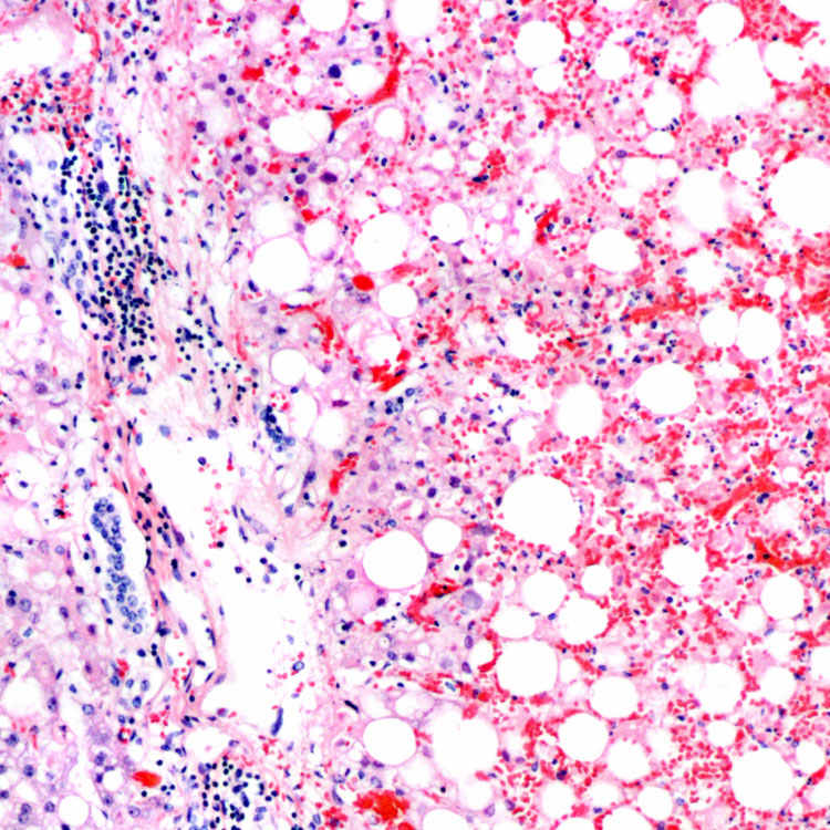
H&E shows extensive hemorrhagic necrosis with negligible portal and lobular inflammation.
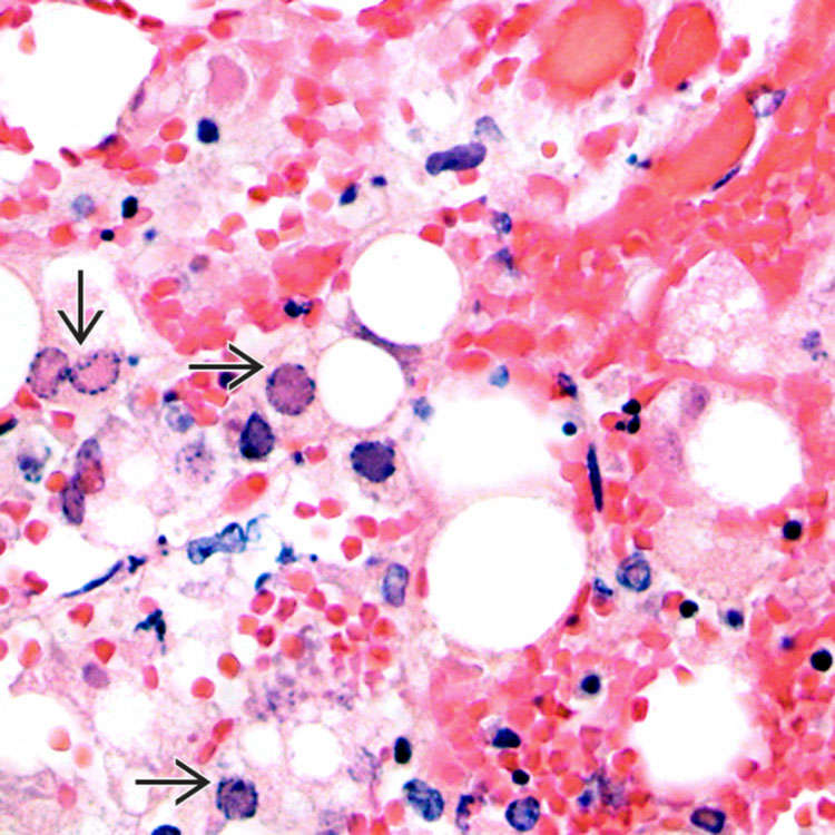
H&E shows herpes simplex virus (HSV) inclusions at the interface of necrotic and viable areas. In most cells, the inclusions appear as eosinophilic ground-glass areas in the nucleus with margination of nuclear chromatin
 .
.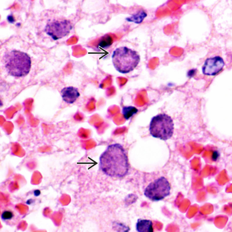
H&E shows viral inclusions
 with smudged hepatocyte nuclei and margination of nuclear chromatin. The inclusions are typically located at the edge of necrotic zones. A prominent owl-eye like appearance typical of CMV inclusions is not present in most HSV inclusions.
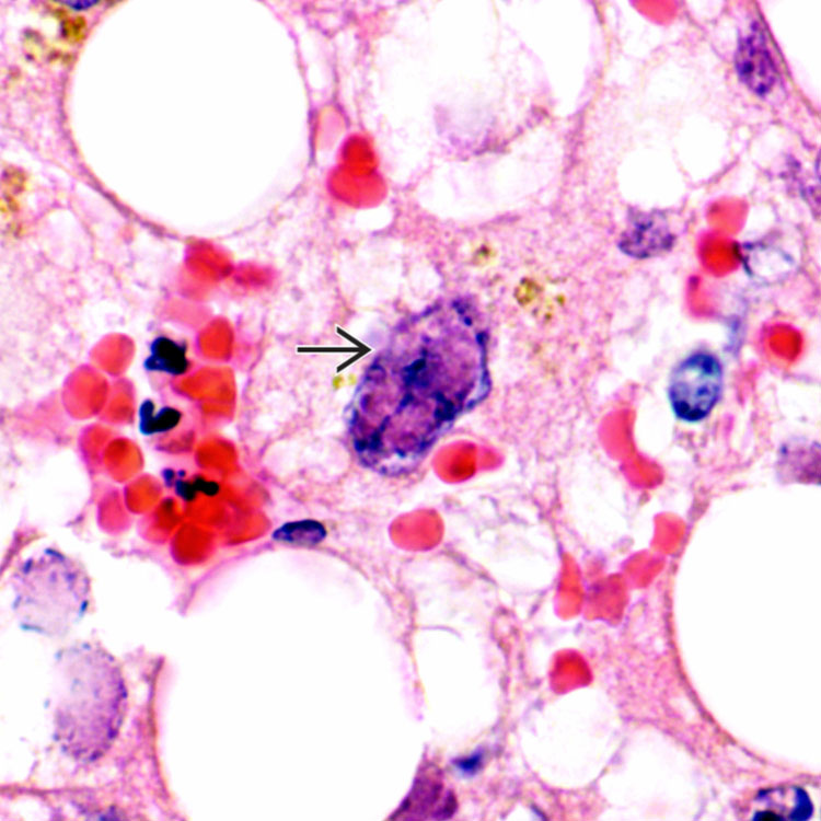
 with nuclear molding. These cells are commonly observed in herpetic mucosal lesions but are uncommon in HSV hepatitis.


