24
Hand Muscles
The hand is the most distal part of the upper limb. It can carry out complex movements because of the unique arrangement of four digits with the opposing thumb. The digits (fingers) are numbered, with the thumb being digit I and the little finger being digit V. The digits can also be referred to as the thumb, index finger, middle finger, ring finger, and little finger. There are 19 bones in the hand: 5 metacarpals and 14 phalanges. The wrist contains 8 bones (carpal bones) and is described in Chapter 23.
Dorsum of the Hand
The skin on the dorsal (posterior) surface of the hand is thin and contains visible superficial veins that make up the dorsal venous network of the hand. The medial end of the dorsal venous network coalesces to form the basilic vein, and the veins on the lateral end form the cephalic vein. Deep to the veins are the long extensor tendons from the forearm, which are bound down as they cross the wrist by the extensor retinaculum. The dorsum (dorsal surface) of the hand contains only four dorsal interossei muscles between the metacarpal bones (Table 24.1).
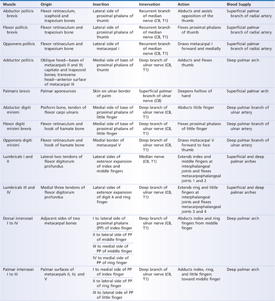
TABLE 24.1 Intrinsic Hand Muscles
Cutaneous innervation of the dorsum of the hand is from the radial, median, and ulnar nerves (Fig. 24.1). Blood is supplied by branches of the radial artery. At the distal lateral aspect of the wrist the radial artery gives off the princeps pollicis artery to the thumb and the dorsalis indicis artery to the index finger. From this point, the radial artery continues as the dorsal carpal arch and gives rise to the dorsal metacarpal arteries, which in turn become the dorsal digital arteries to each finger.
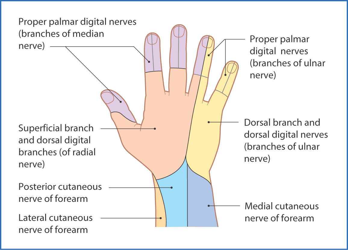
FIGURE 24.1 Cutaneous nerve distribution of the posterior (dorsal) surface of the hand.
Palmar Surface of the Hand
The skin on the palmar (anterior) surface of the hand is thick and hairless and displays numerous longitudinal and transverse flexion creases. The palm is unique in that its ridged surface combined with the many intrinsic muscles of the hand enables it to participate in grasping, lifting, and fine movements.
The deep fascia of the palm is a continuation of the antebrachial fascia of the forearm. In the middle of the palm the fascia becomes thickened as the palmar aponeurosis, which provides distal attachment for the palmaris longus muscle from the anterior compartment of the forearm and protects and contains the tendons and muscles of the hand. Two septa arising from the internal (deep) surface of the palmar aponeurosis, one medial and one lateral, divide the hand into three compartments—medial, central, and lateral. The flexor retinaculum proximal to the palmar aponeurosis is a strong transverse band of fascia that confines the flexor tendons of the forearm as they pass through the hand toward the fingers.
Cutaneous innervation of the palmar surface of the hand is from the radial, median, and ulnar nerves (Fig. 24.2).
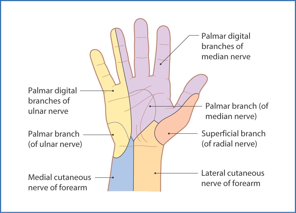
FIGURE 24.2 Cutaneous nerve distribution of the anterior (palmar) surface of the hand.
Muscles of the Hand
The intrinsic muscles of the hand are divided into three groups named after the three major (medial, central, and lateral) compartments of the hand. The intrinsic hand muscles originate and have insertion points on the hand, whereas the extrinsic hand muscles (e.g., flexor digitorum longus) originate outside the hand.
The medial or hypothenar compartment contains four intrinsic muscles—abductor digiti minimi, flexor digiti minimi brevis, opponens digiti minimi, and palmaris brevis (Fig. 24.3)—that take their general proximal origin from the carpal bones and extend distally toward the little finger. These muscles are innervated by the deep branch of the ulnar nerve (the palmaris brevis by the superficial branch) and, as a group, flex, abduct, and oppose the little finger. The first three muscles create the small bulge (hypothenar eminence) on the medial aspect of the palm.
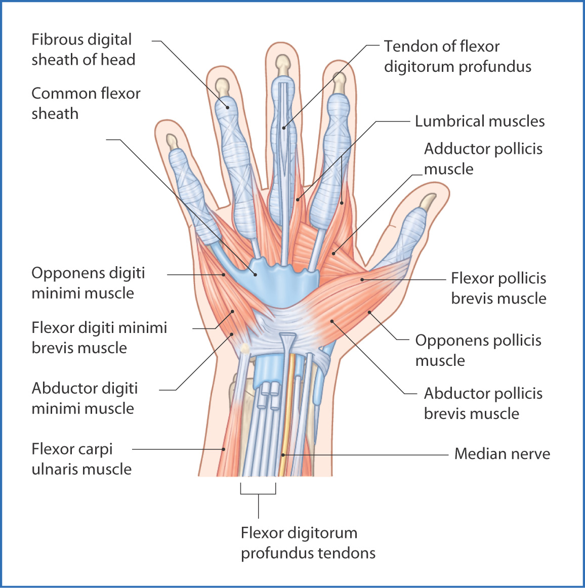
FIGURE 24.3 Muscles of the anterior (palmar) surface of the hand—intermediate layer.
The central compartment of the hand contains three short intrinsic muscle groups—four lumbricals (I to IV), three palmar interossei, and four dorsal interossei. These small muscles are oriented so that digit III (the middle finger) delineates the central axis of the hand.
- The lumbrical muscles are four small hand muscles that originate proximally on the tendons of the flexor digitorum profundus and insert onto the lateral surface of the extensor expansion. They assist flexion of the metacarpophalangeal joints and extension of the interphalangeal joints.
- The three palmar interossei muscles adduct the fingers toward the central axis created by the middle finger. Each arises from the metacarpal shaft of the digit that it abducts and inserts onto the fascial extensor expansion on the first, third, and fourth fingers.
- The four dorsal interossei muscles abduct the fingers away from the central axis. Each arises by two heads from the adjacent sides of the metacarpal bones and inserts onto the extensor expansion of the first, second, and third fingers.
- The three palmar interossei muscles adduct the fingers toward the central axis created by the middle finger. Each arises from the metacarpal shaft of the digit that it abducts and inserts onto the fascial extensor expansion on the first, third, and fourth fingers.
The lateral or thenar compartment of the hand contains the abductor pollicis brevis, flexor pollicis brevis, and opponens pollicis. These muscles flex, abduct, and oppose the thumb. The first three muscles form the thenar eminence
Stay updated, free articles. Join our Telegram channel

Full access? Get Clinical Tree


