Gynecologic and obstetric surgery

Chapter 34


Gynecologic and obstetric surgery


Key terms and definitions


Abortion 


Termination of pregnancy. Several descriptions are as follows:


Elective voluntary surgical ending of pregnancy 


First trimester: pregnancy is terminated during first 3 months of gestation; Second trimester: pregnancy is terminated during second 3 months of gestation; Late: pregnancy is terminated after the second trimester has ended.


Incomplete natural termination of pregnancy before the age of viability 


Products of conception are partially retained and may need surgical removal to prevent sepsis in the mother.


Missed 


Natural termination of gestation before the age of viability. Products of conception must be surgically removed to prevent sepsis in the mother.


Spontaneous 


Natural termination of pregnancy before age of viability. Products of conception are expelled without surgical intervention.


Threatened 


Pregnancy is diagnosed at risk for natural termination before the age of viability. Medical and/or surgical management may be attempted to prevent full natural termination.


Amenorrhea 


Absence of menstrual periods.


Cesarean birth 


Surgical delivery of a fetus through an abdominal incision. Also known as cesarean section or C-section.


Chromopertubation 


Instillation of dye through the fallopian tubes as a test of patency.


Dilation and curettage (D&C) 


Progressive enlargement of the opening of the uterine cervix is to permit instrumentation for debulking the endometrium and other surgical procedures.


Endometrium 


Lining of the uterus that is normally shed during menstruation.


Fundus 


The round top or dome of uterus.


Leiomyofibroma 


Term for fibroid tumors. Also known as myoma (e.g., uterine muscle tumor).


Graafian follicle 


Mature ovum.


Gravid 


Pregnant.


Menarche 


Beginning of menstruation.


Menometrorrhagia 


Bleeding between menstrual periods in a premenopausal woman that is surgically treated by dilation and curettage, endometrial ablation, or hysterectomy.


Menorrhagia 


Excessive bleeding at menstruation that is surgically treated by dilation and curettage, endometrial ablation, or hysterectomy.


Menstruation 


The shedding of the endometrium during periodic hormonal cycles.


Neoplasia 


New tissue overgrowth. Potentially cancerous.


Retained secundus 


Products of conception retained either at delivery of a viable fetus or at the time of incomplete abortion. Placental remnants.


Tanner’s stages 


Incremental measurement of sexual development from first signs of puberty to maturity in both sexes.


Thelarche 


The beginning of breast development that is measured in stages.


Anatomy and physiology of the female reproductive system


The female genitourinary system comprises the organs, glands, secretions, and other elements of reproduction referred to as the pudendum. Components of the female reproductive system are both external and internal organs.


External female genitalia


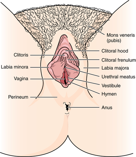
The term vulva is used collectively for the female external genitalia (Fig. 34-1). This sensitive, delicate area is highly vascular, with an extensive superficial and deep lymph supply and rich cutaneous sensory innervation. It includes the following:


Perineum


The perineum is a diamond-shaped wedge of fibromuscular tissue between the vagina and the anus. It is divided by a transverse septum into an anterior urogenital triangle and a posterior anal triangle. It consists of the perineal body and perineal musculature. With fibers of six muscles converging at its central point, the perineum forms the base of the pelvic floor and helps support the posterior vaginal wall.


These muscles are the bulbocavernosus (vaginal sphincter), the two superficial transverse perineal muscles, the two levator ani muscles, and the external anal sphincter. The levator ani muscles are the largest muscles and, in contrast to the other superficial muscles, are deep. The most important muscles in the pelvis, the levator ani muscles form a hammock-type suspension (pelvic diaphragm) from the anterior to the posterior pelvic wall, beneath the pelvic viscera. These muscles retain the organs within the pelvis by offering resistance to repeated increases in intraabdominal pressure, such as coughing, bearing down in labor, and straining at stool.


Internal female reproductive organs


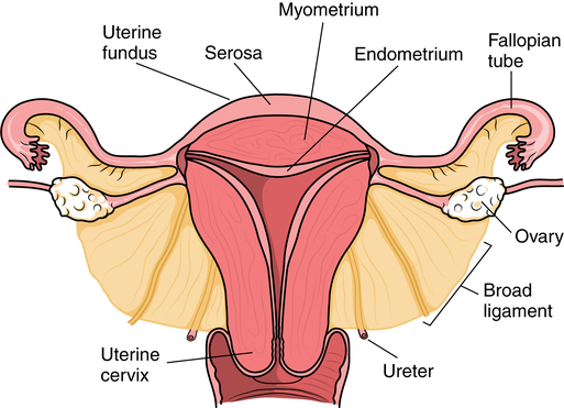
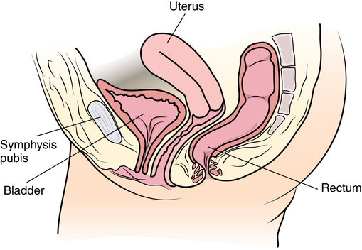
The internal female reproductive organs (Fig. 34-2) lie within the pelvic cavity, protected by the bony pelvis. Bones and ligaments form the pelvic outlet. The dilated cervix of the uterus and the vagina constitute the birth canal.


Vagina


The vagina is a thin-walled, 8-cm fibromuscular tube extending from the vestibule obliquely backward and upward to the uterus, where the cervix projects into the top of the anterior wall. The vagina is elastic and capable of distention during intercourse and parturition. The bladder lies anteriorly; the rectum lies posteriorly. It is lined with mucous membrane and contains glands that produce a cleansing acid secretion.


The anterior vaginal wall is shorter than the posterior wall. The upper third of the posterior wall is covered by peritoneum reflected onto the rectum. Normally the anterior and posterior walls relax and are in contact. However, the lateral walls remain rigid because of the pull of the muscles and therefore are in close contact with pelvic tissues.


A rich venous plexus in the muscular walls makes the vagina highly vascular. Uterine and vaginal arteries supplying the area are branches of the internal iliac artery. Branches of the vaginal artery extend to the external genitalia and the adjacent bladder and rectum. Lymphatic drainage is extensive. The upper two thirds of the vagina drain into the external and internal iliac nodes; the lower third drains into the superficial inguinal nodes.


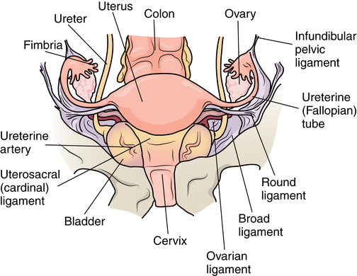
The vault (upper part of the vagina) is divided into four fornices, or arches (Fig. 34-3). During digital pelvic examination, pelvic organs can be palpated through the thin walls of the vault. The anterior fornix, in front of the cervix, is adjacent to the base of the bladder and distal ends of the ureters.


The pouch of Douglas (retrouterine cul-de-sac) directly behind the larger posterior fornix lies behind the cervix. This pouch separates the back of the uterus from the rectum: anteriorly by the uterine peritoneal covering, which continues down to cap the posterior vaginal fornix; and posteriorly by the anterior wall of the rectum. Lateral uterosacral ligaments embrace the lower third of the rectum. The floor of the pouch, about 7 cm above the anus, is formed by reflection of the peritoneum from the rectum to the upper vagina and uterus.


The posterior cul-de-sac is the route of entry for a number of diagnostic or surgical procedures because the pouch of Douglas (the lowest part of the peritoneal cavity) is separated from the vagina only by the thin vaginal wall and peritoneum.


The lateral fornices lie on either side of the cervix, in contact with anterior and posterior sheets of the broad ligaments surrounding the uterus. Proximal structures are the uterine artery, ureters, fallopian tubes, ovaries, and sigmoid colon.


Uterus


The uterus is the female organ of gestation. It receives and holds the fertilized ovum during development of the fetus and expels it during childbirth. Resembling an inverted pear in shape, this hollow muscular retroperitoneal organ is situated in the bony pelvis. It lies between the bladder anteriorly and the sigmoid colon posteriorly. The uterus is divided transversely by a slight constriction into a wider upper part (the body or corpus uteri) and a narrower lower part (the 2- to 3-cm cervix uteri or ectocervix that protrudes into the vagina).


The corpus meets the cervix at the internal os. Peritoneum covers the corpus externally; the endometrium, ranging in thickness from 2 to 10 mm, lines it internally. This mucous membrane is uniquely adapted to receive and sustain the fertilized ovum. The fundus, or rounded top portion of the uterus, lies above the uterine cavity.


The shape of the uterine cavity (endometrial cavity), flattened from front to back, is roughly triangular in the nonpregnant female. The upper lateral angles extend out toward openings of the fallopian tubes, which enter bilaterally through the uterine walls at the cornua. The apex of the triangle is directed downward to the cervix. The cavity within the cervix, the endocervical canal, narrows to a slit at the distal orifice, where the cervix communicates with the vagina via the external os. The endocervix is the glandular mucous membrane of the cervix. The corpus of the uterus and the cervix are considered individually in relation to disease and therapy because they differ in structure and function.


The uterus is capable of expansion to accommodate a growing fetus. Much of the bulk of the corpus consists of involuntary muscle, the myometrium, composed of three layers. The inner layer prevents reflux of menstrual flow into the tubes and peritoneal cavity, which could result in endometriosis. It also contributes to the competency of the internal os sphincter to prevent premature expulsion of the fetus. The middle layer encloses large blood vessels. These muscle fibers act as living ligatures for hemostasis after delivery. The outer layer has expulsive action, ejecting menstrual flow and clots, an aborted embryo, or the fetus at term.


Usually the uterus lies forward at a right angle to the vagina and rests on the bladder. Although the cervix is anchored laterally by ligaments, the fundus may pivot about the cardinal ligaments widely in an anteroposterior plane. Mobility rather than position is the criterion for normality.


Fallopian tubes (salpinges)


The fallopian tubes (small, hollow musculomembranous tubes, sometimes called oviducts or uterine tubes) run bilaterally like arms from each side of the upper part of the uterus to the ovaries. Near each ovary, the open end of each tube expands into the infundibulum, which divides into fimbriae, or fingerlike projections that sweep up the ovum (the female reproductive cell) as it is expelled from the ovary. Ciliated cells move the ovum toward the uterus.


The lumen of the tube becomes very narrow where it penetrates the uterine wall to reach the uterine cavity. Contractions in the muscular walls change the shape and position of the tubes. At ovulation, they move the fimbriated ends into close apposition with ovarian surfaces. The fallopian tubes serve as a continuous passage from the external environment into the abdominal cavity via the vagina, cervix, and uterine body. Infectious diseases and other substances can enter the peritoneal cavity through this route.


Ovaries


The ovaries are oval glandular gonads located in shallow peritoneal fossae on the lateral pelvic walls, from which they are suspended by the infundibulopelvic ligaments. They are attached to the posterior layer of the broad ligament by the mesovarium (a peritoneal fold) and to the uterus by the ovarian ligament (a fibromuscular cord).


The ovaries, the counterpart of the male testes, contain ova. Each ovary measures 2 × 3 cm and consists of a center of cells and vessels surrounded by the cortex. This main portion contains the stroma or fibrous framework in which the ovarian follicles are embedded. Of the approximately 200,000 primordial follicles present at birth, fewer than 400 are likely to produce a mature ovum (graafian follicle) during the reproductive years. A serous covering derived from peritoneum surrounds the ovaries. In addition to protecting maturing ova, the ovaries, which atrophy after menopause, produce female sex hormones.


The ureters course along the retroperitoneum bilaterally and lie close to the ovarian blood supply just anterior to the common iliac arteries. The ovaries, when diseased, may be adherent to the ureters. Some surgeons will have a urologist place ureteral catheters to make the structural identification easier during dissection.


Muscles and ligaments


Muscles and ligaments support and suspend the uterus and fallopian tubes in the normal position in the center of the pelvic cavity.


Physiology


The function of the female reproductive organs is to conceive, nurture, and produce offspring. The development and function of these organs are influenced by the hormonal secretions of the ovaries and adrenal, thyroid, and pituitary glands. These hormonal relationships also affect primary and secondary sex characteristics.


The physiologic hormonal cycle prepares the uterus for the fertilized ovum. Hormone production stimulates the endometrium and breasts, resulting in thickening of tissue and increased blood supply.


Each month during the years from puberty to menopause, one (or both) of the ovaries matures a follicle from within. When the matured graafian follicle ruptures, it discharges the enclosed ovum, which enters the fallopian tube at the fimbriated end. The process of maturation and discharge of the egg is called ovulation, resulting in a fertile period that lasts several days. Changes in cervical mucus and vaginal epithelium also accompany ovulation.


Union of the ovum with a viable mature male germ cell (spermatozoon), which has ascended to the fallopian tube from the vagina, results in fertilization within 12 hours of ovulation. The union takes place in the outer third of the tube and travels toward the uterus over a period of 3 days. The fertilized ovum normally proceeds to the cornu of the uterus, where it enters to implant in the endometrium within 14 days of fertilization. By day 17, the blood supply of the fetal and maternal blood vessels is functional. The placental circulation is established. The ensuing pregnancy will last approximately 266 days, or 9 calendar months, if carried to full term.


The ovarian hormone estrogen, together with progesterone, causes a sequence of changes in the endometrial lining of the uterus to prepare for implantation of the fertilized ovum. Estrogen also produces the development of secondary sexual characteristics. Progesterone is responsible for maintaining pregnancy until hormones from the placenta assume this role.


Menstruation is the periodic discharge of blood, mucus, disintegrated ovum, and uterine mucosa formed during the hormonal cycle if pregnancy does not take place. The duration of the menstrual period varies but averages 3 to 5 days. The amount of blood lost varies greatly. The menstrual cycle, the time between the onset of each period, is approximately 28 days.


Regularity of menstruation can be disturbed by disease conditions and emotions, in addition to the onset of pregnancy. This physiologic cycle continually recurs throughout the reproductive life of the woman. Assessment of the female patient should include documentation of the age of menarche (first menses), the date of the last menstrual period (LMP), use of contraception, and sexual history. The possibility of pregnancy should be considered if the female patient is within childbearing age, regardless of age, social status, or vocation. A surgical procedure and/or the administration of anesthetic agents could be hazardous to a developing embryo.


Gynecology: general considerations


The emotional preparation of gynecologic patients presents a special challenge to the OR team. Anticipation of physical exposure, potential loss of sexual function, infertility problems, or termination of pregnancy can create severe anxiety. Some surgical procedures terminate reproductive capability and produce menopause. Patients must be able to express concerns, ask questions, and receive reassurance and support.


Special features of gynecologic surgery


Diagnostic and surgical procedures may be carried out through a vaginal or an abdominal approach, or the two approaches may be combined. Each requires a different position and different preparation, drapes, and setup. Surgical techniques for general abdominal procedures apply to gynecologic surgery. Diagnostic and definitive surgical procedures are often combined and done in one surgical procedure.


The following apply to both vaginal and abdominopelvic procedures:


1. Spinal, epidural, or (more commonly) general anesthesia is used. An epidural catheter may be inserted preoperatively for postoperative pain control.


2. A Foley catheter may be inserted after the administration of the anesthetic agent to prevent the bladder from becoming distended during the procedure and to record urinary output. The circulating nurse and the anesthesia provider should check the urinary drainage bag frequently and report urine volume or evidence of blood. This could indicate injury to the bladder or ureters. Percutaneous insertion of a cannula directly into the bladder through the abdominal wall (suprapubic cystostomy) provides an alternative indwelling urinary drainage system in select patients.


3. An electrosurgical unit (ESU), with either monopolar or bipolar electrodes, is frequently employed.


4. Argon, CO2, and neodymium:yttrium aluminum garnet (Nd:YAG) lasers are used, generally in conjunction with a colposcope or laparoscope and the operating microscope for some procedures.


5. Closed-wound suction drainage, or another type of drain, may be used to prevent hematoma or serum accumulation in the pelvis and/or the wound.


6. Prophylactic preoperative anticoagulation with subcutaneous heparin, antiembolic stockings, sequential compression devices, and early ambulation are especially important in pelvic surgery because of the potential for deep vein thrombosis (DVT) and subsequent pulmonary emboli (PE).


Vaginal approach


1. The patient is in the lithotomy position.


2. Instrumentation must be of sufficient length for use within the vaginal canal and uterine cavity. In addition to retracting, cutting, holding, clamping, and suturing instruments, vaginal setups include dilation and curettage (D&C) instruments for intrauterine procedures (Table 34-1).


3. A laser, ESU, or cryosurgical unit may be used to remove hypertrophied tissue or certain benign neoplasms.


4. A suction system, including a Poole suction tip with guard, tubing, and collection canister, is part of the setup.


5. Raytec sponges are rolled and secured on sponge forceps in deep areas (also referred to as sponge sticks). Long, narrow, 4 × 18-inch sponges with radiopaque markers on the end are used for packing off abdominal viscera in vaginal procedures. All counts are very important in these procedures.


6. Vaginal packing is inserted after certain procedures for hemostasis and/or therapeutic purposes. Antibiotic or hormone cream may be applied to the packing during insertion. Impregnated vaginal packing is commercially available. The packing should be recorded on the patient’s chart and removed at the surgeon’s order.


7. At the completion of the surgical procedure, a sanitary pad is placed against the perineum between the patient’s legs.


Abdominal approach


1. The supine or Trendelenburg’s position is used for abdominopelvic procedures to displace pelvic organs cephalad. Preparation and drapes are the same as for abdominal laparotomy. Abdominal incisions commonly used in gynecologic open procedures are shown in Figure 34-4.


2. When a large abdominal mass is present or the pelvic organs are pushed from their normal relationships, ureteral catheters may be inserted via cystoscopy before the surgical procedure to facilitate identification of the ureters to prevent inadvertent dissection. Severing of the ureters during the procedure greatly increases postoperative morbidity and mortality if the injury is not immediately detected and corrected.


3. Instrumentation includes the basic laparotomy setup with the addition of long instruments for deep manipulations within the pelvis and uterine graspers, such as a Somer uterine elevator or a tenaculum.


Combined vaginal-abdominal approach


If vaginal and abdominal surgery is indicated, a combined procedure is planned. For example, the patient may be scheduled for a total abdominal hysterectomy with anterior vaginal colporrhaphy. In such instances the vaginal procedure is performed first. The patient is then removed from the lithotomy position and repositioned in the supine position, and the abdominal preparation and surgical procedure are carried out. Care is taken to ensure that the dispersive electrode has not been displaced, and a new one should be applied when the patient is repositioned.


The following considerations apply:


Diagnostic techniques


The gynecologist employs both noninvasive and invasive diagnostic techniques. Some are performed as office or ambulatory procedures, especially those using the vaginal approach.


A pelvic examination includes inspection and palpation of the external genitalia; bimanual abdominovaginal and abdominorectal palpation of the uterus, fallopian tubes, and ovaries; and speculum examination of the vagina and cervix. This inspection is augmented by a cytologic study of smears of cervical and endocervical tissue obtained by scrapings.


The Papanicolaou (Pap) smear has significantly facilitated diagnosis of cervical cancer and premalignant lesions. Characteristic cellular changes in cervical epithelial cells are identified. Cytologic aspiration from within the endocervical canal may reveal unsuspected carcinoma of the endometrium, fallopian tubes, or ovaries and occult cervical cancer.


The Schiller test involves staining the vaginal vault and cervical squamous epithelium with Lugol’s solution. Glycogen in normal epithelium takes up the iodine. Abnormal tissues, with little or no glycogen, do not stain brown and thereby pinpoint sites for biopsy. Abnormal cytologic findings are an indication for further evaluation by histologic tissue study. Use of iodine products is contraindicated in patients with systemic iodine allergy.


Uterine cancer consists of two entities: cervical cancer and endometrial cancer. These differ by age groups, types, and consequences. Cancer of the cervix appears most often in association with coitus at an early age, multiple partners, nonbarrier contraceptives, poor sexual hygiene, a chronically infected cervix, or a history of sexually transmitted diseases. These include infection with herpes simplex virus (HSV), human papillomavirus (HPV), cytomegalovirus (CMV), Chlamydia trachomatis, and Trichomonas vaginalis. A higher frequency of abnormal Pap smear findings is associated with these infections.


HPV produces condylomata (venereal or genital warts), which have been identified as a possible cause of cervical carcinoma. HPV screening is part of the routine gynecologic examination. HPV vaccination, a series of three injections given over a 6-month period, is recommended for all girls and women from 11 to 26 years of age as a cervical cancer preventive. The Centers for Disease Control and Prevention (CDC) speculate that 50% of sexually active men and women will experience HPV infection (genital warts) during their lifetime.


Cancer of the endometrium, more common than cervical cancer, occurs primarily in postmenopausal women. It is often associated with obesity, low parity, late menopause, hypertension, and diabetes mellitus.


Biopsy of the cervix


Cervical cancer may not present symptoms in the early stage and may progress to invasion before discovery. Spotting, postmenopausal bleeding, or chronic cervicitis may be the first visible sign. The condition may be suspected by results of cytologic examination or visual inspection, but diagnosis is made by biopsy.


Cone biopsy


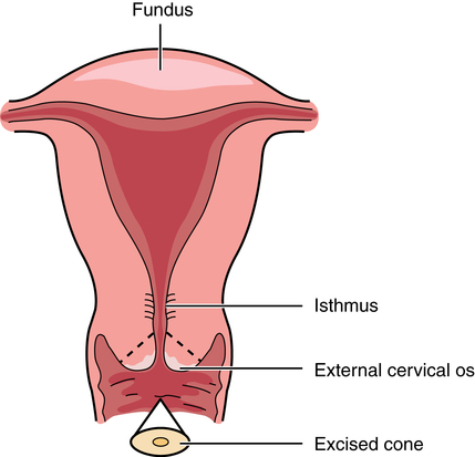
Patients diagnosed by Pap smear as having severe cervical dysplasia or intraepithelial carcinoma of the cervix require conization to remove the lesion and rule out invasive carcinoma. The biopsy, obtained with a laser, scalpel, or cervitome (cold knife conization), includes the squamocolumnar junctions of the ectocervix (transformation zone) and is tapered to include the endocervical canal to the level of the internal os (Fig. 34-5). Most of the lesions categorized as cervical intraepithelial neoplasia (CIN), dysplasia, or carcinoma in situ are found in this area. Conization of the cervix provides the most comprehensive specimen to diagnose a premalignant or malignant lesion. Multiple blocks and sections are examined by the pathologist to determine the extent of invasive disease.


Complications include hemorrhage, infection, cervical stenosis, an incompetent cervix, and infertility. The CO2 laser used for conization minimizes these risks. Hemostasis is secured with sutures as needed.


Cervical dysplasia can occur in sexually active females aged 12 years and older; the peak incidence is between the ages of 25 and 35 years. Employed therapeutically for chronic inflammation and for premalignant lesions in women of childbearing age, conization may be performed by scalpel, electrosurgery, or laser. Laser conization is used to treat severe dysplasia and carcinoma in situ.


Loop electrosurgical excision procedures (LEEPs) use a stainless steel or tungsten loop electrode to excise a central core of tissue from the transformation zone of the endocervical canal. Large loop excision of the transformation zone (LLETZ) is performed with minimal bleeding and few complications. These procedures may be performed in the OR or office setting with local anesthesia. Future childbearing is unaffected. Although the specimen is comparable in size to that obtained with cold knife conization, the specimen has superficial desiccation and may be inferior in quality. Wide conization may result in scarring and obstruction of the os.


Fractional curettage


Tissue is obtained for histologic examination by scraping the uterine cavity. Fractional curettage differentiates specimens between the endocervix and the endometrium in a series of two steps. A biopsy specimen may be taken from the cervix, if indicated by the Schiller test, in association with endocervical curettage. A small curette is introduced into the endocervical canal, which is scraped from the internal to the external os. The specimen is placed on a Telfa pad, and both are put into a container. This scraping precedes cervical dilation to avoid dislodging tissue from above the internal os.


After cervical dilation, a different curette is inserted into the uterine cavity for curettage of the endometrium. The endometrial specimen is placed in a separate container from the one used for the specimens obtained by endocervical curettage.


Colposcopy


Illumination and binocular magnification afforded by the colpomicroscope permit identification of abnormal epithelium to target for biopsy. The colposcope has a cool, intense white light that can be fitted with a green filter to improve visualization of the vascular pattern. With the colposcope positioned in front of the vulva, without touching the patient, the colposcopist can focus light through a speculum on the ectocervix, the lower part of the cervical canal, and the vaginal wall.


The cervix is swabbed with 3% acetic acid to eradicate mucus and to facilitate viewing the surface and vasculature. Biopsies are taken for histologic confirmation of the diagnosis. Endocervical curettage also may be performed. A video or still camera can be attached to a colposcope to photograph lesions.


Vaginal condylomata from HPV and adenoses, preinvasive lesions of the cervix, cervical dysplasia, CIN, and other vaginal and cervical lesions can be treated by laser with the colpomicroscope. Visualization, unobstructed by instruments, is excellent. Condylomata can incubate for 3 weeks to 6 months and will appear as white, raised areas when exposed to 3% acetic acid. Full-strength acetic acid solutions will burn the patient’s tissues.


The CO2 laser permits selective destruction of large areas of vaginal epithelium without vaginal or cervical stenosis. It cuts, coagulates, seals, and sterilizes simultaneously. This results in less blood loss, a shorter period of vaginal discharge after treatment of the cervix, and a lower incidence of infection than in other surgical treatment modalities.


Electrosurgery and cryosurgery are other options to ablate lesions. Care is taken to avoid contact with the plume from viral lesion ablation.


Culdocentesis and colpotomy


Culdocentesis


In culdocentesis, blood, fluid, or pus in the cul-de-sac is aspirated by needle via the posterior vaginal fornix for suspected intraperitoneal bleeding, ectopic pregnancy, or tuboovarian abscess. Clear, straw-colored peritoneal fluid is a negative finding. Blood can indicate an ectopic pregnancy, trauma, or tumor. Bloody fluid should be sent to the laboratory in a heparinized specimen tube. A flat plate of the kidneys, ureters, and bladder (KUB) (x-ray examination without contrast medium) may show air under the diaphragm, which is indicative of a ruptured organ.


Fallopian tube diagnostic procedures


Tubal perfusion


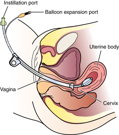
To test tubal patency, chromopertubation is performed. Methylene blue or indigo carmine dye in a solution of sterile normal saline is introduced into the uterine cavity via a 50-mL syringe or intravenous (IV) tubing attached to a cervical cannula. The surgeon views the ends of the fallopian tubes through a laparoscope. Dye seen coming from one or both tubes indicates patency.


Tubal insufflation


Uterotubal insufflation may be used to test the patency of the fallopian tubes. It usually is done as an office procedure to study infertility. The test may be therapeutic in relieving minor obstructions. Contraindications include genital tract infection, possible pregnancy, and uterine bleeding.


A cannula with a seal (i.e., Rumi, Humi, Kahn, Kronner, or Jarco) is inserted into the cervicouterine canal and connected to an insufflation apparatus. CO2 is introduced slowly under controlled flow. A relationship exists between tubal patency and the pressure required to force gas through the fallopian tubes into the peritoneal cavity.


Resistance to flow (i.e., backpressure) is measured on a mercury manometer. To prevent gas embolism, the Rubin test is done before, never after, curettage. Open vessels could introduce an air embolus. Intraabdominal irritation before total absorption of carbon dioxide from the peritoneal cavity causes referred pain in the shoulders. The patient may refer to the feeling as “gas pains.”


Pediatric and adolescent gynecology


Pelvic examination of an infant or child is rarely performed unless there is disease or trauma. Any indication of physical or sexual abuse must be reported to legal authorities. The facility should have a procedure in place for reporting child abuse.


The preadolescent has no hormonal stimulation and has not developed sexual characteristics such as rounded labia or pubic hair. In infants, the uterus regresses in size until the age of 6 years, when it regains the size it was at birth. The cervix is not palpable, and the uterine body is difficult to differentiate from surrounding tissues.


From the age of 7 years, the body begins to respond to estrogen stimulation. The mons thickens, and the vagina begins to elongate. The uterus has a growth spurt at age 9 to 10 years and begins to have the pear shape of the adult uterus. The endometrium develops and proliferates. The vagina extends to its full length of 10 to 12 cm, and the external genitalia resemble those of an adult. Thelarche (first breast development) begins as nipple buds at about 10 years of age.


Menarche (first menstruation) is common between the ages of 12 and 14 years and averages 3 years from the onset of breast development and within 6 months of the appearance of axillary hair. The first routine gynecologic examination is recommended at age 16 years or when a girl becomes sexually active.


Routine exams will be individualized according to the needs and sexual activity of the girl. Sexually transmitted disease (STD) testing is performed. Incidences of STD must be reported to the health department. The presence of STD may indicate an abusive situation.


Females who have not developed secondary sex characteristics and menstruated by age 16 years should seek a gynecology consult. Menarche can be delayed by anorexia nervosa or other dietary practices that diminish body fat stores below 17%. Menstruation can be obstructed by an imperforate hymen.


Pelvic endoscopy


Pelvic endoscopy is an established part of the gynecologist’s diagnostic and therapeutic regimen. It permits detailed intraperitoneal inspection of the pelvic organs without laparotomy. These procedures are not without danger, however. Inadvertent perforation of vessels or a hollow viscus and/or infection are major potential hazards. Operator expertise, careful patient selection, adequate anesthesia, and safe equipment are essential. Pelvic endoscopy is a sterile procedure. Vaginal and abdominal approaches are employed for direct visualization of pelvic organs and adjacent structures.


Culdoscopy


A culdoscope is introduced into the peritoneal cavity via the posterior vaginal fornix and pouch of Douglas. Some gynecologists prefer culdoscopy to investigate ovaries or posterior surfaces in the lower pelvis. Local or caudal anesthesia is used. The patient is placed in the knee-chest or lithotomy position. With the posterior lip of the cervix held by a tenaculum and retracted anteriorly, the uterus is elevated while counterpressure is applied to the posterior vaginal wall by the speculum.


This maneuver stretches the posterior vaginal fornix while a trocar and cannula or sheath penetrate the thin wall and enter the pelvis between the uterosacral ligaments. When the trocar is removed with the cannula in place, air enters the cul-de-sac because of the negative intraabdominal pressure produced by the knee-chest position. Air displaces the bowel to create a working space, and the scope may be inserted through the cannula.


At the completion of the procedure, the culdoscope is removed. Before the cannula is removed, the operating bed is straightened and the patient is flattened while as much air as possible is evacuated by hand pressure on the abdomen into low suction. Care is taken not to aerosolize body substances. Some surgeons place a suture in the puncture site.


Hysteroscopy


A rigid fiberoptic hysteroscope, introduced vaginally through the uterine cervix, provides direct inspection of the interior of the uterus to diagnose disease or treat conditions such as menorrhagia and uterine fibroids. The hysteroscope may also be used to identify and remove polyps, lost intrauterine devices (IUDs), or intrauterine adhesions. Adequate expansion of the uterine cavity is a prerequisite for viewing endometrial surfaces and tubal orifices. Fluid is instilled to expand the uterine cavity to create a working space.


Hysteroscopy systems have been developed that use sterile normal saline as an expansion medium to create the working space. The fluid should be delivered through a pressure-controlled infusion pump that tracks volume instilled. Intrauterine pressures should be maintained at or below the mean arterial pressure. Use of a continuous-flow pump makes it difficult to monitor intrauterine pressures.


The circulating nurse monitors inflow and outflow of the uterine expansion fluid medium and informs the surgeon and the anesthesia provider of any discrepancy in excess of 1500 mL.15 Visibility is further enhanced with a video camera and monitor screen. The procedure may be videotaped.


Hysteroscopy is used to perform endometrial ablation using a laser (Nd:YAG, argon, or potassium titanyl phosphate [KTP]) or ESU to stop or decrease uterine bleeding.


The Nd:YAG laser is the laser of choice for deep photocoagulation, causing endometrial destruction and scarring the uterine lining. The entire endometrial lining is treated from the fundus to about 4 cm above the external cervical os. The tip of the laser fiber can be held away from tissue (blanching technique) or in contact with endometrium (dragging technique). A specialized electrosurgical rollerball electrode is an alternative method to using a laser. This can provide relief from menorrhagia (i.e., excessively heavy menses). The destruction of the endometrium causes the woman to have amenorrhea, thereby causing sterility. Hormonal activity is unchanged.


Air or gas is not used for uterine insufflation or laser fiber cooling during hysteroscopy because of the risk for air or gas embolism. Also, 32% dextran 70 in dextrose (Hyskon) is not used as an irrigant or uterine expansion medium for endometrial laser ablation because of the systemic effects of fluid absorption through open capillaries. Precise measurements of intake and output are critical to patient safety and prevention of congestive heart failure. Hysteroscopy can be performed as an ambulatory surgery or office procedure.


Laparoscopy


Procedures using a 10- or 12-mm fiberoptic laparoscope with a 0- or 30-degree-angle lens inserted into the peritoneal cavity permit direct observation of pelvic and abdominal organs and peritoneal surfaces. Single-port or multiple-port laparoscopic methods can be used.20 Endoscopic techniques may be used to diagnose and treat ectopic pregnancy; inspect the ovaries for evidence of follicular activity and retrieve ova for in vitro fertilization; visualize and reduce pelvic masses; and determine the cause of infertility, endocrinopathies, or amenorrhea. Many pelvic diseases, such as endometriosis, adhesions, and ovarian cysts, may be identified and treated through the laparoscope and its accessory instrumentation.


The gynecologist can perform myomectomy, salpingectomy, oophorectomy, hysterectomy,20 and other procedures such as tuboplasty and incidental appendectomy without the need for a large abdominal incision. Surgical procedures such as tubal sterilization by electrocoagulation with or without partial resection, placement of a clip or silicone ring on the tube, or biopsy can be performed.


Usually a general anesthetic agent is administered. The patient is placed in a modified lithotomy position with the stirrups adjusted so that the legs are at 45-degree angles to the axis of the operating bed. Improper positioning can result in lower leg neuropathy.5


The vaginal area is prepped, followed by the abdominal prep. The patient is straight-catheterized, or an indwelling Foley catheter is inserted to monitor output and keep the bladder decompressed. The patient is draped for a combined abdominovaginal procedure. The sterile drapes must provide two exposures (i.e., an abdominal opening and a perineal opening) and cover the legs.


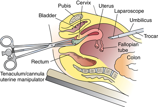
A tenaculum is placed on the cervix. A cannula is inserted into the uterine cervix for instillation of dye and for manipulation of the uterus during the procedure to provide greater visibility (Fig. 34-7). A D&C may be performed as part of the procedure after the injection of dye or contrast media.


Insertion of the scope is preceded by a pneumoperitoneum of CO2 to produce a working space in the abdomen and pelvis. The infraumbilical midline area is most commonly used if no scars, with possible adherent viscera beneath, are present. This area is preferred because it has no abdominal wall vessels that might be injured. The firm attachment of the fascia to the peritoneum facilitates entry. Great care is taken to avoid injury to the great vessels or intraabdominal organs. Insufflation can be performed with a Veress needle or a blunt trocar, such as a Hasson (open laparoscopy).


The patient is placed in a 10-degree Trendelenburg’s position to shift the abdominal organs cephalad. A small infraumbilical skin incision is made in the anterior abdominal wall. Through it a sharp trocar and sheath are introduced into the peritoneal cavity via blind puncture if a blunt Hasson is not used. Some sharp trocars and sheaths have spring-loaded end guards that cover the sharp tip and protect the underlying structures after penetration of abdominal tissue layers, although studies have not shown this to be an advantage. The trocar is removed, and a telescope of the same caliber is inserted through the trocar sheath, which remains in the cavity. Some sheaths have threads that are used to anchor it into position.


Additional secondary trocars and sheaths can be placed in the suprapubic hairline (see Fig. 34-12, later). These trocars may be inserted into the peritoneal cavity under direct vision through the endoscope, which offers good transillumination for the puncture when the room lights are dimmed, or visualization on the video monitor. A secondary trocar should have a gas port to which the CO2 tubing can be attached to prevent the cold gas from causing the scope to fog. The suction-irrigation tubing is attached to a side port on the secondary sheath.


Before use, warming the tip of the telescope in a warm moist towel or normal saline solution can prevent fogging of the distal lens of the endoscope caused by the intraperitoneal temperature and moisture. (Sterile antifog solution is commercially available.) The telescope is connected to the light source by a fiberoptic cable. Because of the potential fire hazard to drapes, the light source is not activated until the cable is attached to the telescope. The video camera is draped and connected to the telescope.


At the completion of the surgical procedure, the carbon dioxide, video monitor, and light source are turned off, accessory instruments and sheaths are removed, and the patient is leveled into a flat supine position. Hemostasis is surveyed before the telescope is removed from the primary trocar site.


The valve of its sheath closes as the telescope is withdrawn to prevent escape of CO2 gas into the room air. The pneumoperitoneum contains aerosolized blood and body fluids and should be evacuated through the suction tubing into the suction canister. Electrosurgical plume also should be suctioned from the peritoneal cavity because it binds with hemoglobin and causes the arterial oxygenation to decrease. The patient can become hypoxic.


The large fascial and skin incisions are sutured, and small dressings or adhesive bandages (Band-Aids or Steri-Strips) are applied.


The patient requires close monitoring by the anesthesia provider because increased intraabdominal pressure may lead to cardiovascular disturbances from vagal reflex. This reflex is caused by stretching of the peritoneum, retention of CO2, or compression of the inferior vena cava.


Postoperative shoulder pain may follow the use of a pneumoperitoneum. This is referred pain caused by pressure on the diaphragm, which is somewhat displaced by CO2 during the procedure. Slight elevation of the head after recovery from anesthesia relieves this pain.


Although numerous complications have been reported, the most common are perforation of the intestine or major blood vessel, hemorrhage from a biopsy site, gas embolism from intravascular injection, and burns of the abdominal wall and bowel. Some injuries, such as viscus puncture or thermal damage, may not be immediately apparent. Symptoms may not be present until 48 to 72 hours postoperatively, when tissue necrosis or sloughing occurs.


Vulvar procedures


Benign growths on the vulva, although rare, consist mainly of fatty and fibrous tumors. These are excised if large. Suspicious lesions should be removed for pathologic examination.17 Cancerous lesions may be multicentric, with the majority found on the labia majora and a lesser percentage on the labia minora, vestibule, clitoris, and posterior commissure. Vaginal smears should be taken to determine the presence of metastatic growth to the vaginal wall. Treatment depends on the size of the primary lesion, involvement of nodes, and extent of metastasis. Mutilative procedures require emotional adjustment to permanent change.


Diseases of the vulva


Wide local excision of a single well-localized area with no premalignant changes elsewhere may be done. Punch biopsies may be obtained. Leukoplakia and preinvasive lesions of the vulva may be treated with a laser or ultrasonic aspirator.17


Stay updated, free articles. Join our Telegram channel

Apr 6, 2017 | Posted by in GENERAL SURGERY | Comments Off on Gynecologic and obstetric surgery

Full access? Get Clinical Tree

Get Clinical Tree app for offline access