Changes are centered around minor papilla and involve “groove” between pancreas and duodenum

Microscopic
• Duodenal wall around minor ampulla is thickened with marked fibrosis involving muscular propria and adjacent head of pancreas


• Nonparaduodenal pancreas frequently shows dilated ducts with inspissated secretions and prominent interlobular fibrosis
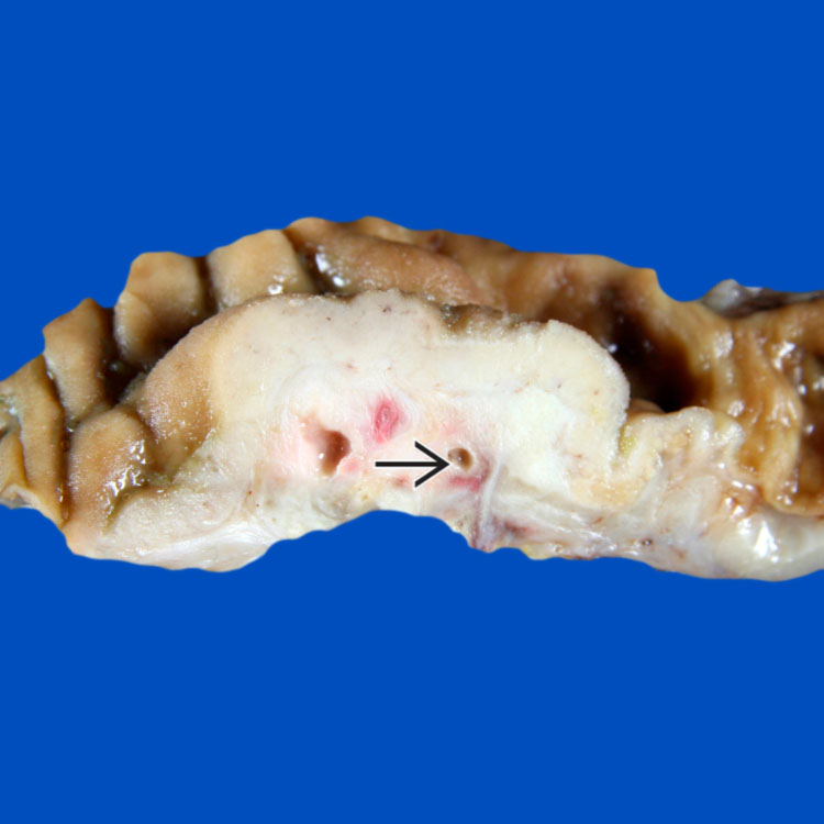
Gross photograph of a pancreaticoduodenectomy specimen shows a mass-like lesion beneath the duodenal mucosa. Note the paraduodenal zone of fibrosis with numerous small cysts
 .
.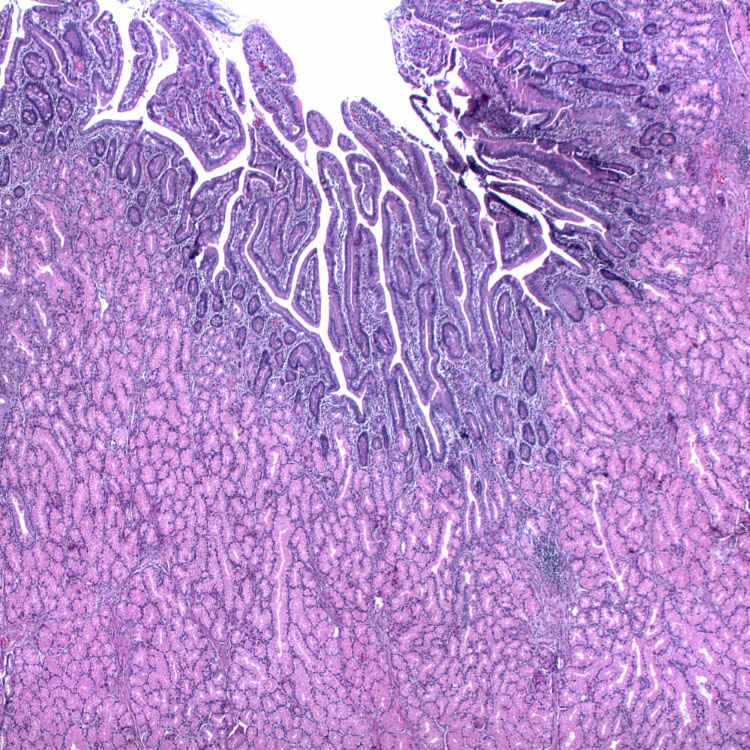
Prominent Brunner gland hyperplasia in the region of the minor ampulla frequently accompanies groove pancreatitis.
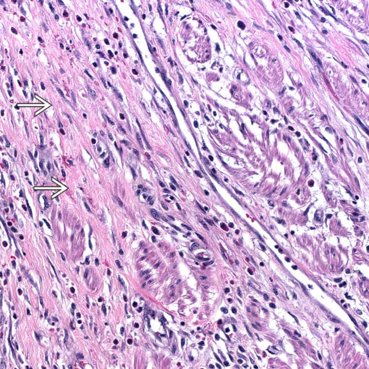
High-power view of groove pancreatitis shows exuberant fibrosis
 within the muscularis propria.
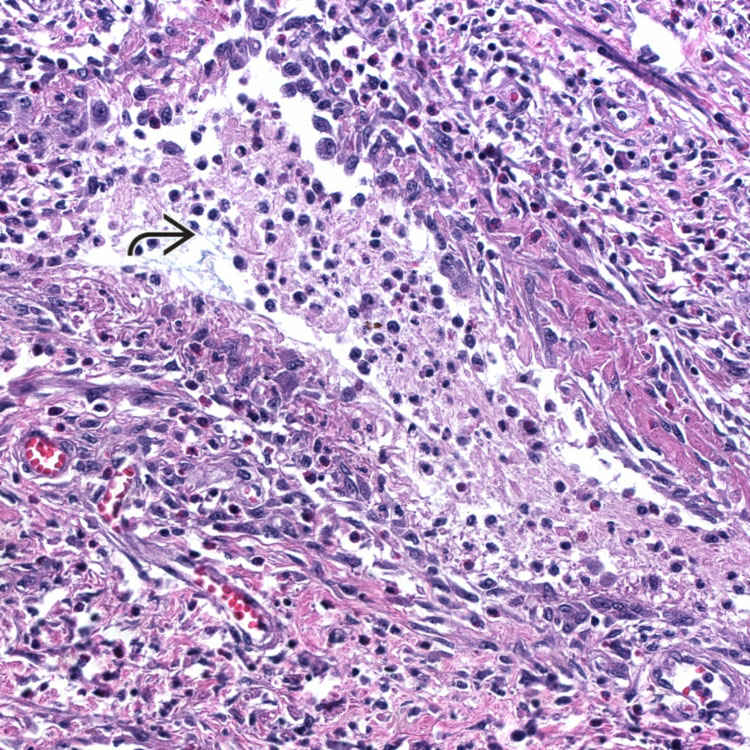
 is frequently seen in groove pancreatitis, accounting for one of the plethora of names used for this entity, “paraduodenal wall cyst.”
ETIOLOGY/PATHOGENESIS
Combination of Factors
• Developmental anomalies and environmental exposure
 Anatomical/functional variations in region of minor papilla, such as pancreatic divisum, predispose to development of groove pancreatitis


Anatomical/functional variations in region of minor papilla, such as pancreatic divisum, predispose to development of groove pancreatitisStay updated, free articles. Join our Telegram channel
Full access? Get Clinical Tree
