Granulosa Cell Tumor
Steven S. Shen, MD, PhD
Mahul B. Amin, MD
Jae Y. Ro, MD, PhD
Key Facts
Terminology
Testicular tumor of granulosa cell differentiation resembling analogous ovarian counterpart
Clinical Issues
Extremely rare; fewer than 2 dozen cases have been well documented
Range: 16-76 years (average: 44 years)
Macroscopic Features
Well-circumscribed, sometimes encapsulated, homogeneous yellow to gray firm mass
Microscopic Pathology
Growth patterns: Microfollicular (most common), solid, trabecular, insular, macrofollicular, gyriform, or cystic
Presence of Call-Exner bodies (eosinophilic materials surrounded by palisading granulosa cells)
Relatively uniform round or ovoid cells (carrot-shaped) with scant, lightly staining cytoplasm
Elongated or angular nuclei with groove (coffee bean-shaped) with 1 or 2 peripherally located nucleoli
Some show focal theca cell differentiation or have smooth muscle or osteoid differentiation
Features seen more often in malignant tumor: Large size (> 7 cm), hemorrhage, necrosis, lymphovascular invasion
Ancillary Tests
Positive for vimentin, inhibin, calretinin, SMA, CD56, and focally positive for PAN-CK(AE1/AE3)
Negative for PLAP, Podoplanin(D2-40), Oct3/4, AFP, HCG, CD30(BerH2)
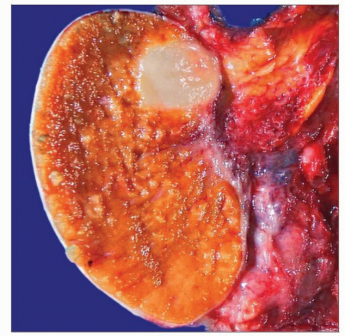 A GCT shows a well-circumscribed, homogeneous, tan-white nodule. The tumor is small and like many sex cord stromal tumors, does not extensively involve the testis. Hemorrhage and necrosis are lacking. |
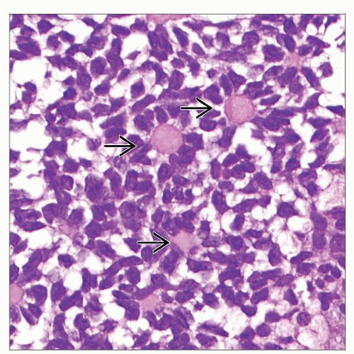 A GCT shows typical Call-Exner bodies  characterized by central eosinophilic material and palisading tumor cells resulting in a rosette appearance. The tumor cells have scant cytoplasm. characterized by central eosinophilic material and palisading tumor cells resulting in a rosette appearance. The tumor cells have scant cytoplasm. |
TERMINOLOGY
Abbreviations
Granulosa cell tumor (GCT)
Definitions
Sex cord stromal tumor of testis occurring in adults and resembling its counterpart of ovarian granulosa cell tumor
CLINICAL ISSUES
Epidemiology
Incidence
Extremely rare; fewer than 2 dozen cases have been documented
Age
Range: 16-76 years (mean: 44 years)
Juvenile GCT occurs in 1st few months of life
Presentation
Painless testicular mass
May be associated with gynecomastia (about 25%)
Treatment
Surgical approaches
Curable by surgical resection in most cases
May be managed by partial orchiectomy
Prognosis
Most have indolent clinical course but have malignant potential
Metastasis has been reported (20% of cases)
Long-term follow-up is recommended for all patients
MACROSCOPIC FEATURES
General Features
Well-circumscribed, sometimes encapsulated, homogeneous yellow to gray firm mass
± small cysts
Hemorrhage or necrosis is unusual
Size
Range: 2-10 cm (average: 5 cm)
MICROSCOPIC PATHOLOGY
Histologic Features
Growth patterns: Microfollicular (most common), solid, trabecular, insular, macrofollicular, gyriform, or cystic
Presence of Call-Exner bodies (eosinophilic material surrounded by palisading granulosa cells)
Relatively uniform round or ovoid cells (carrot-shaped) with scant, lightly staining cytoplasm
Elongated or angular nuclei with grooves (coffee bean-shaped) with 1 or 2 peripherally located nucleoli
Focal cytologic atypia and rare mitoses; mitoses may be high with varying degree of nuclear pleomorphism
May intermingle with seminiferous tubules and infiltrate tunica albuginea
Some show focal theca cell differentiation or have smooth muscle or osteoid differentiation
Rare hemorrhage, necrosis, or angiolymphatic invasion
Features seen more often in tumors with malignant outcome: Large size (> 7 cm), frequent mitoses (> 4/10 HPFs), hemorrhage, necrosis, lymphovascular invasion
Predominant Pattern/Injury Type
Solid and microfollicular
Predominant Cell/Compartment Type
Stay updated, free articles. Join our Telegram channel

Full access? Get Clinical Tree


