Most common nonepithelial tumor of extrahepatic bile ducts
Clinical Issues
• Occur most often in young African American women
Mean age: 34.7 years
• Most frequent location is common bile duct
• Often discovered incidentally
• Cured by adequate surgical excision
Excellent prognosis
Malignant granular cell tumors (GCTs) not reported in biliary tract
Macroscopic
• Often grows concentrically around bile duct, compressing lumen
• Usually < 3 cm in greatest dimension
Microscopic
• Nests or sheets of infiltrating cells
Cells may be separated by collagenous bands
Oval to polygonal cells
Abundant pink granular cytoplasm with small hyperchromatic nuclei
• Can be associated with marked atypia of overlying biliary surface epithelium
May mimic malignancy
Important to recognize underlying GCT
Ancillary Tests
• Strong diffuse immunopositivity with S100
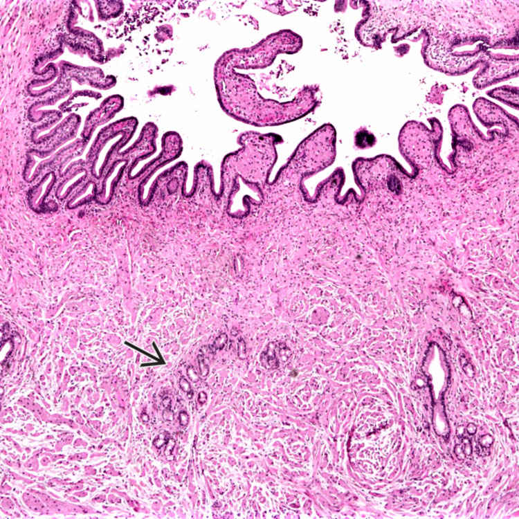
Concentric Growth Pattern This granular cell tumor is growing concentrically around the common bile duct, compressing the lumen. The tumor cells can have a very infiltrative growth pattern and may surround small peribiliary glands .
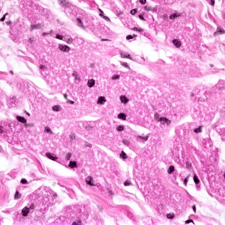
Granular Cytoplasm The tumor cells have abundant pink granular cytoplasm and small hyperchromatic nuclei.
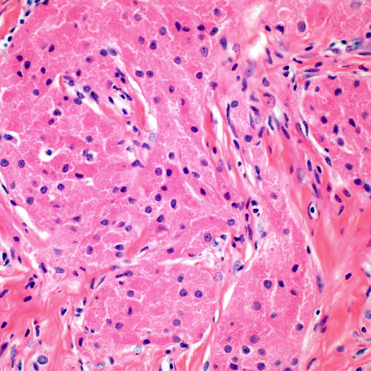
Interspersed Collagen Bands The granular, pink, polygonal tumor cells are often interspersed with bands of collagen.
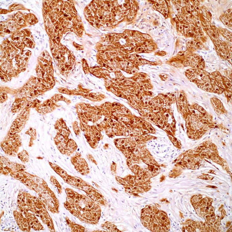
S100 S100 is strongly and diffusely positive within tumor cells. (Courtesy J. McKenney, MD.)
TERMINOLOGY
Abbreviations
• Granular cell tumor (GCT)
Synonyms
• Granular cell myoblastoma
Definitions
• Benign tumor composed of large, granular, eosinophilic cells
Immunohistochemistry and electron microscopy have shown schwannian differentiation
Only gold members can continue reading. Log In or Register to continue


 .
.