33
Gastrointestinal Tract
The abdominal cavity contains the accessory abdominal organs (see Chapter 34) and the hollow tube of the gastrointestinal (GI) tract. The GI tract extends from the mouth to the anus and is subdivided structurally and functionally into several organs that specialize in processing ingested food (Fig. 33.1). These GI organs are discussed here in the order that food passes through them.
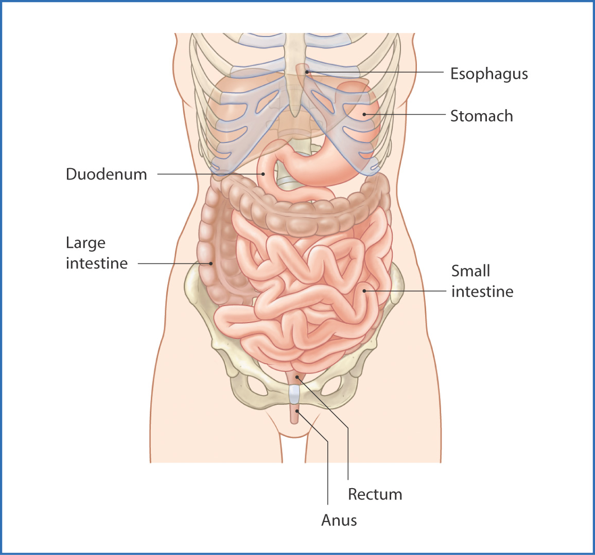
FIGURE 33.1 Gastrointestinal tract.
Esophagus
The first part of the GI tract is the oral cavity (see Chapter 9), where food is formed into a bolus that is propelled posteriorly by the tongue and oral cavity muscles into the oropharynx. From here, the bolus is transferred to the esophagus, which transports food and swallowed substances to the stomach. In the chest the esophagus runs slightly to the left of the vertebral bodies and posterior to the trachea (Fig. 33.2). It enters the abdomen through a hiatus in the diaphragm at level TX. The abdominal esophagus is approximately 2 to 3 cm long and joins the stomach at the esophagogastric sphincter (lower esophageal sphincter). This muscular sphincter prevents regurgitation of stomach contents into the esophagus. Innervation of the esophagus is by
- parasympathetic nerve fibers, which travel with the vagus nerves [X]
- sympathetic nerve fibers, which originate from the greater and lesser splanchnic nerves
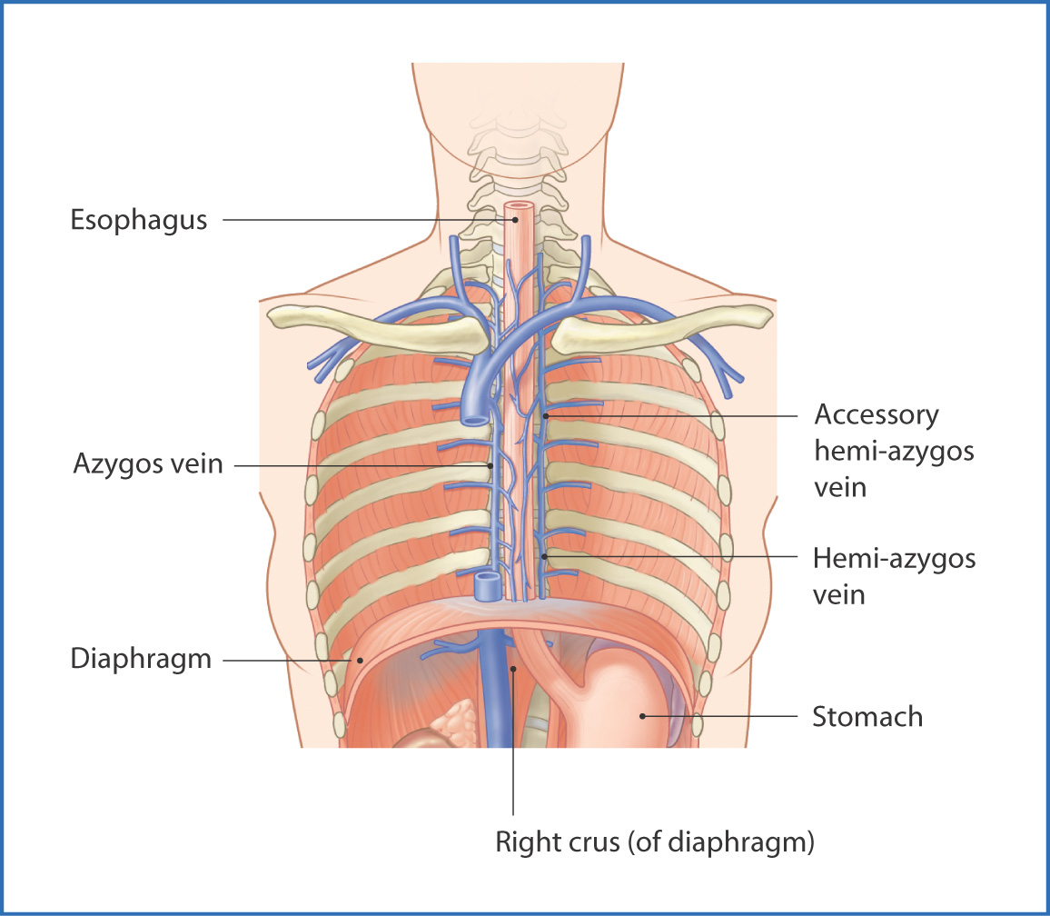
FIGURE 33.2 Esophagus.
Blood is supplied to the esophagus by several vessels—the subclavian artery, thoracic aorta, inferior phrenic artery, and left gastric artery. Venous drainage of the distal thoracic esophagus is to the azygos venous system, whereas the short abdominal esophagus drains to the hepatic portal venous system. This forms clinically significant portal-systemic anastomoses. Lymphatic drainage of the lower part of the esophagus is via the left gastric and celiac nodes.
Stomach
The stomach breaks down large food particles into smaller pieces so that they can be processed more easily (Fig. 33.3). The stomach also secretes digestive enzymes, such as pepsin, that break down proteins and starches. The stomach is entirely intraperitoneal and is located in the left hypochondrium and epigastric regions of the abdomen. It is subdivided into four parts—the cardia, fundus, body, and pylorus. It also has anterior and posterior surfaces covered with peritoneum and two curvatures (the greater and the lesser curvatures).
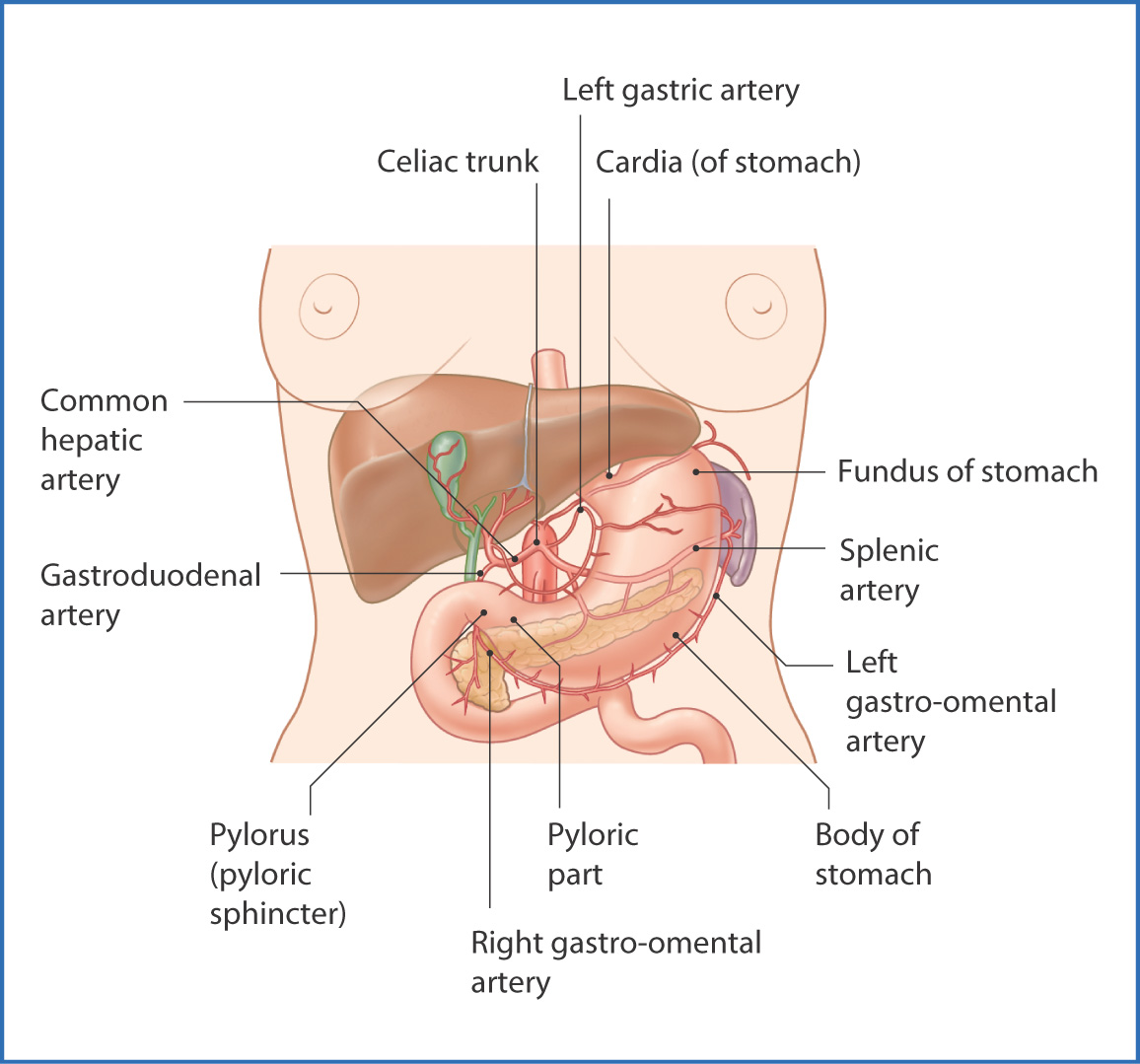
FIGURE 33.3 Stomach.
The gastric cardia is the part of the stomach that receives swallowed food. It contains the esophagogastric sphincter (see earlier). The second part of the stomach is the domed fundus of the stomach; when seen on an upright chest radiograph it usually contains gas. The body of the stomach is the longest part of the stomach and where most of the gastric digestion of proteins and starches occurs. It is rich in glands that produce hydrochloric acid, which facilitates the breakdown of food products. The pyloric part is the distal, funnel-like part of the stomach. Its proximal part, the pyloric antrum, leads into the narrow pyloric canal and pyloric sphincter, which controls the release of stomach contents into the duodenum (small intestine).
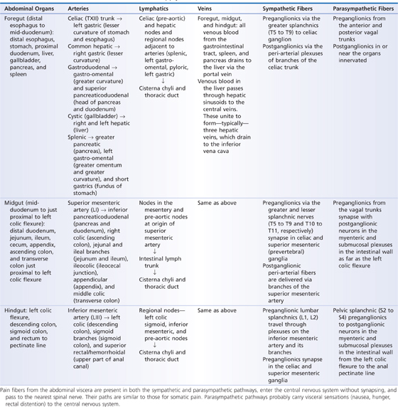
Sensory and autonomic (parasympathetic and sympathetic) innervation of the stomach is provided by the celiac plexus of nerves near the origin of the celiac artery. Blood is supplied by the celiac trunk and its branches—the left gastric, splenic, and common hepatic arteries. Venous drainage of the stomach is to the gastric and gastro-omental veins. Lymphatic drainage is to the celiac nodes and also lymph node groups adjacent to the spleen and pancreas (Table 33.1).
TABLE 33.1 Gastrointestinal Tract: Neurovascular Supply
Small Intestine
The small intestine (small bowel) is the longest part of the GI tract (usually around 5.5 m long) and comprises three major regions—the duodenum, jejunum, and ileum. It joins the stomach at the pyloric sphincter and terminally joins the large intestine at the ileocecal fold or valve (Fig. 33.4).
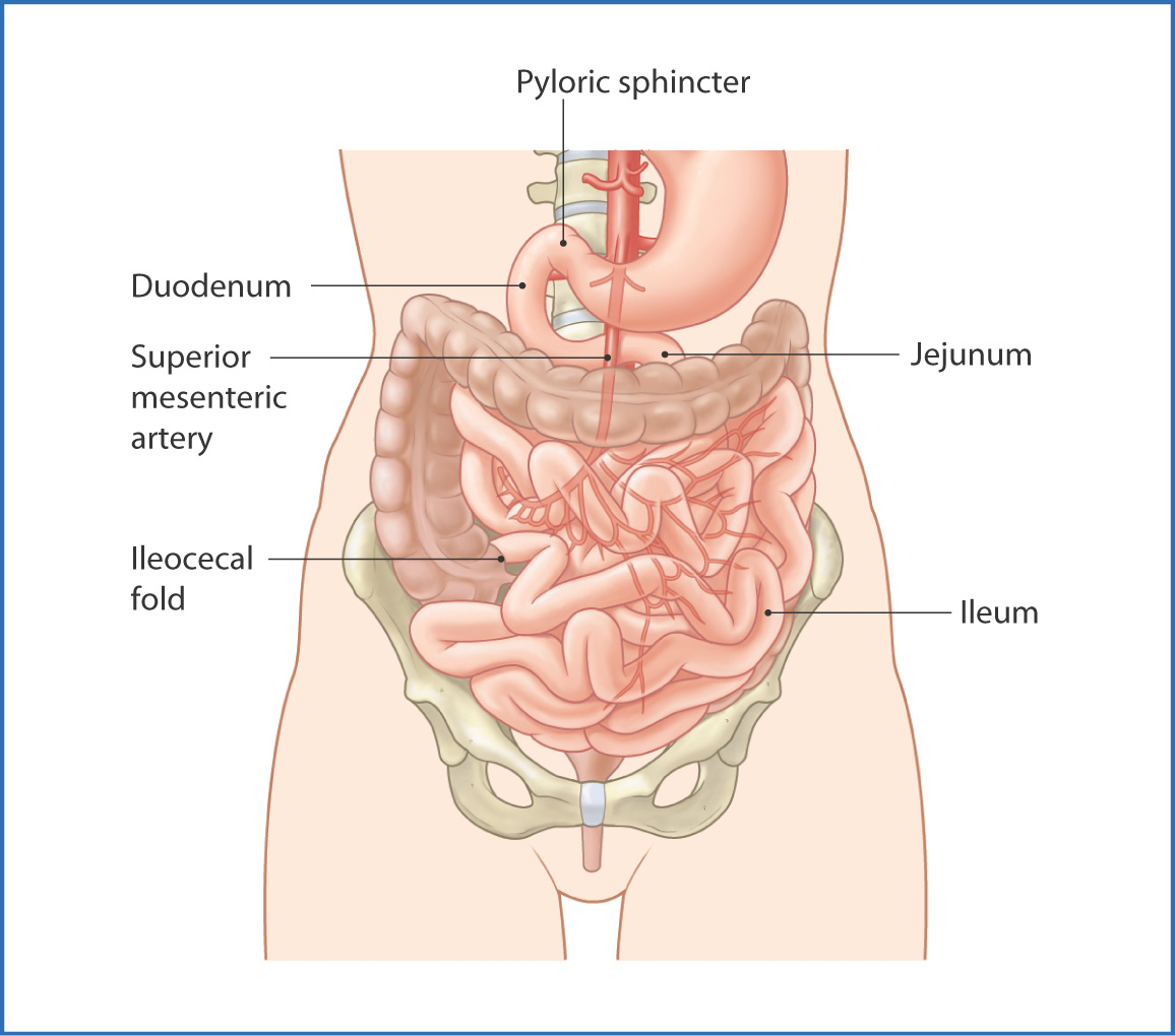
FIGURE 33.4 Small intestine.
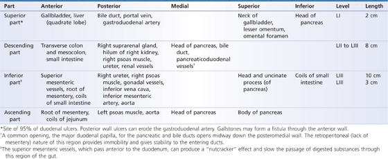
The short duodenum is the first part of the small intestine and has four parts—superior, descending, inferior, and ascending. The middle half is retroperitoneal (descending and inferior parts). The duodenum is approximately 25 to 27 cm long and encircles the head of the pancreas. It is in the epigastric and umbilical regions of the abdomen at vertebral levels LI to LIII just anterior to the inferior vena cava, aorta, and right kidney.
The superior part of the duodenum passes toward the right side of the abdomen from the pylorus. The descending part passes inferiorly to the right of vertebra LII. The hepatic and pancreatic ducts open into the duodenal lumen at the midpoint of the descending section through the major duodenal papilla and minor duodenal papilla. This is important clinically because damage to or pathology involving this part of the duodenum prevents hepatic and pancreatic secretions from entering the digestive system and thus inhibits digestion.
The inferior part of the duodenum then passes to the left and crosses the inferior vena cava and aorta before curving superiorly to become the ascending part. The duodenum terminates at the duodenojejunal flexure, where it joins the jejunum. The duodenojejunal flexure is suspended from the abdominal wall by the suspensory ligament of the duodenum. Abnormalities in embryologic rotation of the gut may occur at this site.
The duodenum is innervated by autonomic nerves from the celiac plexus. Its blood supply is provided by branches of the celiac trunk and superior mesenteric arteries (Table 33.2). Venous drainage is to the hepatic portal system through veins that follow the route of the arteries. Lymphatic drainage is to the lymph node group near the head of the pancreas.
TABLE 33.2 Relationships of the Duodenum
The second part of the small intestine is the jejunum, which is located in the left hypochondrium of the abdomen. The jejunum accounts for approximately 2 m of the small intestine and is held in place by a posterior mesentery. The jejunum is a location for lipid and protein digestion; primary starch digestion occurs in the oral cavity and stomach. Internally, the jejunum has plicae circulares, which are circular rings created by the musculature within the jejunal walls. The jejunum usually has thicker walls and a larger diameter than the ileum (Table 33.3).
TABLE 33.3 Features of the Jejunum and Ileum
| Jejunum | Ileum |
Length (fraction of combined 3-5 m total) |
|
|
Location | Umbilical region, below left side of transverse mesocolon | Pubic region and groin; usually extends into pelvis |
Diameter | 2-4 cm | 2-3 cm |
Wall | Thick and heavy | Thin and light |
Vessels | More, one or two arcades | Less, three to four arcades |
Vasa recta (small mesenteric vessels) |


